. 2012 Jul 1;26(13):1473-85.
doi: 10.1101/gad.193615.112.

Scc1 sumoylation by Mms21 promotes sister chromatid recombination through counteracting Wapl

Affiliations

Scc1 sumoylation by Mms21 promotes sister chromatid recombination through counteracting Wapl

Nan Wu et al. Genes Dev. .

Abstract

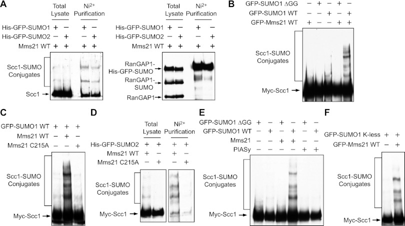
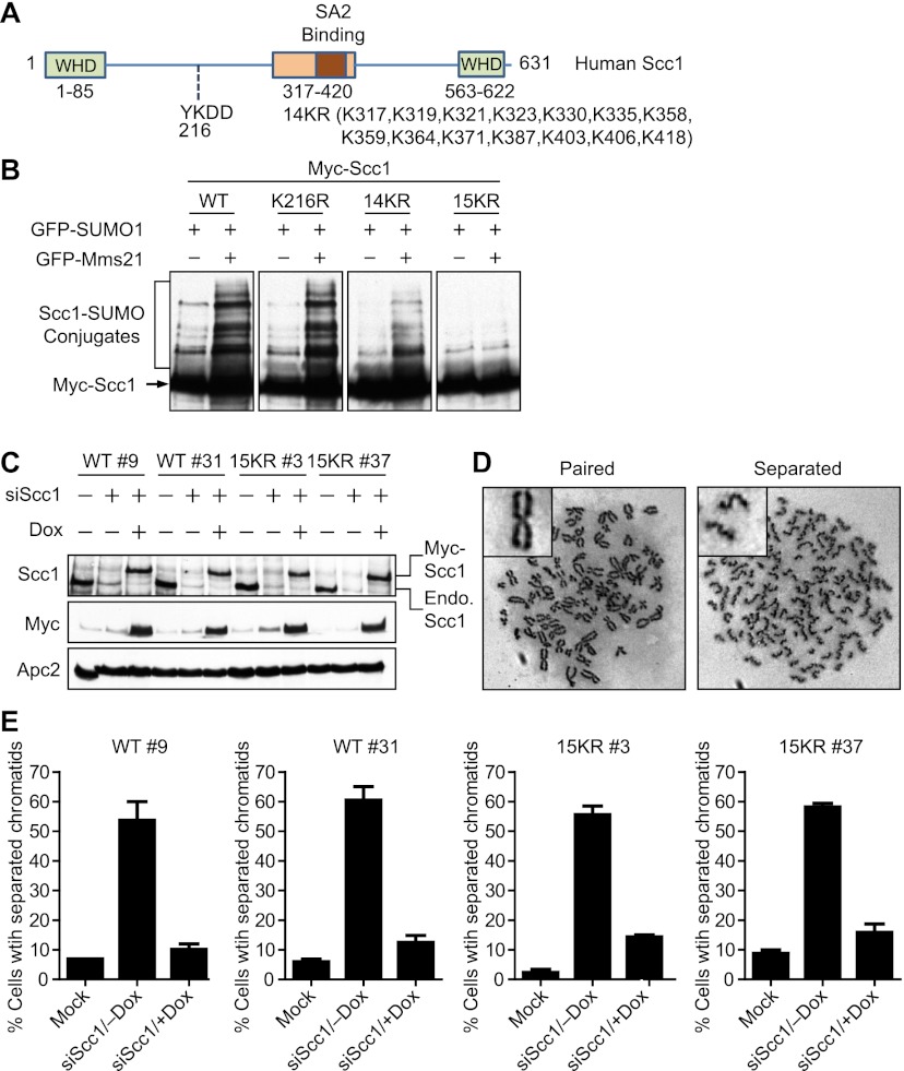
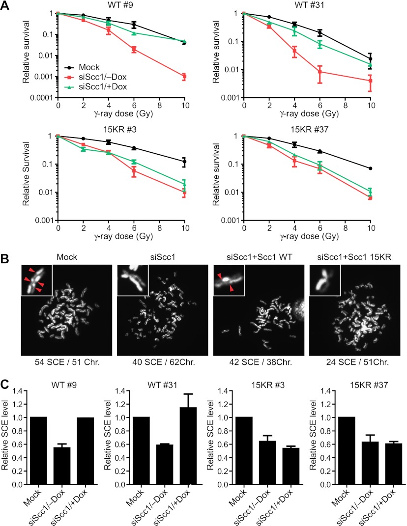
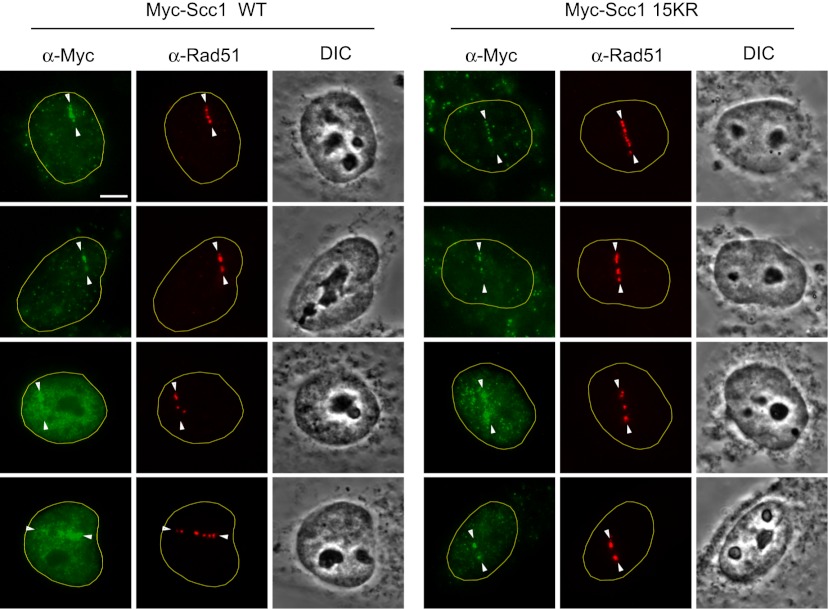
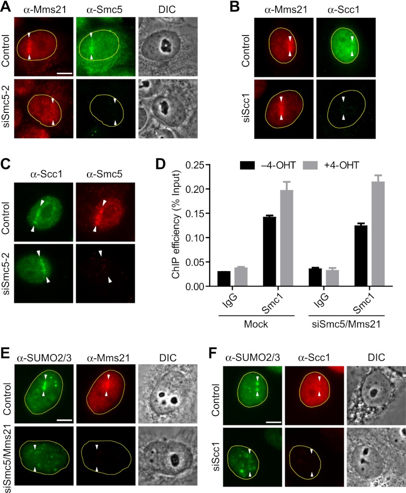
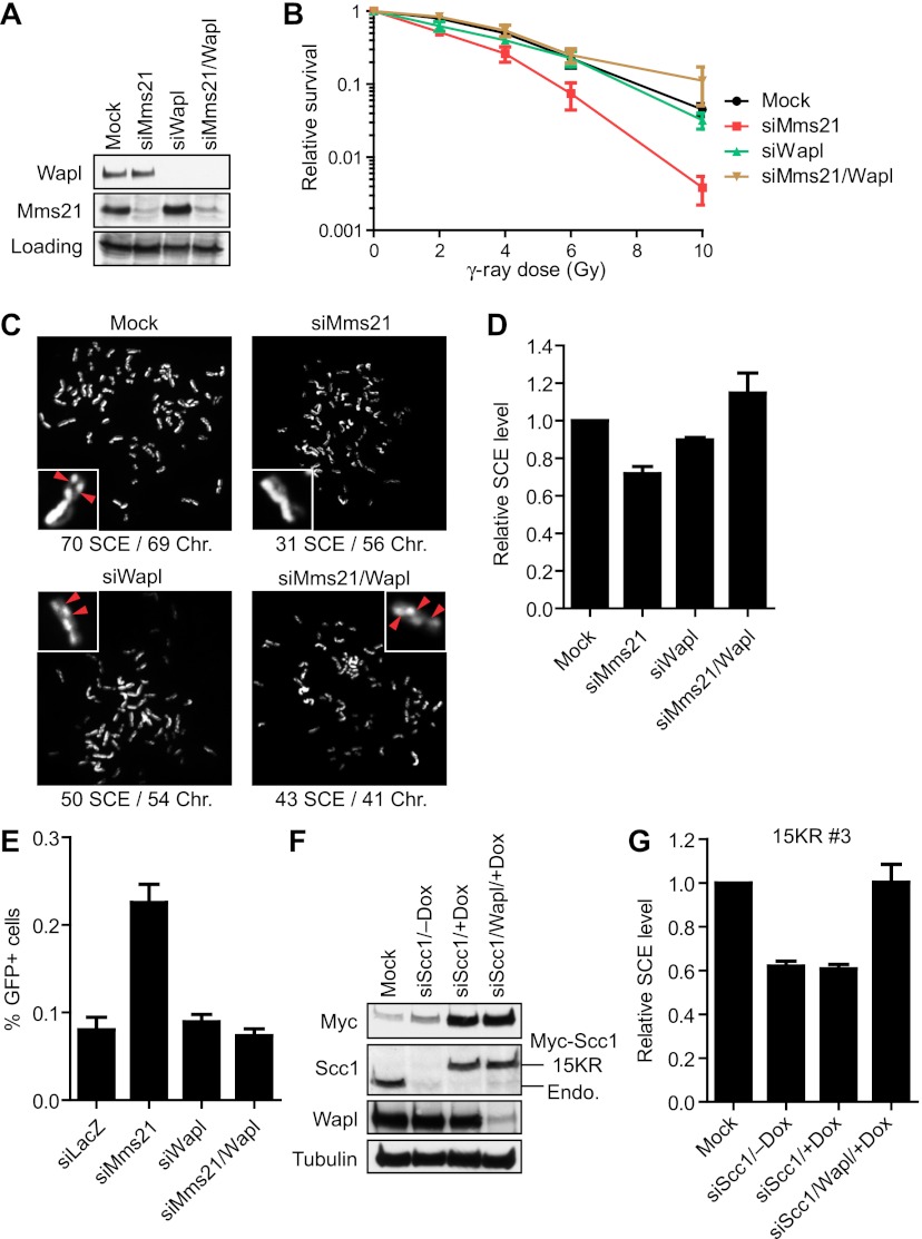
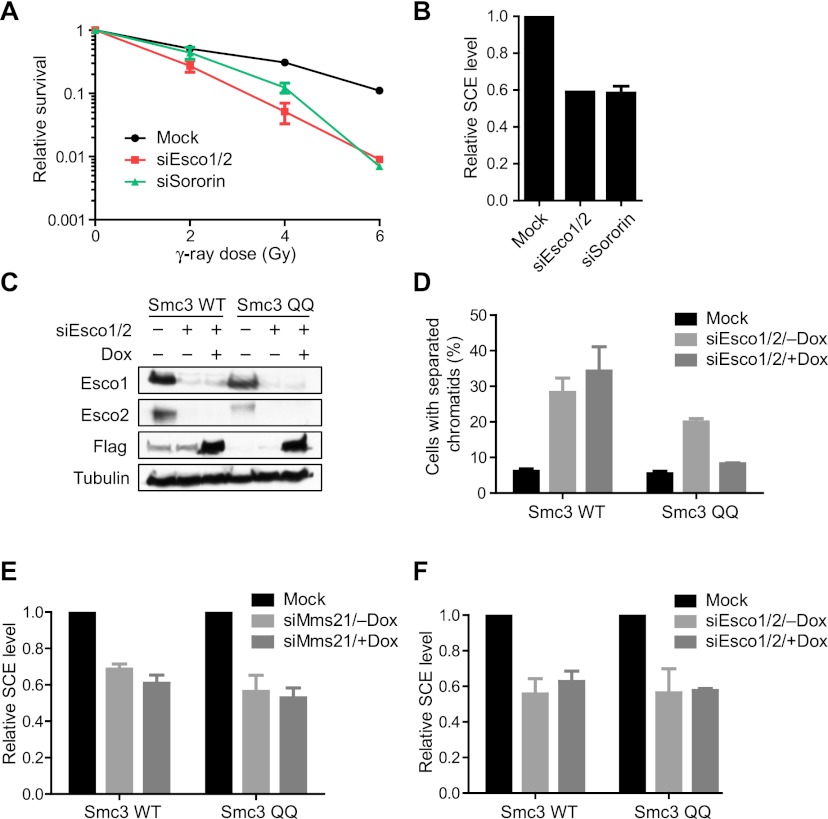
DNA double-strand breaks (DSBs) fuel cancer-driving chromosome translocations. Two related structural maintenance of chromosomes (Smc) complexes, cohesin and Smc5/6, promote DSB repair through sister chromatid homologous recombination (SCR). Here we show that the Smc5/6 subunit Mms21 sumoylates multiple lysines of the cohesin subunit Scc1. Mms21 promotes cohesin-dependent small ubiquitin-like modifier (SUMO) accumulation at laser-induced DNA damage sites in S/G2 human cells. Cells expressing the nonsumoylatable Scc1 mutant (15KR) maintain sister chromatid cohesion during mitosis but are defective in SCR and sensitive to ionizing radiation (IR). Scc1 15KR is recruited to DNA damage sites. Depletion of Wapl, a negative cohesin regulator, rescues SCR defects of Mms21-deficient or Scc1 15KR-expressing cells. Expression of the acetylation-mimicking Smc3 mutant does not bypass the requirement for Mms21 in SCR. We propose that Scc1 sumoylation by Mms21 promotes SCR by antagonizing Wapl at a step after cohesin loading at DSBs and in a way not solely dependent on Smc3 acetylation.


Figures

Figure 1.
Figure 1.
Mms21 stimulates cohesin sumoylation in human cells. (A) HeLa Tet-On cells transfected with Mms21 and His6-GFP-SUMO1/2 plasmids were lysed with a protein-denaturing buffer and subjected to Ni2+ bead pull-down. The total lysates and pull-down were blotted with α-Scc1 (left panel) or α-RanGAP1 (right panel). (B) Mms21 enhances Scc1 sumoylation in human cells. Myc-Scc1 was coexpressed with GFP-SUMO1 wild type (WT) or ΔGG in the presence or absence of GFP-Mms21 in HeLa cells for 24 h. The cell lysates were blotted with α-Myc. The positions of unmodified and sumoylated Scc1 are labeled. (C) The ligase activity of Mms21 is required for Scc1 sumoylation. Lysates of HeLa cells transfected with the indicated plasmids were blotted with α-Myc. The C215A mutation abolished the ligase activity of Mms21. (D) HeLa Tet-On cells transfected with Myc-Scc1, Mms21 wild type/C215A, and His6-GFP-SUMO2 plasmids were lysed with a protein-denaturing buffer and subjected to Ni2+ bead pull-down. The total lysates (left panel) and pull-down (right panel) were blotted with α-Myc. (E) Lysates of HeLa Tet-On cells transfected with the indicated plasmids were blotted with α-Myc. The positions of sumoylated Scc1 are labeled. (F) Mms21 sumoylates multiple lysines of Scc1. HeLa cells were transfected with a plasmid encoding GFP-SUMO1 K-less (the lysine-less SUMO1 mutant that cannot form chains) in the presence or absence of the GFP-Mms21 plasmid. Lysates were blotted with α-Myc.
Figure 2.
Figure 2.
The nonsumoylatable Scc1 15KR mutant is functional in mitotic cohesion. (A) Schematic drawing of the domains and motifs of human Scc1. The positions of two winged helix domains (WHD) required for Smc1/3 binding, the central conserved domain that binds to SA, and a consensus sumoylation motif at K216 are indicated. The Scc1 15KR mutations contain the K216R mutation and mutations of all 14 lysines in the central region (residues 317–420) to arginines (14KR). (B) Myc-Scc1 wild type (WT) and mutants were expressed together with GFP-SUMO1 in the presence or absence of GFP-Mms21 in HeLa cells. The cell lysates were blotted with α-Myc. (C) Construction of stable HeLa Tet-On cell lines that inducibly expressed Myc-Scc1 wild type or 15KR. Two clones of each cell line cultured with or without doxycycline (Dox) were either mock-transfected or transfected with siScc1. Cell lysates were blotted with the indicated antibodies. The positions of Myc-Scc1 or endogenous (Endo.) Scc1 are labeled. (D) Representative metaphase spreads of cells in C with paired (left panel) or separated (right panel) sister chromatids. (E) Quantification of the percentage of mitotic cells in C with separated sister chromatids. More than 200 cells in each sample were counted. The mean and standard deviation of data in two independent experiments are shown.
Figure 3.
Figure 3.
Cells expressing Scc1 15KR are sensitive to IR and defective in SCR. (A) Two clones each of HeLa Tet-On cell lines that inducibly expressed Myc-Scc1 wild type (WT) or 15KR were cultured with or without doxycycline (Dox), mock-transfected or transfected with siScc1, and irradiated with different doses of IR. The relative colony survival numbers are plotted against IR doses. Each data point represents the mean and standard deviation of values in two independent experiments, with duplicate samples in each experiment. The differences in the IR sensitivity seen in different clones of the Myc-Scc1-expressing lines after Scc1 RNAi are likely due to clonal variation. (B) Representative images of the SCE assays on cells in A. The numbers of SCE events and chromosomes in each image are shown below. A pair of sister chromatids for each image is magnified and shown in the inset, with the SCE events marked by red arrowheads. (C) Quantification of the relative SCE levels of cells described in A. The mean and standard deviation of data from two experiments are shown. About 30 cells were counted in each experiment.
Figure 4.
Figure 4.
Scc1 15KR is recruited to laser-induced DNA damage sites. HeLa Tet-On cells expressing Myc-Scc1 wild type (WT) or 15KR were cultured in the presence of doxycycline, synchronized at S/G2 by thymidine arrest and release, and subjected to laser irradiation along a straight line. At 1 h after laser cutting, cells were fixed and stained with α-Myc (green) and α-Rad51 (red). (DIC) Differential interference contrast. We irradiated 20 Myc-Scc1 wild-type-expressing and 20 Myc-Scc1 15KR-expressing cells. Among them, 16 Myc-Scc1 wild-type and 15 Myc-Scc1 15KR cells were Myc-positive. All Myc-positive cells in both groups exhibited enrichment of Myc signals at DNA damage sites, with 12 of 16 in the wild-type group and 11 of 15 in the 15KR group showing strong recruitment of Myc-Scc1 proteins to damage sites. Four representative cells of each cell line are shown. Bar, 10 μm.
Figure 5.
Figure 5.
The Smc5/6 complex is dispensable for cohesin loading at DNA damage sites. (A) HeLa cells transfected with siControl or siSmc5-2 were synchronized at S/G2 by thymidine arrest and release and subjected to laser irradiation along a straight line. At 1 h after laser cutting, cells were fixed and stained with α-Smc5 (green) and α-Mms21 (red). (DIC) Differential interference contrast. Bar, 10 μm. (B) HeLa cells transfected with siControl or siScc1 were synchronized at S/G2 and subjected to laser irradiation along a straight line. At 1 h after laser cutting, cells were fixed and stained with α-Scc1 (green) and α-Mms21 (red). (C) HeLa cells transfected with siControl or siSmc5-2 were synchronized at S/G2 by thymidine arrest and release and subjected to laser irradiation along a straight line. At 1 h after laser cutting, cells were fixed and stained with α-Scc1 (green) and α-Smc5 (red). (D) HeLa Tet-On cells were transfected with siControl or siSmc5-2/siMms21-6 and then transfected with the ER-I-PpoI plasmid. Cells were then treated with or without 4-OHT for 12 h and subjected to ChIP by IgG and α-Smc1 followed by qPCR analysis. The mean and standard deviation of two independent experiments are shown. (E) HeLa cells transfected with siControl or siSmc5-2/siMms21-6 were synchronized at S/G2 and subjected to laser irradiation along a straight line. At 1 h after laser cutting, cells were fixed and stained with α-SUMO2/3 (green) and α-Mms21 (red). (DIC) Differential interference contrast. Bar, 10 μm. (F) HeLa cells transfected with siControl or siScc1 were synchronized at S/G2 and subjected to laser irradiation along a straight line. At 1 h after laser cutting, cells were fixed and stained with α-SUMO2/3 (green) and α-Scc1 (red). (DIC) Differential interference contrast. Bar, 10 μm. Note that the Scc1 accumulation was not as prominent as in C because a different Scc1 antibody was used for this experiment to allow the costaining of SUMO2/3 and Scc1.
Figure 6.
Figure 6.
Wapl depletion rescues IR sensitivity and SCR defects of Mms21-RNAi or Scc1 15KR-expressing cells. (A) Lysates of HeLa cells transfected with the indicated siRNAs were blotted with α-Wapl and α-Mms21. A cross-reacting band in the α-Mms21 blot served as the loading control. (B) IR colony survival assay of cells described in A. Each data point represents the mean and standard deviation of values in three independent experiments, with duplicate samples in each experiment. (C) Representative images from the SCE assay on cells in A. The numbers of SCE events and chromosomes in each image are shown below. A pair of sister chromatids for each image is magnified and shown in the inset, with the SCE events marked by red arrowheads. (D) Quantification of the relative SCE levels of cells in A. The mean and standard deviation of data from three experiments are shown. More than 30 cells were counted in each experiment. (E) Quantification of the normalized percentage of GFP-positive cells in the I-SceI-based gene targeting assay of 293/A658 cells transfected with the indicated siRNAs. The cells were cotransfected with siRNAs and the repair plasmid. Three days later, cells were harvested and analyzed by FACS. (F) HeLa Tet-On cells expressing Myc-Scc1 15 KR (clone 3) cultured in the absence or presence of doxycycline (Dox) were transfected with the indicated siRNAs. Lysates were blotted with the indicated antibodies. The positions of Myc-Scc1 and the endogenous (Endo.) Scc1 are labeled. (G) Quantification of the relative SCE levels of cells described in F. The mean and standard deviation of data from two experiments are shown. About 30 cells were counted in each experiment.
Figure 7.
Figure 7.
Expression of the Smc3 acetylation-mimicking mutant does not bypass the requirement for Mms21 in SCR. (A) IR colony survival assay of HeLa cells transfected with the indicated siRNAs. Each data point represents the mean and standard deviation of values in two independent experiments, with duplicate samples in each experiment. (B) Quantification of the relative SCE levels of cells in A. The mean and standard deviation of data from two experiments are shown. About 30 cells were counted in each experiment. (C) 293T cell lines inducibly expressing Flag-Smc3 wild type (WT) or K105Q/K106Q (QQ) were cultured in the absence or presence of doxycycline (Dox) and then either mock-transfected or transfected with siEsco1/2. Lysates were blotted with the indicated antibodies. (D) Quantification of the percentage of mitotic cells in C with separated sister chromatids. The mean and standard deviation of three independent experiments are shown. (E) 293T cell lines inducibly expressing Flag-Smc3 wild type or K105Q/K106Q (QQ) were cultured in the absence or presence of doxycycline (Dox) and then either mock-transfected or transfected with siMms21. The relative SCE levels of these cells were quantified. The mean and standard deviation of data from two experiments are shown. About 30 cells were counted in each experiment. (F) Quantification of the relative SCE levels of cells in C. The mean and standard deviation of data from two experiments are shown. About 30 cells were counted in each experiment.

Similar articles

Cited by

References

    1. Behlke-Steinert S, Touat-Todeschini L, Skoufias DA, Margolis RL 2009. SMC5 and MMS21 are required for chromosome cohesion and mitotic progression. Cell Cycle 8: 2211–2218 - PubMed
    1. Berkovich E, Monnat RJ Jr, Kastan MB 2007. Roles of ATM and NBS1 in chromatin structure modulation and DNA double-strand break repair. Nat Cell Biol 9: 683–690 - PubMed
    1. Berkovich E, Monnat RJ Jr, Kastan MB 2008. Assessment of protein dynamics and DNA repair following generation of DNA double-strand breaks at defined genomic sites. Nat Protoc 3: 915–922 - PubMed
    1. De Piccoli G, Cortes-Ledesma F, Ira G, Torres-Rosell J, Uhle S, Farmer S, Hwang JY, Machin F, Ceschia A, McAleenan A, et al. 2006. Smc5–Smc6 mediate DNA double-strand-break repair by promoting sister-chromatid recombination. Nat Cell Biol 8: 1032–1034 - PMC - PubMed
    1. De Piccoli G, Torres-Rosell J, Aragon L 2009. The unnamed complex: What do we know about Smc5–Smc6? Chromosome Res 17: 251–263 - PubMed

Publication types

MeSH terms