. 2012 Sep;32(17):3500-12.
doi: 10.1128/MCB.00298-12. Epub 2012 Jul 2.

Loss of the androgen receptor cofactor p44/WDR77 induces astrogliosis

Affiliations

Loss of the androgen receptor cofactor p44/WDR77 induces astrogliosis

Bryce Vincent et al. Mol Cell Biol. 2012 Sep.

Abstract

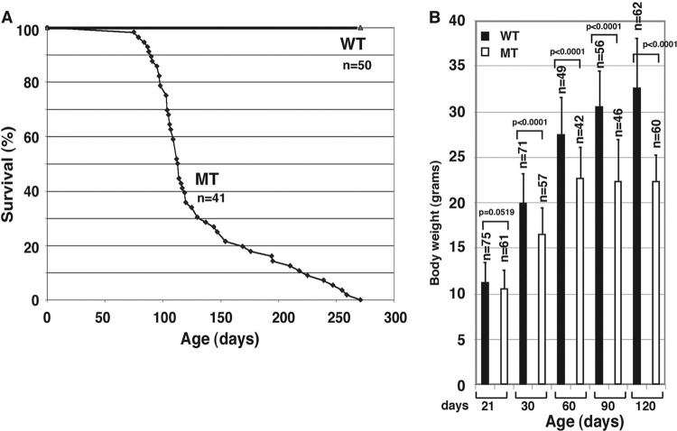
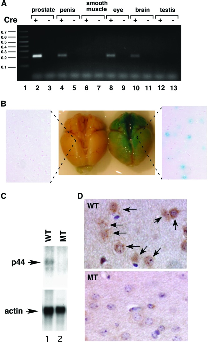
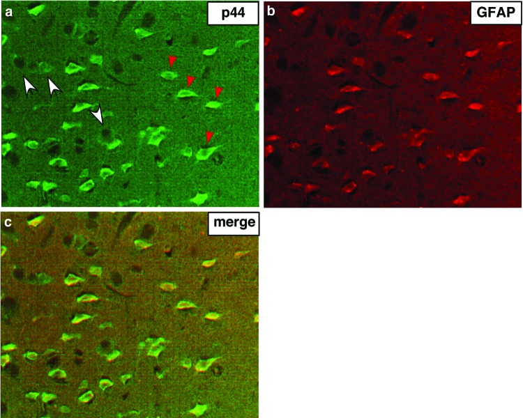
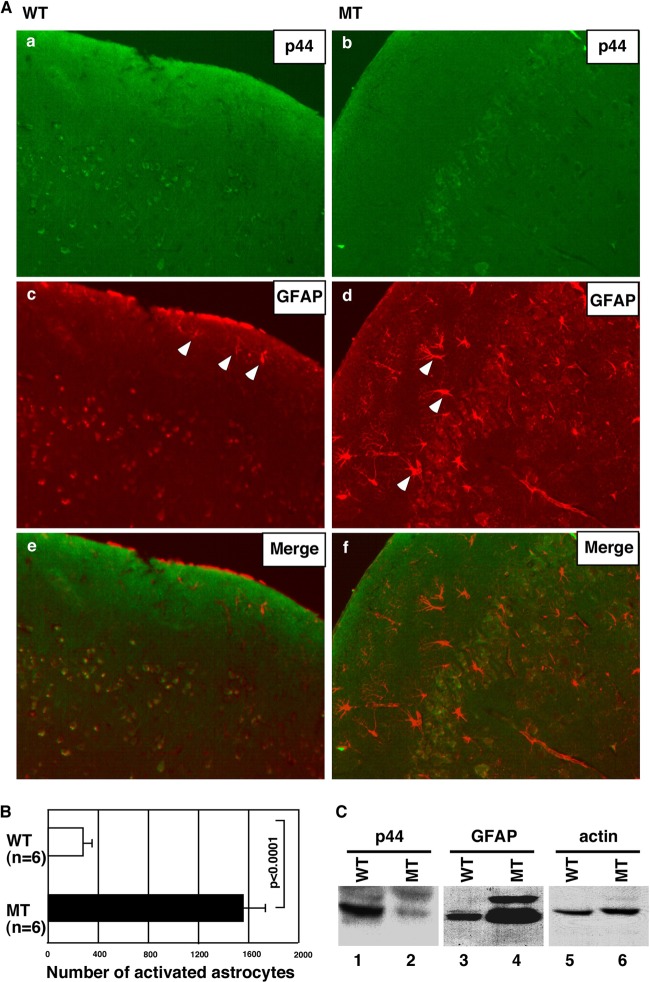
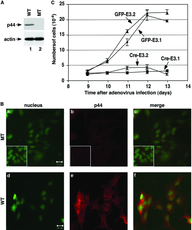
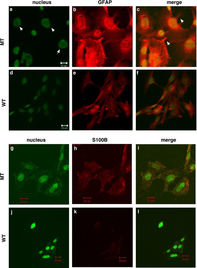
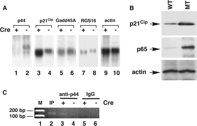
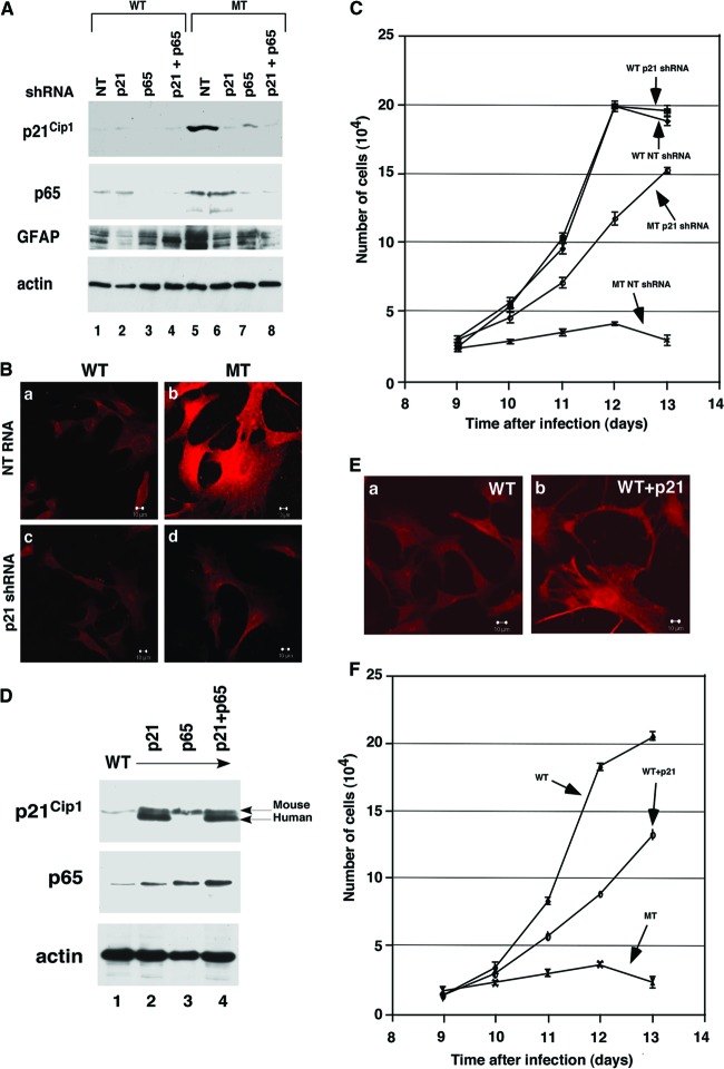
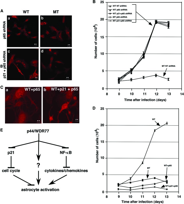
Astrogliosis is induced by neuronal damage and is also a pathological feature of the major aging-related neurodegenerative disorders. The mechanisms that control the cascade of astrogliosis have not been well established. In a previous study, we identified a novel androgen receptor (AR)-interacting protein, p44/WDR77, that plays a critical role in the proliferation and differentiation of prostate epithelial cells. In the present study, we found that deletion of the p44/WDR77 gene caused premature death with dramatic astrogliosis in mouse brain. We further found that p44/WDR77 is expressed in astrocytes and that loss of p44/WDR77 expression in astrocytes leads to growth arrest and astrogliosis. The astrocyte activation induced by deletion of the p44/WDR77 gene was associated with upregulation of p21(Cip1) expression and NF-κB activation. Silencing p21(Cip1) or NF-κB p65 expression with short hairpin RNA (shRNA) abolished astrocyte activation and rescued the astrocyte growth inhibition induced by deletion of the p44/WDR77 gene. Our results reveal a novel role for p44/WDR77 in the control of astrocyte activation through p21(Cip1) and NF-κB signaling.


Figures

Fig 1
Fig 1
Deletion of the p44/WDR77 gene leads to decreased life span in mice. (A) Survival data are presented for p44/WDR77loxP/loxP; Cre (MT) and p44/WDR77loxP/loxP (WT) mice. The median life span was 120 days for MT mice. (B) Comparison of mean body weights of WT and MT mice. The error bars indicate SEM.
Fig 2
Fig 2
The p44/WDR77 gene was deleted in the mouse brain. (A) PCR analysis of deletion of the p44/WDR77 gene in various organs in the MT mouse. Genomic DNA was isolated from organs of WT and MT mice and subjected to PCR analysis. Excision of exons 2 to 5 of the p44/WDR77 gene generated the 220-bp DNA fragment in the PCR. Lane 1, 0.1-kb DNA ladder. (B) Expression of Cre recombinase in the brains of ARR2PPbi-Cre transgenic mice. The ARR2PPbi-Cre transgenic mouse was crossed with the B6; 129-Gt(ROSA)26Sor/J mouse. Brains from 2-month-old B6; 129-Gt(ROSA)26Sor/J (left) or B6; 129-Gt(ROSA)26Sor/J-ARR2PPbi-Cre (right) mice were stained with β-Gal (middle) and then formalin fixed and paraffin embedded. The slides were counterstained with eosin (left and right). (C) Loss of p44/WDR77 gene expression in the MT mouse brain. Shown is Northern blot analysis of p44/WDR77 and β-actin mRNAs in the mouse brain. mRNA was isolated from the brains of 2-month-old WT (n = 5; lane 1) or MT (n = 5; lane 2) mice, fractionated by electrophoresis, and transferred to a Hybond N+ membrane. The membrane was hybridized with probes as indicated. (D) Loss of the p44/WDR77 protein in the brain of the MT mouse. Tissues from the cerebral cortices of WT (top) and MT (bottom) mouse brains were immunostained with anti-p44/WDR77 antibody. Representative photomicrographs are shown. The p44/WDR77 staining seen in WT mouse brain cells (indicated by the arrows) was dramatically decreased in MT mouse brain cells.
Fig 3
Fig 3
p44/WDR77 expression in astrocytes of the mouse brain. (a and b) Tissues from cerebral cortices of WT mouse brains were double immunostained for p44/WDR77 (a) and GFAP (b). (c) p44/WDR77 staining merged with GFAP staining.
Fig 4
Fig 4
Loss of the p44/WDR77 gene leads to reactive astrocytes in the mouse brain. (A) Reactive astrocytes were observed in the MT mouse brain. Sections at the 2.00-mm lateral plane of cerebral cortices from WT (left) or MT (right) brains were stained with anti-p44/WDR77 (top) or anti-GFAP (middle) antibodies. (Bottom) p44/WDR77 staining merged with GFAP staining. Some reactive astrocytes are indicated by arrowheads. (B) Counts of reactive astrocytes in WT and MT brains. The error bars indicate SEM. (C) Loss of p44/WDR77 expression in astrocytes isolated from MT mouse brains. Whole-cell lysates (20 μg of protein) were prepared from WT (lanes 1, 3, and 5) and MT (lanes 2, 4, and 6) astrocytes. Western blot analysis was performed with the indicated antibodies.
Fig 5
Fig 5
p44/WDR77 is essential for growth of astrocytes. (A and B) Loss of p44/WDR77 expression in p44/WDR77-null astrocytes. (A) Whole-cell lysates (10 μg of protein) were isolated from WT (lane 1) and MT (lane 2) astrocytes. Western blot analysis was performed with the indicated antibodies. (B) Astrocytes were infected with Ad-GFP (WT) or Ad-Cre-GFP (MT) and stained for the nucleus (a and d) and p44/WDR77 expression (b and e). (c and f) p44/WDR77 staining merged with nuclear staining. The insets (a to c) depict staining of WT astrocytes without the p44/WDR77 antibody. (C) Growth curves of WT and MT astrocytes after infection with adenovirus. The error bars indicate SEM.
Fig 6
Fig 6
Deletion of the p44/WDR77 gene led to reactive astrocytes. Astrocytes were infected with Ad-GFP (WT) or Ad-Cre-GFP (MT) and stained for the nucleus (a, d, g, and j) and GFAP (b and e) or S100B (h and k). (c, f, i, and l) GFAP or S100B staining merged with nuclear staining. The arrowheads in panel a indicate hypertrophy of cell nuclei, and those in panel c indicate thin processes protruding from the cell body.
Fig 7
Fig 7
Loss of the p44/WDR77 gene induced p21Cip1 expression and NF-κB activation. (A) Loss of the p44/WDR77 gene induced p21Cip1 expression in mouse brains. Shown is Northern blot analysis of p44/WDR77, p21Cip1, Gadd45A, RGS16, and β-actin mRNAs in mouse brains. mRNA was isolated from the brains of 2-month-old WT (n = 5; lanes 2, 4, 6, 8, and 10) or MT (n = 5; lanes 1, 3, 5, 7, and 9) mice, fractionated by electrophoresis, and transferred to a Hybond N+ membrane. The membrane was hybridized with probes as indicated. (B) Loss of the p44/WDR77 gene induced p21Cip1 and p65 expression in astrocytes. Whole-cell lysates (20 μg of protein) were prepared from WT (lane WT) and MT (lane MT) astrocytes. Western blot analysis was performed with anti-p21Cip1 (top), anti-p65 (middle), or antiactin (bottom) antibody. (C) Chromatin immunoprecipitation assay to identify p21Cip1 as a direct target gene of p44. Immunoprecipitation was performed with antigen-purified anti-p44 (lanes 3 and 4) antibodies or rabbit IgG (lanes 5 and 6). The purified DNA was amplified by PCR with two specific primers derived from the promoter region (bp −93 to −215) of p21Cip1. The same set of PCRs was performed with chromatin DNA used for immunoprecipitation (IP) (lane 2).
Fig 8
Fig 8
p21Cip1 mediated the astrocyte activation induced by loss of p44/WDR77. (A) Silencing p21Cip1 and/or p65 expression in WT and MT astrocytes. Whole-cell lysates (20 μg of protein) were prepared from WT (lanes 1 to 4) and MT (lanes 5 to 8) astrocytes expressing NT (lanes 1 and 5), p21Cip1 (lanes 2 and 6), p65 (lanes 3 and 7), or p21Cip1 plus p65 (lanes 4 and 8) shRNA. Western blot analysis was performed with the indicated antibodies. (B) Silencing p21Cip1 expression abolished the astrocyte activation induced by loss of p44/WDR77. WT (left) and MT (right) astrocytes expressing NT (top) or p21Cip1 (bottom) shRNA were stained for GFAP. (C) Growth curves of WT and MT astrocytes expressing NT or p21Cip1 shRNA. The error bars indicate SEM. (D) Shown is overexpression of p21Cip1 and/or p65 in astrocytes. Whole-cell lysates (20 μg of protein) were prepared from control astrocytes (lane 1) or astrocytes expressing p21Cip1 (lane 2), p65 (lane 3), or p21Cip1 plus p65 (lane 4). Western blot analysis was performed with the indicated antibodies. Endogenous (mouse) and exogenous (human) p21Cip1 are indicated by the arrows on the right. (E) Overexpression of p21Cip1 induced astrocyte activation. Control astrocytes (a) and astrocytes expressing p21Cip1 (b) were stained for GFAP. (F) Growth curves of control astrocytes or astrocytes expressing p21Cip1.
Fig 9
Fig 9
NF-κB mediated the astrocyte activation induced by loss of p44/WDR77. (A) Silencing p65 expression abolished the astrocyte activation induced by loss of p44/WDR77. WT (left) and MT (right) astrocytes expressing p65 (top) or p21Cip1 plus p65 (bottom) shRNA were stained for GFAP. (B) Growth curves of WT and MT astrocytes expressing NT, p65, or p21Cip1 plus p65 shRNA. The error bars indicate SEM. (C) Overexpression of p65 or p21Cip1 plus p65 induced astrocyte activation. Astrocytes expressing p65 (a) or p21Cip1 plus p65 (b) were stained for GFAP. (D) Growth curves of control astrocytes or astrocytes expressing p65 or p21Cip1 plus p65. (E) Model of how p44/WDR77 induces astrocyte activation by inhibiting p21Cip1, NF-κB, and other (unknown [?]) signals.

Similar articles

Cited by

References

    1. Altuwaijri S, et al. 2003. Interruption of nuclear factor kappaB signaling by the androgen receptor facilitates 12-O-tetradecanoylphorbolacetate-induced apoptosis in androgen-sensitive prostate cancer LNCaP cells. Cancer Res. 63:7106–7112 - PubMed
    1. Bancroft J. 2005. The endocrinology of sexual arousal. J. Endocrinol. 186:411–427 - PubMed
    1. Barreto G, Veiga S, Azcoitia I, Garcia-Segura LM, Garcia-Ovejero D. 2007. Testosterone decreases reactive astroglia and reactive microglia after brain injury in male rats: role of its metabolites, oestradiol and dihydrotestosterone. Eur. J. Neurosci. 25:3039–3046 - PubMed
    1. Besson A, Dowdy SF, Roberts JM. 2008. CDK inhibitors: cell cycle regulators and beyond. Dev. Cell 14:159–169 - PubMed
    1. Coers S, Tanzer L, Jones KJ. 2002. Testosterone treatment attenuates the effects of facial nerve transection on glial fibrillary acidic protein (GFAP) levels in the hamster facial motor nucleus. Metab. Brain Dis. 17:55–63 - PubMed

Publication types

MeSH terms

LinkOut - more resources