. 2012 Sep;32(17):3464-78.
doi: 10.1128/MCB.00125-12. Epub 2012 Jul 2.

Oxidative processing of latent Fas in the endoplasmic reticulum controls the strength of apoptosis

Affiliations

Oxidative processing of latent Fas in the endoplasmic reticulum controls the strength of apoptosis

Vikas Anathy et al. Mol Cell Biol. 2012 Sep.

Abstract

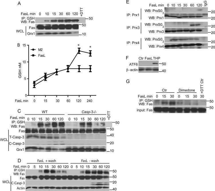
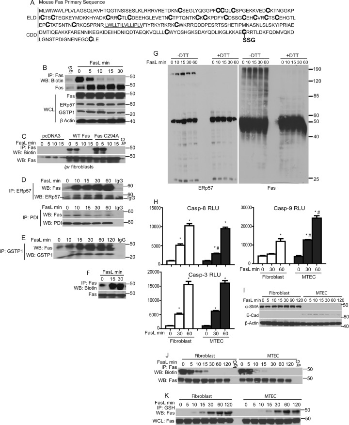
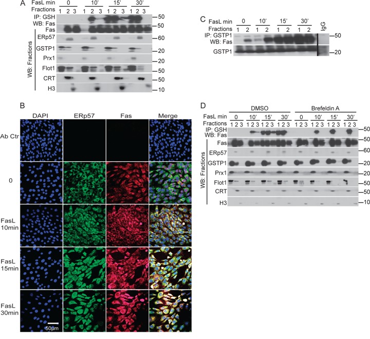
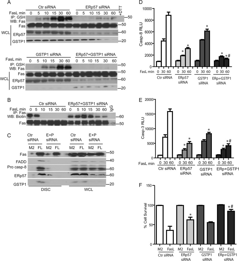
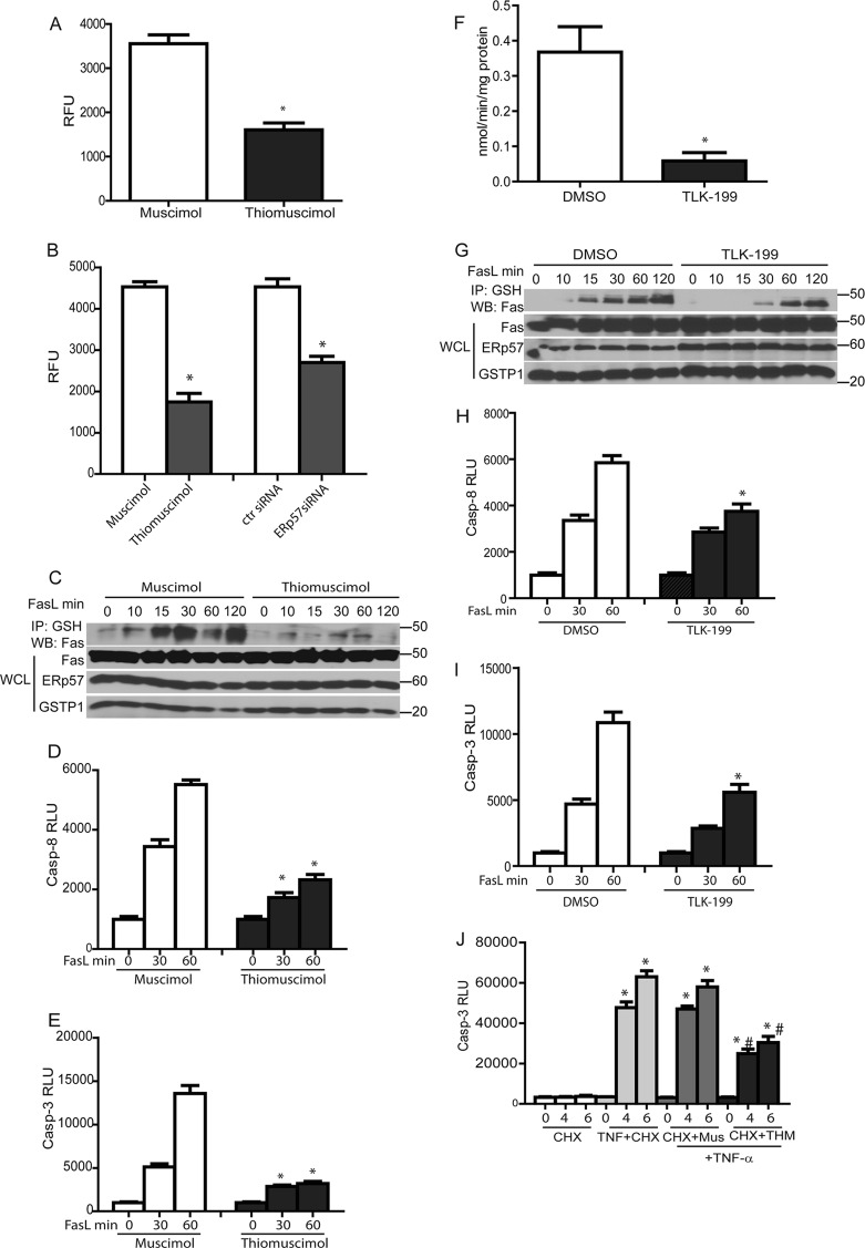
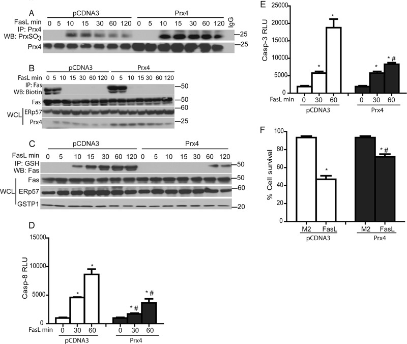
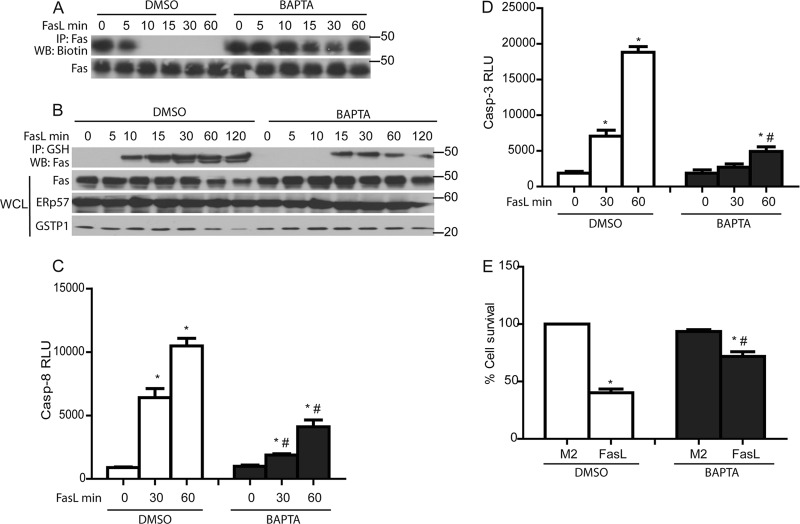
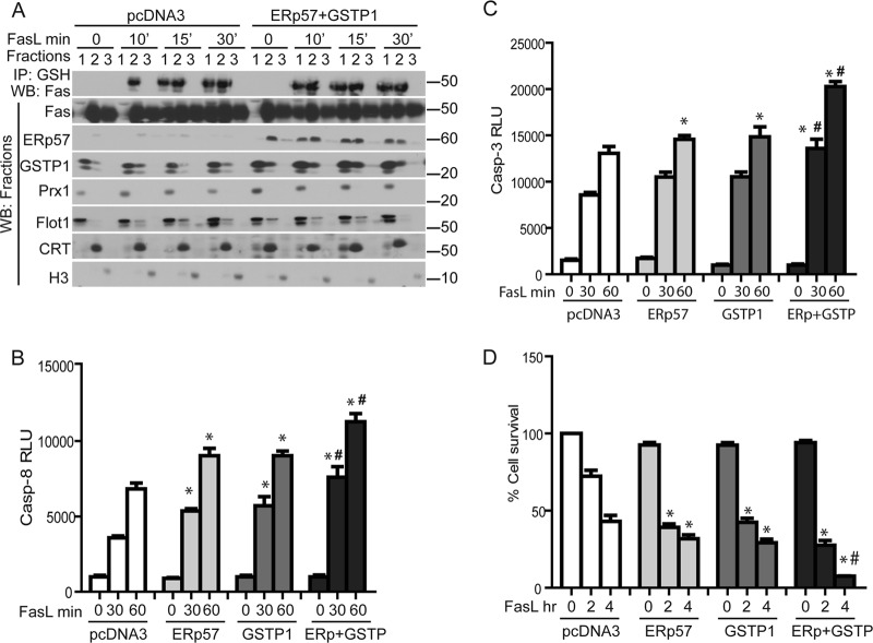
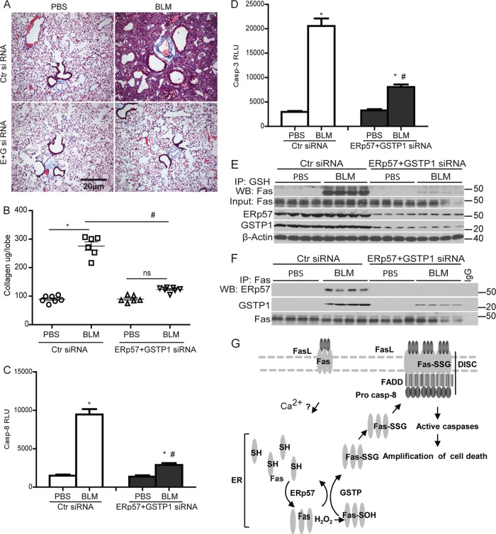
We recently demonstrated that S-glutathionylation of the death receptor Fas (Fas-SSG) amplifies apoptosis (V. Anathy et al., J. Cell Biol. 184:241-252, 2009). In the present study, we demonstrate that distinct pools of Fas exist in cells. Upon ligation of surface Fas, a separate pool of latent Fas in the endoplasmic reticulum (ER) underwent rapid oxidative processing characterized by the loss of free sulfhydryl content (Fas-SH) and resultant increases in S-glutathionylation of Cys294, leading to increases of surface Fas. Stimulation with FasL rapidly induced associations of Fas with ERp57 and glutathione S-transferase π (GSTP), a protein disulfide isomerase and catalyst of S-glutathionylation, respectively, in the ER. Knockdown or inhibition of ERp57 and GSTP1 substantially decreased FasL-induced oxidative processing and S-glutathionylation of Fas, resulting in decreased death-inducing signaling complex formation and caspase activity and enhanced survival. Bleomycin-induced pulmonary fibrosis was accompanied by increased interactions between Fas-ERp57-GSTP1 and S-glutathionylation of Fas. Importantly, fibrosis was largely prevented following short interfering RNA-mediated ablation of ERp57 and GSTP. Collectively, these findings illuminate a regulatory switch, a ligand-initiated oxidative processing of latent Fas, that controls the strength of apoptosis.


Figures

Fig 1
Fig 1
Early increases in Fas S-glutathionylation (Fas-SSG) occur independently of efflux of GSH or caspase activation and are associated with enhanced oxidation in the ER. (A) Rapid S-glutathionylation of Fas in response to FasL. C10 lung epithelial cells were stimulated with Flag-tagged FasL plus anti-Flag cross-linking antibody (M2) or M2 alone (0), and at the indicated times, lysates were prepared and immunoprecipitated (IP) using an anti-GSH antibody. The subsequent Western blot (WB) was probed with anti-Fas antibody (top). The +DTT control (Ctr) reflects lysates prepared from FasL-stimulated cells at 120 min and treated with 25 mM dithiothreitol (DTT) prior to IP with the anti-GSH antibody. Bottom panels show the content of Fas and Grx1 in whole-cell lysates (WCL). (B) FasL induces GSH efflux. C10 lung epithelial cells were stimulated with FasL plus M2 cross-linking antibody or M2 alone, and at the indicated times supernatants were collected. Free GSH was measured. *, P < 0.05 (by ANOVA) compared to M2 controls at the same time points. (C) Early formation of Fas-SSG does not require caspase-3. WT and caspase3−/− lung fibroblasts were treated with FasL as indicated. Lysates were subjected to IP with anti-GSH antibody, and subsequent Western blots were probed with Fas. Bottom panels show contents of Fas, total full-length caspase-3 (T-Casp-3), cleaved caspase-3 (active; C-Casp-3), and Grx1 in WCL. (D) Continuous presence of FasL is needed for sustained Fas-SSG and caspase-3 activation. C10 cells were treated with FasL in the cold for 20 min. FasL was washed away (+wash) or left in the dishes (−wash), and cells were then returned to 37°C for the indicated times. Lysates were processed as described for panel A. (E) Assessment of overoxidation of Prx in response to FasL. Prx1, Prx3, and Prx4 were immunoprecipitated following stimulation with FasL. Western blots were probed for overoxidized Prx (PrxSO3) or with the respective immunoprecipitated Prx proteins as a control. (F) Stimulation of cells with FasL does not induce ER stress. C10 lung epithelial cells were stimulated with FasL plus M2, thapsigargin (THP), or M2 antibody/DMSO as a control (Ctr). Cells were lysed after 4 h. Western blots were probed for the ER stress marker, ATF6, and β-actin as a loading control. (G) A sulfenic acid intermediate (SOH) of Fas precedes its S-glutathionylation. Cells were incubated with the sulfenic acid-trapping agent, dimedone, for 2 h prior to stimulation of cells with FasL. Lysates were subjected to IP as described for panel A. Western blots were probed for Fas. The bottom panel shows input Fas in the WCL.
Fig 2
Fig 2
FasL induces oxidative processing of latent Fas and a rapid interaction between ERp57, GSTP1, and Fas. (A) Primary sequence of murine Fas (NP_032013; www.ncbi.nlm.nih.gov/protein/NP_032013.2), showing cysteines (boldface) in the ligand binding domain predicted to form disulfide bridges, and the death domain cysteines, including Cys294, which is S-glutathionylated (SSG). The transmembrane domain is underlined. ELD, extracellular ligand binding domain; CDD, cytoplasmic death domain. (B) FasL induces rapid oxidative processing of latent Fas. C10 lung epithelial cells were treated with FasL. Lysates were labeled with MPB and subjected to IP using an anti-Fas antibody. Western blots were probed sequentially with streptavidin-conjugated HRP and anti-Fas antibody (top panels). The bottom gels are WCL showing contents of Fas, ERp57, GSTP1, and β-actin. (C) FasL induces rapid oxidative processing of wild-type Fas or Cys294A mutant Fas. WT and lpr mutant mouse lung fibroblasts were treated with FasL. Lysates were processed as described for panel B. (D) FasL induces rapid association of latent Fas with ERp57. Cells were treated with FasL as indicated, and lysates were subjected to IP using anti-ERp57 and anti-PDI antibodies or preimmune IgG as a control. Western blots were probed sequentially with anti-Fas and anti-ERp57 (top) or anti-Fas and anti-PDI (bottom) antibodies. (E) FasL induces a rapid association of Fas with GSTP1. Cells were treated with FasL, and lysates were subjected to IP using anti-GSTP1 antibody or control IgG. Blots were probed sequentially with anti-Fas and GSTP1 antibodies. (F) FasL increases membrane Fas localization. C10 cells were stimulated with FasL for the indicated times. Prior to harvest, cells were incubated with biotinylated DTSSP, and lysates were subjected to IP with anti-Fas antibody. Blots were probed sequentially with streptavidin-conjugated HRP and anti-Fas antibody. (G) FasL induces DTT-sensitive high-molecular-mass forms of ERp57 and Fas. Cells were treated with FasL and lysates subjected to nonreducing (−DTT) and reducing (+DTT) SDS-PAGE. Blots were probed with ERp57 or Fas antibodies. Approximate molecular sizes (in kDa) are indicated. (H) Measurement of caspase activities in primary lung fibroblasts and tracheal epithelial cells (MTEC) following stimulation with FasL. (I) Confirmation of purity of primary fibroblasts and MTEC via Western blotting for the epithelial marker E-cadherin (E-cad) or the fibroblast marker α-smooth muscle actin (α-SMA). Assessment of oxidative processing (J) and S-glutathionylation of Fas (K) in fibroblasts and epithelial cells stimulated with FasL is also shown. Lysates were labeled and processed as described for panel B and Fig. 1A, respectively.
Fig 3
Fig 3
Localization of ERp57, Fas, and GSTP1 and S-glutathionylation of Fas in the ER. (A) C10 cells were stimulated with FasL, and cells were fractionated into cytosolic/plasma membrane (lanes 1), endoplasmic reticulum (lanes 2), and nucleus (lanes 3). Proteins from each fraction were subjected to IP using anti-GSH antibody (Ab). Western blots were probed for Fas. Twenty-five μg of total protein from each fraction was separated on an SDS-PAGE and probed for Fas, ERp57, GSTP1, Prx1 (cytosolic protein), flotillin1 (Flot1; plasma membrane protein), calreticulin (CRT; an ER restricted protein), and histone H3 (nuclear marker). (B) Fas colocalizes with the ER protein ERp57. Cells were treated with FasL and stained with ERp57 (green), Fas (red), and the nuclear marker DAPI (blue). Yellow staining in merged images indicates colocalization of Fas and ERp57. (C) Stimulation with FasL causes an enhanced interaction between GSTP1 and Fas. Proteins from fractions characterized in panel A were subjected to IP using anti-GSTP1 antibodies or control IgG. (D) Fas is S-glutathionylated in the ER and then translocated to the cytosol/PM fraction. Epithelial cells were treated with FasL in the presence or absence of brefeldin A. Cells were fractionated as described for panel A, and proteins from each fraction were subjected to IP using anti-GSH antibody. Western blots were probed for Fas. Twenty-five μg of total protein from each fraction was separated by SDS-PAGE and probed for Fas, ERp57, GSTP1, Prx1 (cytosol), Flot1 (plasma membrane), CRT (ER), and H3 (nucleus).
Fig 4
Fig 4
Knockdown of ERp57 and GSTP1 decreases FasL-induced S-glutathionylation of Fas and increases cell survival. (A) Cells were transfected with control (Ctr), ERp57 (top), GSTP1, or ERp57 and GSTP1 (bottom) siRNAs. Cells were exposed to FasL, and cell lysates were processed as described in the legend to Fig. 1A. Western blots from WCL were sequentially probed for Fas, ERp57, and GSTP1. (B) FasL-induced oxidative processing of Fas was attenuated in cells lacking ERp57 and GSTP1. Cells were treated with FasL, and lysates were processed as described in the legend to Fig. 2B. (C) FasL-induced formation of the death-inducing signaling complex (DISC) is attenuated in cells lacking ERp57 and GSTP1. Cells were transfected with ERp57 and GSTP1 siRNA (E+P), and 24 h later they were exposed to FasL+M2 cross-linking antibody (FL) or M2 alone for 30 min. Cell lysates were subjected to IP of the DISC. Western blots were sequentially probed for Fas, FADD, procaspase-8, ERp57, and GSTP1. WCL indicates the assessment of the same proteins in whole-cell lysates as a control. Knockdown of ERp57 and GSTP1 decreases caspase-8 (D) and caspase-3 activity (E) and increases cell survival (F) according to MTT assay 4 h following stimulation with FasL. *, P < 0.05 by ANOVA compared to Ctr siRNA groups. #, P < 0.05 compared to ERp57 or GSTP1 siRNA groups.
Fig 5
Fig 5
Inhibition of ERp57 and GSTP1 decreases FasL-induced S-glutathionylation of Fas and caspase activity. (A) Cells were incubated with the PDI inhibitor, thiomuscimol (10 μM), or its inactive analog, muscimol (10 μM), for 1 h prior to determination of PDI activity using an insulin reduction assay. Results are expressed as relative fluorescence units (RFU). *, P < 0.05 (Student t test) compared to muscimol-treated cells. (B) Determination of PDI activity (as described in the legend to panel A) in thiomuscimol- and ERp57 siRNA-treated cells. *, P < 0.05 (by Student t test) compared to control cells. (C) Cells were preincubated with the PDI inhibitor, thiomuscimol (10 μM), or its inactive analog, muscimol (10 μM), for 1 h prior to stimulation with FasL for the indicated times. Cell lysates were processed as described in the legend to Fig. 1A to determine the level of Fas-SSG. (D and E) Inhibition of ERp57 decreases caspase-3 and caspase-8 activities. *, P < 0.05 by ANOVA compared to muscimol-treated cells. (F) Cells were incubated with GSTP inhibitor, TLK-199 (50 μM), or 0.2% DMSO for 2 h prior to determination of the GSTP activity using the CDNB-GST assay. Results are expressed as nmol of CDNB oxidized/min/mg protein. *, P < 0.05 (by Student t test) compared to DMSO controls. (G) TLK199 decreases Fas-SSG. Cells were preincubated with TLK-199 or DMSO for 2 h prior to stimulation with FasL. Lysates were processed as described in the legend to Fig. 1A. Inhibition of GSTP decreases the activities of caspase-8 (H) and -3 (I) induced by FasL. *, P < 0.05 by ANOVA compared to DMSO controls. (J) Effect of thiomuscimol on TNF-α plus cycloheximide (CHX) induced caspase-3 activation. Mus, muscimol control. *, P < 0.05 by ANOVA compared to TNF-α+CHX controls.
Fig 6
Fig 6
Prx4 does not affect Fas-SH but decreases FasL-induced Fas-SSG and apoptosis. (A) Assessment of overoxidation of Prx4 (top) following its overexpression (bottom). (B) Lack of impact of Prx4 overexpression on oxidative processing of Fas. Cells were transfected with pCDNA3 and Prx4 plasmids and subsequently treated with FasL. The lysates were processed as described in the legend to Fig. 2B. The bottom panel shows Prx4 and ERp57 content in WCL. Prx4 overexpression decreases Fas-SSG (C), caspase-3 and -8 activities (D and E), and cell death (F) in response to FasL. *, P < 0.05 by ANOVA compared to M2 controls. #, P < 0.05 compared to pCDNA3.
Fig 7
Fig 7
Chelation of Ca2+ decreases FasL-induced oxidative processing of Fas and Fas-SSG as well as epithelial cell apoptosis. BAPTA inhibits oxidative processing of Fas (A) and decreases Fas-SSG (B), caspase-3 and -8 activities (C and D), and cell death (E) in response to FasL. *, P < 0.05 by ANOVA compared to M2 controls. #, P < 0.05 compared to DMSO-treated cells.
Fig 8
Fig 8
Overexpression of ERp57 and GSTP1 increases the kinetics of translocation of S-glutathionylated Fas from the ER to the cytosolic/plasma membrane fraction and decreases cell survival in response to FasL. (A) Assessment of Fas-SSG in cytosol/plasma membrane fractions (fraction 1), ER (fraction 2), and nucleus (fraction 3) in cells overexpressing ERp57 and GSTP. C10 lung epithelial cells were transfected with pCDNA3 or pERp57 plus pGSTP1 plasmids for 24 h prior to exposure to FasL. Cell fractionations were prepared for IP using anti-GSH antibody and for WB as described in the legend to Fig. 3A. Overexpression of ERp57 and GSTP1 increases caspase-8 (B) and caspase-3 (C) activities and cell death (D). *, P < 0.05 compared to pcDNA3 control groups. #, P < 0.05 compared to cells transfected with ERp57 or GSTP1 individually (ANOVA).
Fig 9
Fig 9
Knockdown of ERp57 and GSTP1 ameliorates bleomycin-induced pulmonary fibrosis in mice. C57BL/6 mice were instilled with control (Ctr) siRNA or ERp57+GSTP1 (E+G) siRNA 1 day prior to and 5 and 10 days after bleomycin (BLM) or PBS instillations. (A) Histological assessment of collagen using Masson's trichrome. (B) Quantitative assessment of collagen content in the upper right lung lobe of mice instilled with siRNAs and bleomycin or PBS by the Sircol assay. Results are expressed as μg collagen/lobe and are representative of 6 to 7 mice/group. *, P < 0.05 compared to PBS groups. #, P < 0.05 compared to BLM-Ctr siRNA-instilled mice (ANOVA). Measurement of caspase-3 (C) and -8 (D) activities in lung homogenates 15 days following instillation of PBS or BLM. *, P < 0.05 compared to PBS groups. #, P < 0.05 compared to the Ctr siRNA group (ANOVA). (E) Fas-SSG in lung tissue 15 days following instillation with bleomycin (BLM). (F) Associations between Fas, ERp57, and GSTP 15 days following instillation with BLM. Lung lysates were subjected to IP using anti-Fas antibody or IgG as a control. WB were probed for Fas, ERp57, and GSTP1. (G) Model depicting initial FasL-Fas signaling to the ER by an undetermined Ca2+-dependent mechanism. FasL-triggered oxidative processing of a latent pool of Fas in the ER mediates its S-glutathionylation (Fas-SSG) via the coordinated actions of ERp57 and GSTP. This in turn increases surface Fas, promotes DISC assembly and caspase activation, and amplifies cell death.

Similar articles

Cited by

References

    1. Adler V, et al. 1999. Regulation of JNK signaling by GSTp. EMBO J. 18: 1321– 1334 - PMC - PubMed
    1. Alcorn JF, et al. 2009. c-Jun N-terminal kinase 1 is required for the development of pulmonary fibrosis. Am. J. Respir. Cell Mol. Biol. 40: 422– 432 - PMC - PubMed
    1. Anathy V, et al. 2009. Redox amplification of apoptosis by caspase-dependent cleavage of glutaredoxin 1 and S-glutathionylation of Fas. J. Cell Biol. 184: 241– 252 - PMC - PubMed
    1. Aoshiba K, Yasui S, Tamaoki J, Nagai A. 2000. The Fas/Fas-ligand system is not required for bleomycin-induced pulmonary fibrosis in mice. Am. J. Respir. Crit. Care Med. 162: 695– 700 - PubMed
    1. Appenzeller-Herzog C, Ellgaard L. 2008. The human PDI family: versatility packed into a single fold. Biochim. Biophys. Acta 1783: 535– 548 - PubMed

Publication types

MeSH terms