. 2012 Sep;32(17):3570-84.
doi: 10.1128/MCB.00636-12. Epub 2012 Jul 2.

Regulation of insulin signaling by the phosphatidylinositol 3,4,5-triphosphate phosphatase SKIP through the scaffolding function of Pak1

Affiliations

Regulation of insulin signaling by the phosphatidylinositol 3,4,5-triphosphate phosphatase SKIP through the scaffolding function of Pak1

Takeshi Ijuin et al. Mol Cell Biol. 2012 Sep.

Abstract

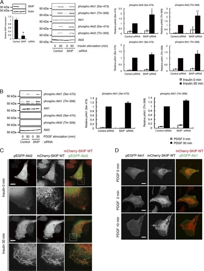
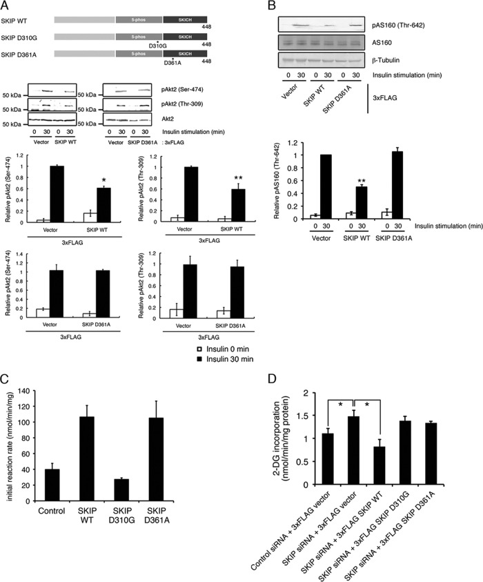
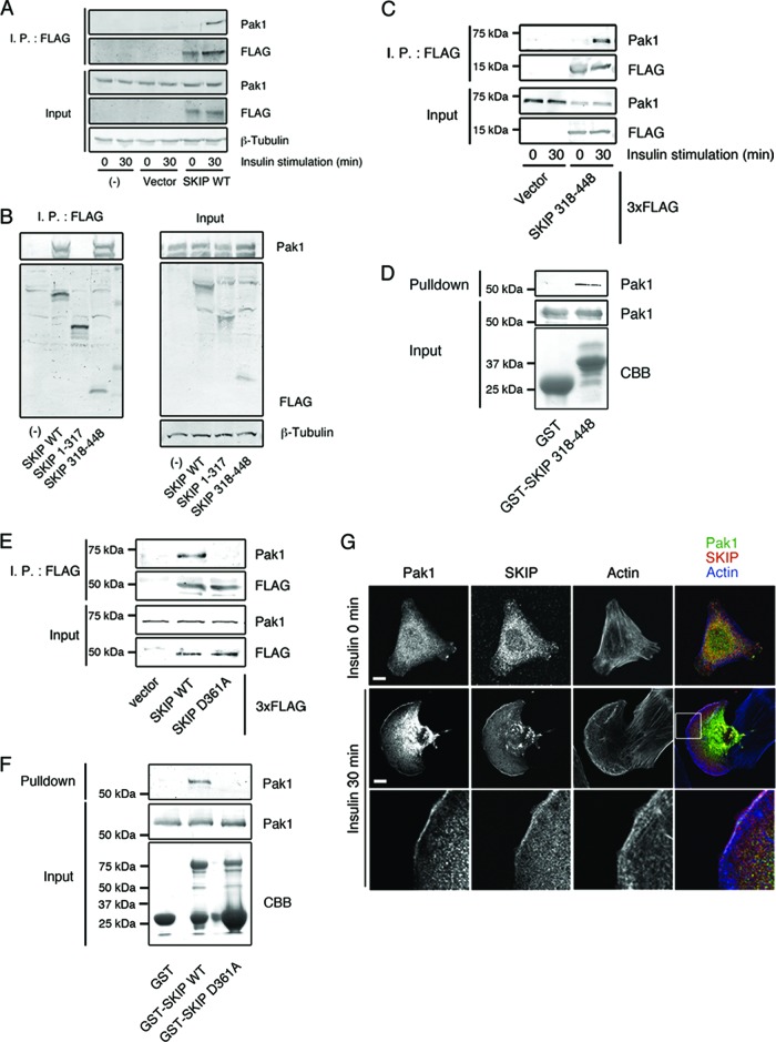
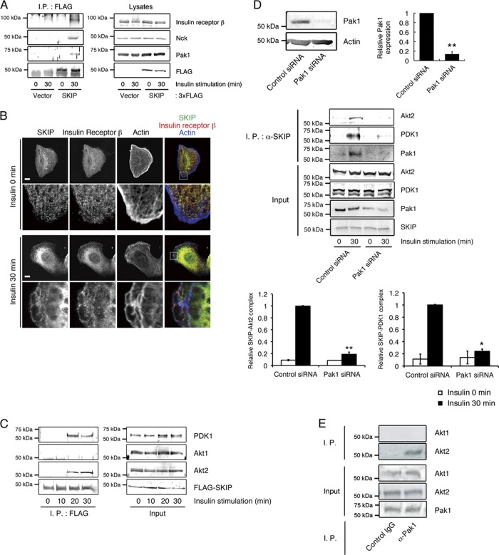
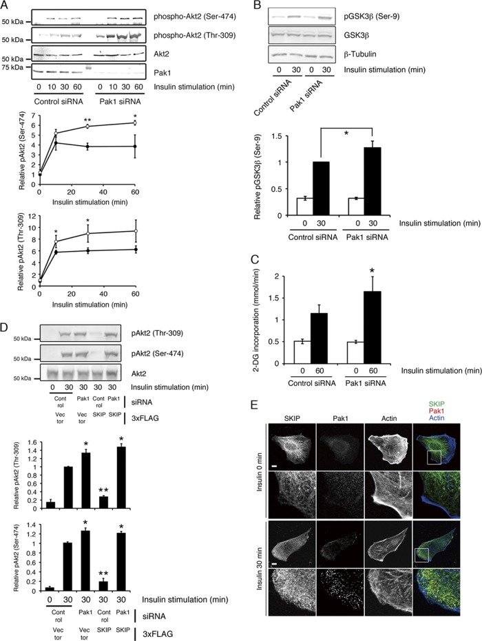
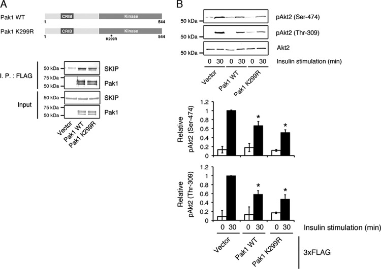
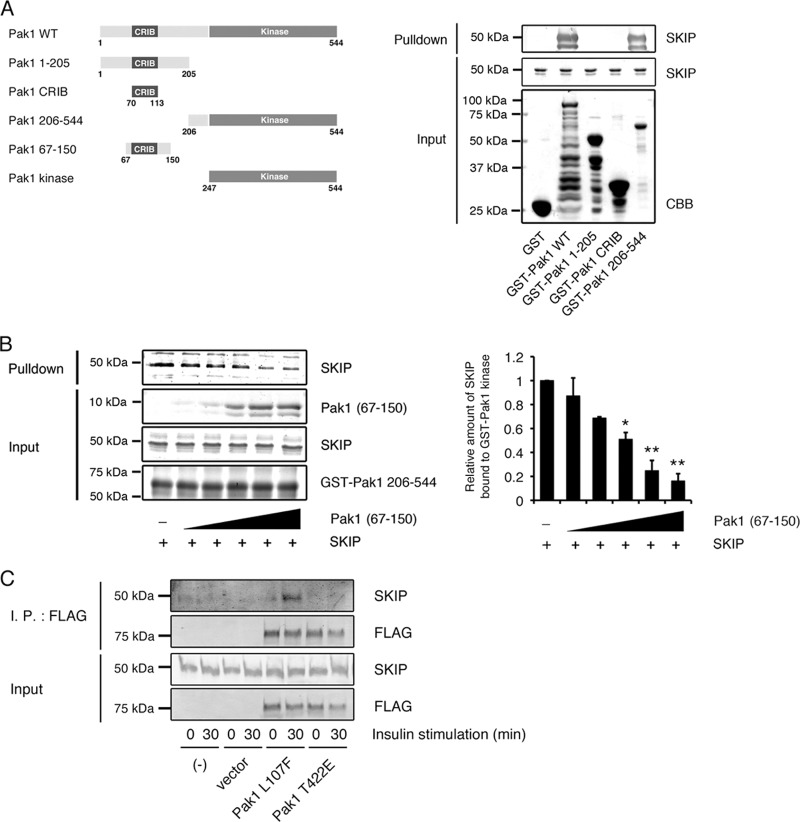
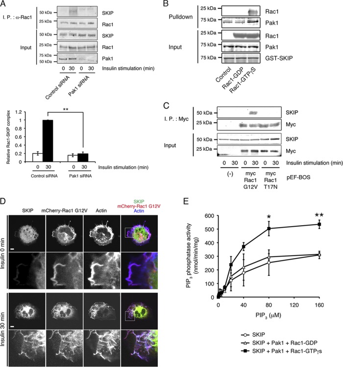
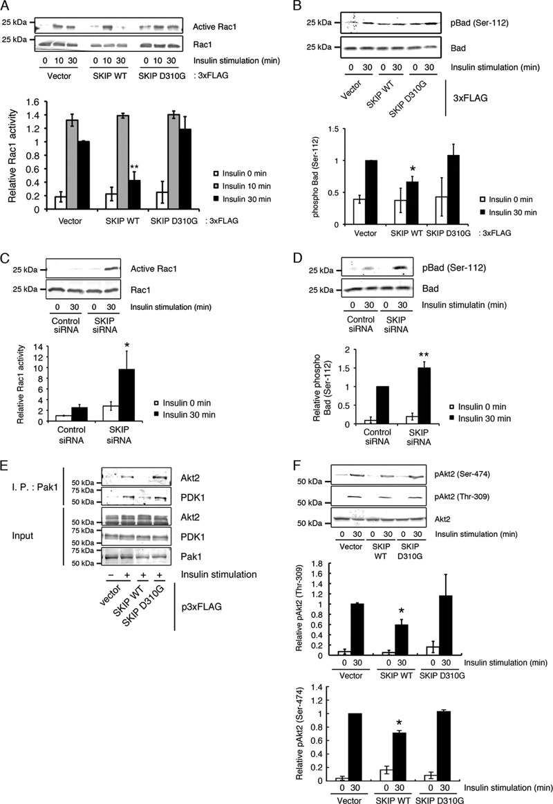
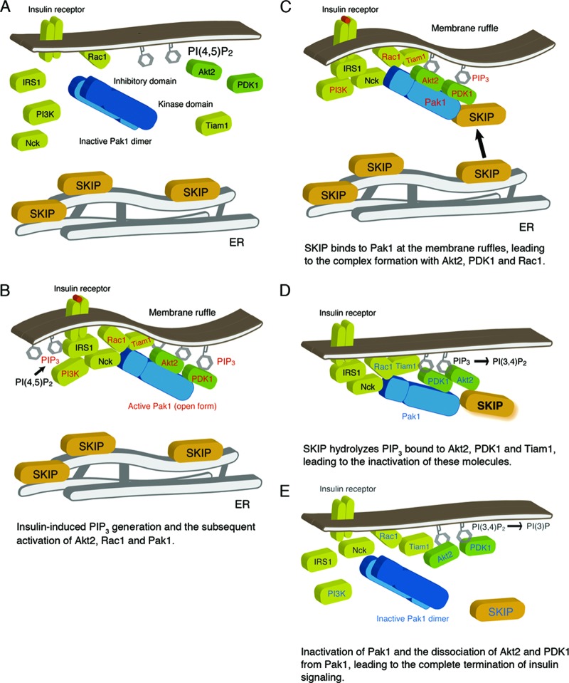
Skeletal muscle and kidney-enriched inositol polyphosphate phosphatase (SKIP) has previously been implicated in the regulation of insulin signaling in skeletal muscle. Here, we present the first report of the mechanisms by which SKIP specifically suppresses insulin signaling and the subsequent glucose uptake. Upon insulin stimulation, SKIP is translocated to the membrane ruffles, where it binds to the active form of Pak1, which mediates multiple protein complex formation with phosphatidylinositol 3,4,5-triphosphate (PIP(3)) effectors such as Akt2, PDK1, and Rac1; this leads to inactivation of these proteins. SKIP also promotes the inhibition of Rac1-dependent kinase activity and the scaffolding function of Pak1, which results in the dissociation of Akt2 and PDK1 from Pak1. Thus, specific suppression of insulin signaling is achieved via the spatiotemporal regulation of SKIP through the scaffolding function of Pak1. These interactions are the foundation of the specific and prominent role of SKIP in the regulation of insulin signaling.


Figures

Fig 1
Fig 1
Negative regulation of insulin-dependent Akt2 phosphorylation by SKIP. (A) Effect of SKIP silencing on the phosphorylation of Akt1 at Ser-473 and Thr-308 and of Akt2 at Ser-474 and Thr-309 after insulin stimulation for 30 min. Values shown are normalized to total Akt1 or Akt2. Results are presented as the means ± SDs of 3 independent experiments. *, P < 0.05; **, P < 0.01. (B) Effect of SKIP knockdown on PDGF-mediated phosphorylation of Akt1 in C2C12 cells. C2C12 cells transfected with control or SKIP siRNA were stimulated with PDGF for the indicated times. PDGF-induced phosphorylation of Akt2 was not detected. Results are presented as the means ± SDs of 3 independent experiments. (C) Insulin-dependent colocalization of SKIP with Akt2 at the membrane ruffles in C2C12 cells is shown in the upper panel. The scale bar indicates 10 μm. Enlarged images of boxed areas in the upper panel are shown in the lower panel. Yellow indicates regions of colocalization. (D) Lack of SKIP mobilization to the membrane ruffles in response to PDGF stimulation. C2C12 cells were transfected with EGFP-Akt1 and mCherry-SKIP WT for 48 h. Cells were stimulated with PDGF for the indicated times. The scale bar indicates 20 μm.
Fig 2
Fig 2
SKICH domain mutant did not inhibit insulin signaling. (A) Role of the SKIP SKICH domain in the negative regulation of insulin signaling. Shown is a schematic representation of the SKIP constructs used in this study (upper panel). Also shown is insulin-induced Akt2 phosphorylation in C2C12 cells expressing 3× FLAG-tagged SKIP wild-type (SKIP WT) and the SKICH domain mutant (SKIP D361A) (lower panels). Results are presented as the means ± SDs of 3 independent experiments. *, P < 0.05; **, P < 0.01. (B) Effect of expression of SKIP D361A mutant on insulin-induced AS160 phosphorylation. C2C12 cells expressing SKIP WT or SKIP D361A mutant were stimulated with insulin for the indicated times. Results are presented as the means ± SDs of 3 independent experiments. *, P < 0.05. (C) PIP3 phosphatase activity of the SKIP D310G and the D361A mutants. Results are presented as the means ± SDs of 3 independent experiments. **, P < 0.01. (D) Effect of the expression of the SKIP mutant on insulin-induced glucose uptake in L6 cells. Cells were transfected with SKIP siRNA in combination with the indicated human SKIP expression constructs. Cells were serum deprived for 24 h, and the insulin-induced glucose uptake was measured as described in Materials and Methods. Results are presented as the means ± SDs of 3 independent experiments. *, P < 0.05.
Fig 3
Fig 3
Insulin-dependent interaction between SKIP and Pak1. (A) Coimmunoprecipitation of Pak1 and SKIP in insulin-stimulated cells. C2C12 cells transfected with 3× FLAG SKIP WT for 48 h were stimulated with insulin for 30 min. Lysates were immunoprecipitated (I.P.) with anti-FLAG antibody. (B) Interaction between Pak1 and the SKICH domain of SKIP. Shown is a schematic representation of the SKIP constructs used in this study (upper panel). C2C12 cells transfected with 3× FLAG-tagged SKIP WT, the SKIP 1–317 mutant, and the SKIP 318–448 mutant were stimulated with insulin for 30 min. Lysates were immunoprecipitated with anti-FLAG antibody. (C) Insulin-mediated interaction between Pak1 and the SKICH domain of SKIP. C2C12 cells transfected with the 3× FLAG-tagged SKIP 318–448 mutant were stimulated with insulin for the indicated times. Lysates were immunoprecipitated with anti-FLAG antibody. (D) Binding of Pak1 to the SKICH domain of SKIP. The results of an in vitro binding assay of recombinant Pak1 and GST-SKIP 318–448 protein beads are shown. (E) Interaction between Pak1 and the SKICH domain mutant of SKIP. C2C12 cells transfected with 3× FLAG-tagged SKIP WT or SKIP D361A mutant for 48 h were stimulated with insulin for 30 min. Lysates were immunoprecipitated with anti-FLAG antibody. (F) The SKICH domain mutant does not bind to Pak1. The results of the in vitro binding assay between recombinant Pak1 and GST-tagged SKIP WT or D361A mutant are shown. (G) Immunofluorescence of endogenous SKIP and Pak1 in C2C12 cells. F-actin was visualized with Alexa Fluor 647-labeled phalloidin. Enlarged images of boxed areas are shown. White indicates regions of colocalization of SKIP, Pak1, and F-actin. Scale bar, 20 μm.
Fig 4
Fig 4
Insulin-dependent formation of a SKIP-Akt2 complex. (A) Induction of protein complex formation between SKIP, insulin receptor β, and Nck by insulin stimulation. C2C12 cells were transfected with the indicated constructs and stimulated with insulin for 0 or 30 min. Lysates from these cells were immunoprecipitated (I.P.) with anti-FLAG antibody. (B) Colocalization of endogenous SKIP and insulin receptor β in insulin-stimulated C2C12 cells. Cells were visualized by confocal microscopy. F-actin was visualized with Alexa Fluor 647-labeled phalloidin. Yellow indicates regions of colocalization of SKIP and insulin receptor β; white shows regions of colocalization of SKIP, insulin receptor β, and actin. Scale bar, 20 μm. (C) Insulin-dependent formation of a SKIP-Akt2 complex. C2C12 cells were transfected with 3× FLAG-tagged SKIP WT and then stimulated with insulin for the indicated times. Lysates were immunoprecipitated with anti-FLAG antibodies. (D) Pak1 mediates complex formation between endogenous SKIP and Akt2. C2C12 cells were transfected with control or Pak1 siRNA. Relative expression of Pak1 in these cells is shown (upper panels). These cells were stimulated with insulin for 0 or 30 min. Lysates were immunoprecipitated with anti-SKIP antibody, and the immunoprecipitates were detected with anti-Akt2, -PDK1, or -Pak1 antibodies. The amounts of Akt2 and PDK1 immunoprecipitated with SKIP are shown (lower panels). Results are presented as the means ± SEMs of 5 independent experiments. *, P < 0.05; **, P < 0.01. (E) Pak1 predominantly associated with Akt2 in C2C12 cells. C2C12 cells were serum deprived for 24 h and then stimulated with insulin for the indicated times. Lysates were immunoprecipitated with anti-rabbit IgG and anti-Pak1 antibody. Immunoprecipitates were subjected to Western blot analysis.
Fig 5
Fig 5
SKIP negatively regulates Akt2 phosphorylation through Pak1. (A) Increased insulin-dependent Akt2 phosphorylation caused by Pak1 attenuation. C2C12 cells transfected with control (filled circles) or Pak1 (open circles) siRNA were stimulated with insulin for the indicated times. Results are presented as the means ± SDs of 5 independent experiments. *, P < 0.05; **, P < 0.01. (B) Silencing of Pak1 increased insulin-dependent GSK3β phosphorylation. C2C12 cells transfected with control or Pak1 siRNA were stimulated with insulin for 0 min or 30 min. Phosphorylation of GSK3β at Ser-9 was detected by Western blot analysis. Results are presented as the means ± SDs of 3 independent experiments. *, P < 0.05. (C) Effect of Pak1 silencing on insulin-induced glucose uptake in L6 cells. Results are presented as the means ± SDs of 3 independent experiments. *, P < 0.05. (D) Effect of SKIP expression on insulin-induced Akt phosphorylation in Pak1-silenced cells. C2C12 cells transfected with the p3×FLAG vector or SKIP WT in combination with control or Pak1 siRNA were stimulated with insulin for the indicated times. Results are presented as the means ± SDs of 3 independent experiments. *, P < 0.05. (E) Inhibition of localization of SKIP at the membrane ruffle by the silencing of Pak1. C2C12 cells transfected with control or Pak1 siRNA were stimulated with insulin for 0 min or 30 min. Localization of endogenous SKIP is shown. F-actin was visualized with Alexa Fluor 647-labeled phalloidin. Scale bar, 20 μm.
Fig 6
Fig 6
Effect of Pak1 kinase activity on the interaction with SKIP. (A) Association between SKIP and the WT and K299R mutant of Pak1. Shown is a schematic representation of Pak1 constructs used in this study (upper panel). Lysates from C2C12 cells expressing 3× FLAG-tagged Pak1 WT or 3× FLAG-tagged Pak1 K299R were subjected to immunoprecipitation (I.P.) with anti-FLAG antibody (lower panel). (B) Effects of 3× FLAG-tagged Pak1 WT and kinase-negative mutant (K299R) expression on insulin-induced Akt2 phosphorylation. C2C12 cells expressing 3× FLAG-tagged Pak1 WT or K299R were stimulated with insulin for the indicated times. Results are presented as the means ± SDs of 3 independent experiments. *, P < 0.05.
Fig 7
Fig 7
Insulin-dependent formation of an active Pak1-SKIP complex. (A) Pak1 constructs used in this study (left) and binding of SKIP to the Pak1 kinase domain (right). (B) Competitive binding for the Pak1 kinase domain between SKIP and the Pak1 inhibitory switch region (Pak1 aa 67 to 150). GST-Pak1 kinase domain (1 μg) immobilized to glutathione-Sepharose 4B beads was incubated with SKIP (1 μg) and various amounts of Pak1 aa 67 to 150. *, P < 0.05; **, P < 0.01 (t test). (C) Binding of SKIP to the constitutively active form of Pak1 (Pak1 L107F) in insulin-stimulated C2C12 cells. C2C12 cells transfected with 3× FLAG-tagged Pak1 L107F or Pak1 T422A mutant were stimulated with insulin for the indicated times. Lysates were immunoprecipitated (I.P.) with anti-FLAG antibody.
Fig 8
Fig 8
Complex formation of SKIP and the active form of Rac1. (A) Insulin-dependent interaction between endogenous SKIP and Rac1, which is mediated by Pak1. C2C12 cells transfected with control or Pak1 siRNA were stimulated with insulin for the indicated times. Lysates were immunoprecipitated (I.P.) with anti-Rac1 antibody. Results are presented as the means ± SEMs of 3 independent experiments. **, P < 0.01. (B) SKIP binds to the activated form of Pak1. GST-SKIP domain (0.5 μg) immobilized to glutathione-Sepharose 4B beads was incubated with Pak1 (0.5 μg) in the presence of GDP-Rac1 or GTPγS-Rac1 (2 μg). (C) Complex formation of SKIP with the active form of Rac1 (pEF-BOS myc Rac1-G12V) but not with the dominant negative mutant (pEF-BOS myc Rac1-T17N). (D) Localization of SKIP in C2C12 cells expressing the constitutive active mutant of Rac1 (mCherry-Rac1 G12V). Pak1 activation by the expression of constitutive active Rac1 is not sufficient for the membrane ruffle localization of SKIP. F-actin was visualized with Alexa Fluor 647-labeled phalloidin. Scale bars, 20 μm. (E) Active Pak1 activated PIP3 phosphatase activity of SKIP. Recombinant SKIP (50 ng) in the presence or absence of Pak1 (70 ng) and GTPγS-Rac1 (100 ng) was used for the PIP3 phosphatase activity assay. Recombinant protein was incubated with various concentrations of PIP3 (0 to 160 μM). Results are presented as the means ± SEMs of 5 independent experiments. *, P < 0.05; **, P < 0.01.
Fig 9
Fig 9
Dissociation of Akt2 from the Pak1 complex, as mediated by the PIP3 phosphatase activity of SKIP. (A) Decreased Rac1 activity in insulin-stimulated C2C12 cells caused by expression of SKIP WT but not the D310G mutant. Results are presented as the means ± SDs of 3 independent experiments. **, P < 0.01. (B) Suppression of Pak1 activity caused by expression of SKIP WT but not the D310G mutant. Results are presented as the means ± SDs of 3 independent experiments. *, P < 0.05. (C) Increased Rac1 activity caused by the silencing of SKIP. Results are presented as the means ± SDs of 3 independent experiments. *, P < 0.05. (D) Increased Pak1 activity, measured as insulin-induced phosphorylation of Bad at Ser-112, caused by SKIP attenuation in C2C12 cells. Results are presented as the means ± SDs of 3 independent experiments. **, P < 0.01. (E) Decreased binding of Akt2 and PDK1 to Pak1 caused by the PIP3 phosphatase activity of SKIP. (F) Suppression of Akt2 activity caused by expression of SKIP WT but not the D310G mutant. Results are presented as the means ± SDs of 3 independent experiments. *, P < 0.05.
Fig 10
Fig 10
Working model of SKIP regulation of insulin signaling. (A) Under resting conditions, SKIP is localized to the ER. (B) Insulin-dependent PIP3 generation and Rac1-dependent Pak1 activation. Pak1 activation triggers its recruitment to the insulin receptor complex and subsequent complex formation with Akt2 and PDK1. Activated signaling molecules are highlighted in red. (C) Insulin induces translocation of SKIP to the plasma membrane, where SKIP binds to active Pak1 (the open form). Formation of a complex including SKIP, Pak1, Akt2, and Rac1 is induced at the membrane ruffles. (D) SKIP hydrolyzes PIP3 bound to Akt2. Localization of SKIP in proximity to Akt2 facilitates its inactivation. Signaling molecules inactivated by SKIP are indicated in blue. (E) SKIP negatively regulates Rac1 and Pak1 activity, leading to the inactive conformation of Pak1. Dissociation of Akt2 and PDK1 from inactive Pak1 terminates insulin signaling.

Similar articles

Cited by

References

    1. Bishop AL, Hall A. 2000. Rho GTPases and their effector proteins. Biochem. J. 348(Part 2):241–255 - PMC - PubMed
    1. Carpten JD, et al. 2007. A transforming mutation in the pleckstrin homology domain of AKT1 in cancer. Nature 448: 439– 444 - PubMed
    1. Cho H, et al. 2001. Insulin resistance and a diabetes mellitus-like syndrome in mice lacking the protein kinase Akt2 (PKB beta). Science 292: 1728– 1731 - PubMed
    1. Christoforidis S, Zerial M. 2000. Purification and identification of novel Rab effectors using affinity chromatography. Methods 20: 403– 410 - PubMed
    1. Dugani CB, Klip A. 2005. Glucose transporter 4: cycling, compartments and controversies. EMBO Rep. 6: 1137– 1142 - PMC - PubMed

Publication types

Substances