. 2012 Sep;32(17):3585-93.
doi: 10.1128/MCB.00455-12. Epub 2012 Jul 2.

A novel 4EHP-GIGYF2 translational repressor complex is essential for mammalian development

Affiliations

A novel 4EHP-GIGYF2 translational repressor complex is essential for mammalian development

Masahiro Morita et al. Mol Cell Biol. 2012 Sep.

Abstract

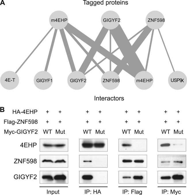
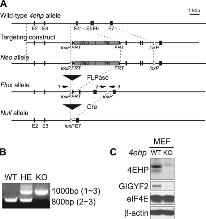
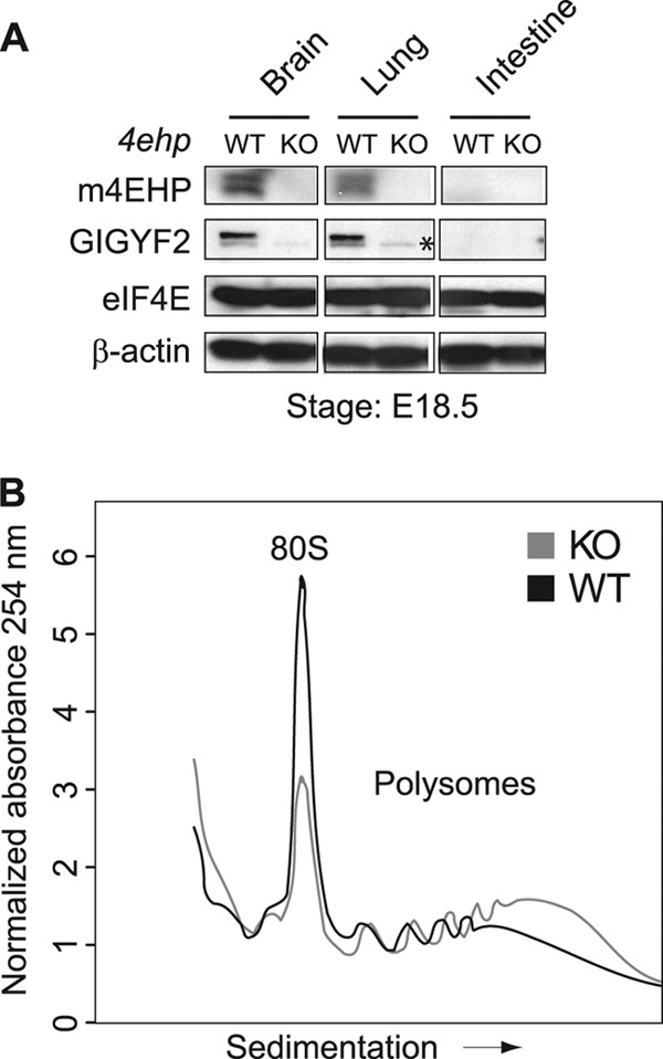
The binding of the eukaryotic initiation factor 4E (eIF4E) to the mRNA 5' cap structure is a rate-limiting step in mRNA translation initiation. eIF4E promotes ribosome recruitment to the mRNA. In Drosophila, the eIF4E homologous protein (d4EHP) forms a complex with binding partners to suppress the translation of distinct mRNAs by competing with eIF4E for binding the 5' cap structure. This repression mechanism is essential for the asymmetric distribution of proteins and normal embryonic development in Drosophila. In contrast, the physiological role of the mammalian 4EHP (m4EHP) was not known. In this study, we have identified the Grb10-interacting GYF protein 2 (GIGYF2) and the zinc finger protein 598 (ZNF598) as components of the m4EHP complex. GIGYF2 directly interacts with m4EHP, and this interaction is required for stabilization of both proteins. Disruption of the m4EHP-GIGYF2 complex leads to increased translation and perinatal lethality in mice. We propose a model by which the m4EHP-GIGYF2 complex represses translation of a subset of mRNAs during embryonic development, as was previously reported for d4EHP.


Figures

Fig 1
Fig 1
Identification of GIGYF proteins as novel m4EHP-interacting partners. (A) Identification of 4EHP-binding proteins by far-Western analysis. Extracts from HeLa S3 cells were resolved by SDS-PAGE and transferred to nitrocellulose membrane, which was incubated with 32P-labeled recombinant 4EHP. Immunoprecipitates were separated by SDS-PAGE, the gel was stained with Coomassie brilliant blue, and the bands of interest were excised for MS analysis. MS identified GIGYF2 and GIGYF1 as candidates for bands 1 and 2, respectively. (B) GIGYF1 and GIGYF2 were depleted by siRNA in HeLa S3 cells. The levels of both proteins were specifically reduced, as demonstrated by immunoblotting (IB; upper panels). β-Actin served as a loading control. The asterisk denotes a nonspecific band. These lysates were subjected to far-Western analysis (FW; lower panels). (C) HeLa S3 cells were transfected with a Myc-GIGYF2 plasmid, with or without an HA-4EHP plasmid. Interactions were examined by coimmunoprecipitation (Co-IP) with anti-HA antibody, followed by immunoblotting (IB) with anti-Myc and anti-HA antibodies. (D) HeLa S3 cells were transfected with an HA-4EHP plasmid, with or without a Myc-GIGYF2 plasmid. Interactions were examined by Co-IP with anti-Myc antibody, followed by IB with anti-Myc, anti-HA, and anti-eIF4E antibodies. For panels C and D, inputs represent 10% of the total lysate used in the IP assay.
Fig 2
Fig 2
Characterization of the interaction between m4EHP and GIGYF2. (A) Schematic representation of human GIGYF1 and GIGYF2 proteins. The m4EHP-binding motif and GYF domain are represented by blue and orange boxes, respectively. (B) Sequence alignment of the m4EHP consensus motif found in GIGYF2, GIGYF1, and the Drosophila 4EHP binding protein Bicoid (dBicoid). (C) HA-m4EHP and the wild-type/mutant Myc-GIGYF2 were ectopically expressed in HeLa S3 cells. Cell lysates were immunoprecipitated with anti-Myc antibody. Immunoblotting was performed with anti-Myc and anti-HA antibodies. Inputs represent 10% of the total lysate used in the IP assay.
Fig 3
Fig 3
GIGYF2 associates with cap-bound m4EHP. (A) HeLa S3 cells were cotransfected with Myc-GIGYF2 and HA-m4EHP. Lysates were incubated with either GDP- or m7GDP-coupled agarose beads (lanes 2 and 4). Cap-bound proteins were eluted from the beads and resolved by SDS-PAGE. Free m7GDP was used to confirm the specific binding of HA-m4EHP and Myc-GIGYF2 to the m7GDP cap (lane 3). (B) HeLa S3 cells were cotransfected with HA-m4EHP and either the wild-type (WT) or m4EHP binding mutant (Mut) Myc-GIGYF2. Cell lysates were incubated with m7GDP-coupled agarose beads. Immunoblotting was performed with anti-Myc and anti-HA antibodies. Inputs represent 5% of the total lysate used in the pulldown assay.
Fig 4
Fig 4
The rate of protein synthesis is increased in m4EHP- and GIGYF2-depleted cells. (A) Immunoblotting of 4EHP and GIGYF2 in HeLa cells transfected with control, m4EHP, or GIGYF2 siRNAs. β-Actin was used as a loading control. The doublet observed on the topmost Western blot corresponds to m4EHP (33). (B) Effects of m4EHP or GIGYF2 silencing on protein synthesis. Protein synthesis was measured by [35S]methionine incorporation normalized to the total amount of protein. The value of the control cells was adjusted to 100%. [35S]methionine labeling was carried out in triplicates and in three independent experiments (n = 3). All values represent means ± standard deviations (SD). (C) Control or siRNA-treated HeLa cells were pulse-labeled with [35S]methionine. Newly synthesized proteins were visualized by SDS-PAGE and autoradiography. Equal amounts of sample were loaded. Numbers to the right indicate molecular masses (kDa).
Fig 5
Fig 5
The mammalian m4EHP interaction network. (A) Cytoscape (38) figure displaying the high-confidence interaction partners by AP-MS. The top row represents bait proteins that were tagged and purified. The bottom row shows the protein hits identified by MS. The thickness of the connectors reflects the total number of spectra acquired for each bottom protein. (B) HEK293T cells were triply transfected with HA-4EHP, Flag-ZNF598, and Myc-GIGYF2-WT or Myc-GIGYF2-Mut expression vectors. Interactions were examined by coimmunoprecipitation (Co-IP) with anti-HA, anti-FLAG, and anti-Myc antibodies followed by immunoblotting (IB) with anti-HA, anti-FLAG, and anti-Myc antibodies. Input represents 10% of the total lysate used in the IP assay.
Fig 6
Fig 6
Disruption of the 4ehp gene in mice. (A) Schematic representation of the targeting construct used for the generation of 4ehp knockout mice. FRT and loxP sequences are indicated by black and white triangles, respectively. Negative (HSV-TK) and positive (PGK-neo) selection markers are indicated by gray boxes (TK, thymidine kinase; Neo, neomycin). Numbered black boxes represent exons in the 4ehp gene. The 4ehp neo allele was produced by homologous recombination. FLPase was used to generate the 4ehp flox allele. 4ehp null allele mice were produced by mating flox allele males with CMV-Cre females. (B) PCR genotyping of genomic DNA from mice with the 4ehp wild-type (WT), heterozygous (HE), and knockout (KO) genotypes. Arrows (numbered 1 to 3) in panel A denote annealing positions of oligonucleotides used for genotyping. (C) Immunoblotting of m4EHP, GIGYF2, and eIF4E proteins in 4ehp WT and KO MEFs. β-Actin was used as a loading control.
Fig 7
Fig 7
Tissue distribution of m4EHP-GIGYF2 proteins and polysome profile of wild-type and 4ehp knockout mice. (A) Immunoblotting of m4EHP, GIGYF2, and eIF4E proteins in 4ehp WT and KO tissues isolated from mice at E18.5. β-Actin was used as a loading control. An asterisk denotes a nonspecific band. (B) Polysome profiles of WT and 4ehp KO whole-brain lysates. Brain lysates were sedimented on 10% to 50% sucrose gradients. A254 was continuously recorded. Polysome profiles were normalized with the area under the curve. 80S denotes the monosome peak.
Fig 8
Fig 8
Perinatal lethality in 4ehp knockout mice. (A) Summary of 4ehp heterozygous intercrosses. Genotyping of embryos and pups at indicated stages was performed by PCR. (B and C) Gross appearance of 4ehp WT and KO pups at postnatal day 0. The arrow indicates a 4ehp KO pup. (D) Body weight of 4ehp WT, heterozygous (HE), and KO pups at E18.5. All values represent means ± SD. n = 5 for each genotype.

Similar articles

Cited by

References

    1. Ash MR, et al. 2010. Conserved beta-hairpin recognition by the GYF domains of Smy2 and GIGYF2 in mRNA surveillance and vesicular transport complexes. Structure 18:944–954 - PubMed
    1. Chekulaeva M, Hentze MW, Ephrussi A. 2006. Bruno acts as a dual repressor of oskar translation, promoting mRNA oligomerization and formation of silencing particles. Cell 124:521–533 - PubMed
    1. Chen GI, et al. 2008. PP4R4/KIAA1622 forms a novel stable cytosolic complex with phosphoprotein phosphatase 4. J. Biol. Chem. 283:29273–29284 - PMC - PubMed
    1. Cho PF, et al. 2006. Cap-dependent translational inhibition establishes two opposing morphogen gradients in Drosophila embryos. Curr. Biol. 16:2035–2041 - PMC - PubMed
    1. Cho PF, et al. 2005. A new paradigm for translational control: inhibition via 5′-3′ mRNA tethering by Bicoid and the eIF4E cognate 4EHP. Cell 121:411–423 - PubMed

Publication types

MeSH terms