

Characterization of gap junction proteins in the bladder of Cx43 mutant mouse models of oculodentodigital dysplasia
- PMID: 22752022
- PMCID: PMC3726213
- DOI: 10.1007/s00232-012-9455-1
Characterization of gap junction proteins in the bladder of Cx43 mutant mouse models of oculodentodigital dysplasia
Abstract
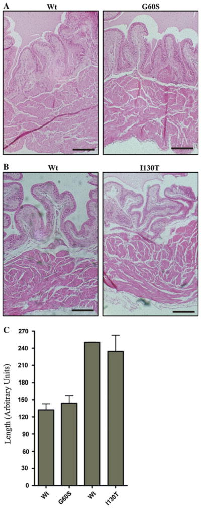
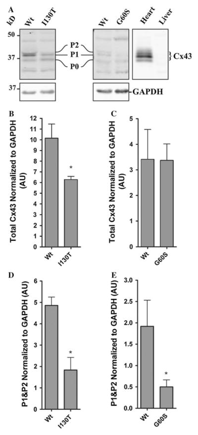
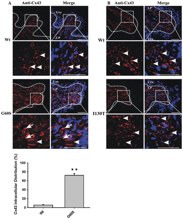
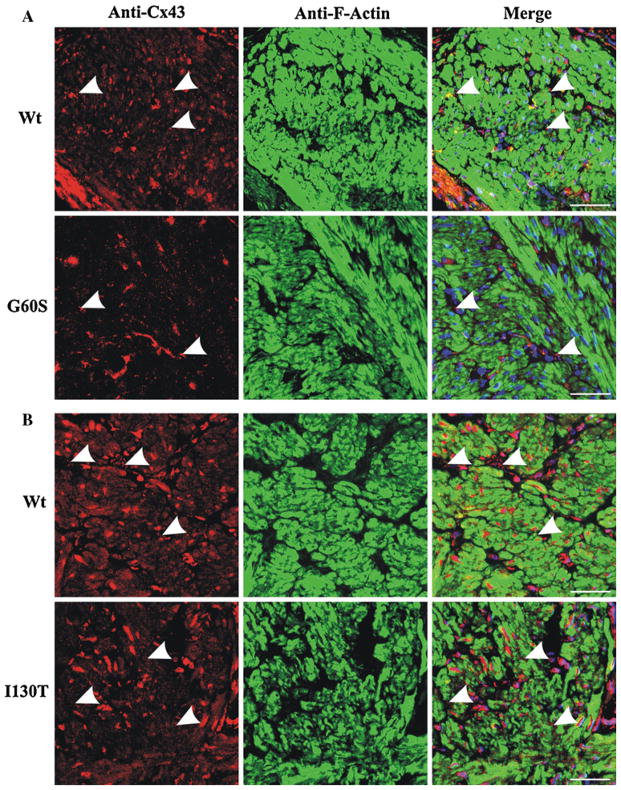
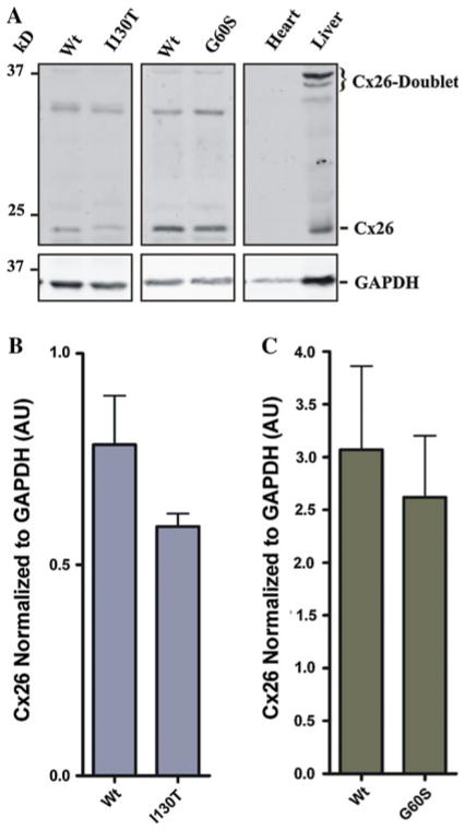
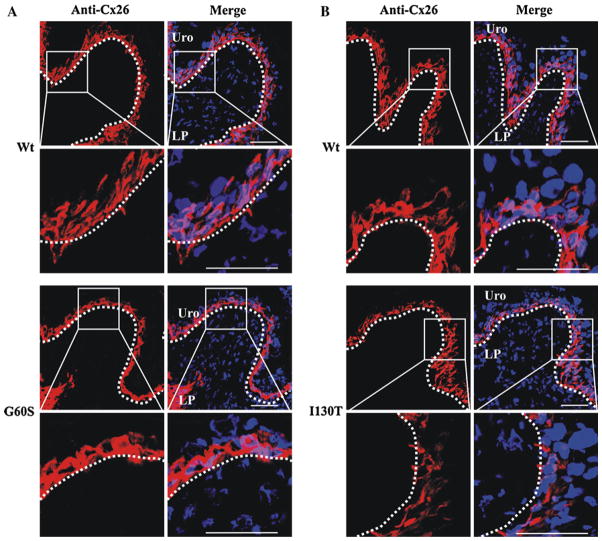
Oculodentodigital dysplasia (ODDD) is a rare developmental disease resulting from germline mutations in the GJA1 gene that encodes the gap junction protein connexin43 (Cx43). In addition to the classical ODDD symptoms that affect the eyes, teeth, bone and digits, in some cases ODDD patients have reported bladder impairments. Thus, we chose to characterize the bladder in mutant mouse models of ODDD that harbor two distinct Cx43 mutations, G60S and I130T. Histological assessment revealed no difference in bladder detrusor wall thickness in mutant compared to littermate control mice. The overall localization of Cx43 in the lamina propria and detrusor also appeared to be similar in the bladders of mutant mice with the exception that the G60S mice had more instances of intracellular Cx43. However, both mutant mouse lines exhibited a significant reduction in the phosphorylated P1 and P2 isoforms of Cx43, while only the I130T mice exhibited a reduction in total Cx43 levels. Interestingly, Cx26 levels and distribution were not altered in mutant mice as it was localized to intracellular compartments and restricted to the basal cell layers of the urothelium. Our studies suggest that these two distinct genetically modified mouse models of ODDD probably mimic patients who lack bladder defects or other factors, such as aging or co-morbidities, are necessary to reveal a bladder phenotype.
Figures
Similar articles
-
Myogenic bladder defects in mouse models of human oculodentodigital dysplasia.Biochem J. 2014 Feb 1;457(3):441-9. doi: 10.1042/BJ20130810. Biochem J. 2014. PMID: 24228978 Free PMC article.
-
Autosomal recessive GJA1 (Cx43) gene mutations cause oculodentodigital dysplasia by distinct mechanisms.J Cell Sci. 2013 Jul 1;126(Pt 13):2857-66. doi: 10.1242/jcs.123315. Epub 2013 Apr 19. J Cell Sci. 2013. PMID: 23606748
-
Effects of reduced connexin43 function on skull development in the Cx43I130T/+ mutant mouse that models oculodentodigital dysplasia.Bone. 2020 Jul;136:115365. doi: 10.1016/j.bone.2020.115365. Epub 2020 Apr 19. Bone. 2020. PMID: 32320893
-
Syndromic and non-syndromic disease-linked Cx43 mutations.FEBS Lett. 2014 Apr 17;588(8):1339-48. doi: 10.1016/j.febslet.2013.12.022. Epub 2014 Jan 14. FEBS Lett. 2014. PMID: 24434540 Review.
-
GJA1 mutations, variants, and connexin 43 dysfunction as it relates to the oculodentodigital dysplasia phenotype.Hum Mutat. 2009 May;30(5):724-33. doi: 10.1002/humu.20958. Hum Mutat. 2009. PMID: 19338053 Review.
Cited by
-
Myogenic bladder defects in mouse models of human oculodentodigital dysplasia.Biochem J. 2014 Feb 1;457(3):441-9. doi: 10.1042/BJ20130810. Biochem J. 2014. PMID: 24228978 Free PMC article.
-
Connexin 43 Mutations Lead to Increased Hemichannel Functionality in Skin Disease.Int J Mol Sci. 2019 Dec 7;20(24):6186. doi: 10.3390/ijms20246186. Int J Mol Sci. 2019. PMID: 31817921 Free PMC article. Review.
-
Connexins in Cardiovascular and Neurovascular Health and Disease: Pharmacological Implications.Pharmacol Rev. 2017 Oct;69(4):396-478. doi: 10.1124/pr.115.012062. Pharmacol Rev. 2017. PMID: 28931622 Free PMC article. Review.
-
Towards a Better Understanding of Genotype-Phenotype Correlations and Therapeutic Targets for Cardiocutaneous Genes: The Importance of Functional Studies above Prediction.Int J Mol Sci. 2022 Sep 15;23(18):10765. doi: 10.3390/ijms231810765. Int J Mol Sci. 2022. PMID: 36142674 Free PMC article. Review.
-
Connexin expression patterns in diseased human corneas.Exp Ther Med. 2014 Apr;7(4):791-798. doi: 10.3892/etm.2014.1530. Epub 2014 Feb 10. Exp Ther Med. 2014. PMID: 24669234 Free PMC article.
References
-
- Abrams P, Cardozo L, Fall M, Griffiths D, Rosier P, Ulmsten U, Van Kerrebroeck P, Victor A, Wein A. The standardisation of terminology in lower urinary tract function: report from the standardisation sub-committee of the International Continence Society. Urology. 2003;61:37–49. - PubMed
-
- Alexander DB, Goldberg GS. Transfer of biologically important molecules between cells through gap junction channels. Curr Med Chem. 2003;10:2045–2058. - PubMed
-
- Apodaca G. The uroepithelium: not just a passive barrier. Traffic. 2004;5:117–128. - PubMed
-
- Brading AF. A myogenic basis for the overactive bladder. Urology. 1997;50:57–73. - PubMed
-
- Christ GJ, Day NS, Day M, Zhao W, Persson K, Pandita RK, Andersson KE. Increased connexin43-mediated intercellular communication in a rat model of bladder overactivity in vivo. Am J Physiol Regul Integr Comp Physiol. 2003;284:R1241–R1248. - PubMed
Publication types
MeSH terms
Substances
Grants and funding
LinkOut - more resources
Full Text Sources
Molecular Biology Databases
Miscellaneous
