. 2012 Nov;69(22):3881-93.
doi: 10.1007/s00018-012-1056-8. Epub 2012 Jul 4.

VRK2 anchors KSR1-MEK1 to endoplasmic reticulum forming a macromolecular complex that compartmentalizes MAPK signaling

Affiliations

VRK2 anchors KSR1-MEK1 to endoplasmic reticulum forming a macromolecular complex that compartmentalizes MAPK signaling

Isabel F Fernández et al. Cell Mol Life Sci. 2012 Nov.

Abstract

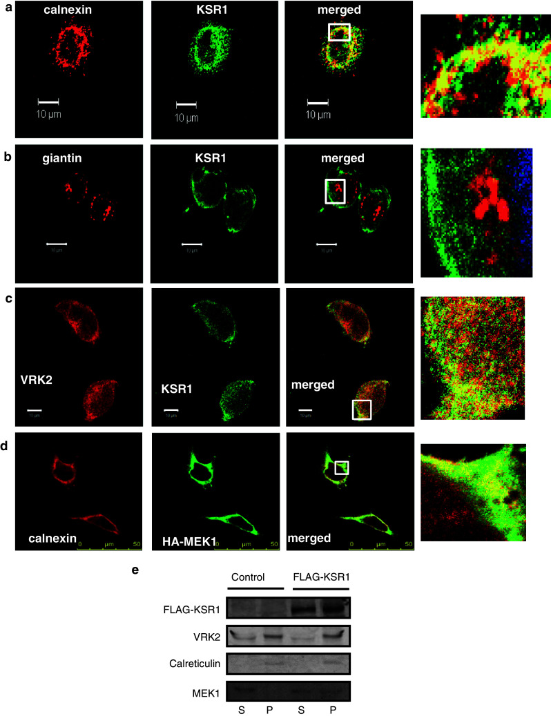
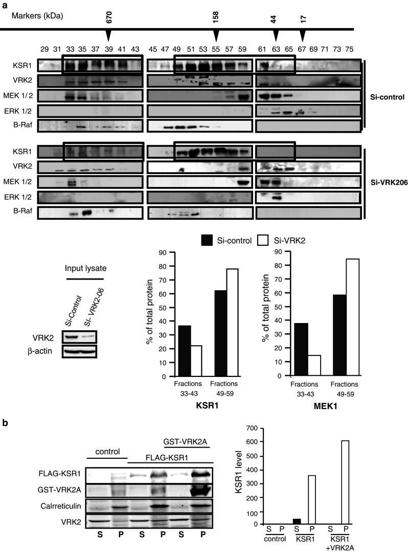
The spatial and temporal regulation of intracellular signaling is determined by the spatial and temporal organization of complexes assembled on scaffold proteins, which can be modulated by their interactions with additional proteins as well as subcellular localization. The scaffold KSR1 protein interacts with MAPK forming a complex that conveys a differential signaling in response to growth factors. The aim of this work is to determine the unknown mechanism by which VRK2A downregulates MAPK signaling. We have characterized the multiprotein complex formed by KSR1 and the Ser-Thr kinase VRK2A. VRK2A is a protein bound to the endoplasmic reticulum (ER) and retains a fraction of KSR1 complexes on the surface of this organelle. Both proteins, VRK2A and KSR1, directly interact by their respective C-terminal regions. In addition, MEK1 is also incorporated in the basal complex. MEK1 independently interacts with the CA5 region of KSR1 and with the N-terminus of VRK2A. Thus, VRK2A can form a high molecular size (600-1,000 kDa) stable complex with both MEK1 and KSR1. Knockdown of VRK2A resulted in disassembly of these high molecular size complexes. Overexpression of VRK2A increased the amount of KSR1 in the particulate fraction and prevented the incorporation of ERK1/2 into the complex after stimulation with EGF. Neither VRK2A nor KSR1 interact with the VHR, MKP1, MKP2, or MKP3 phosphatases. The KSR1 complex assembled and retained by VRK2A in the ER can have a modulatory effect on the signal mediated by MAPK, thus locally affecting the magnitude of its responses, and can explain differential responses depending on cell type.


Conflict of interest statement

The authors declare they have no competing interests.

Figures

Fig. 1
Fig. 1
Subcellular localization of VRK2 and KSR1 in membrane compartments in HeLa cells. a Colocalization of KSR1 (green) and calnexin (red), an endoplasmic reticulum marker. b Lack of colocalization of Giantin (red), a Golgi marker and KSR1 (green). c Colocalization of KSR1 (green) and VRK2 (red). VRK2 is located in endoplasmic reticulum [30]. Bar 10 μm. d Colocalization of calnexin with MEK1. HeLa cells were transfected with plasmid HA-MEK1 and its colocalization with endogenous calnexin determined by confocal microscopy. e HeLa cells were transfected with plasmid control or Flag-KSR1. Extracts were fractionated to separate membrane bound proteins from the soluble S100 fraction containing cytosolic proteins soluble fraction. In gels, one-tenth of the total soluble fraction and the total particulate fraction were loaded. The gel was used for immunoblot detection of VRK2, KSR1, MEK1, and calreticulin (ER marker) with the antibodies indicated in “Materials and methods”
Fig. 2
Fig. 2
Interaction of members of the VRK family with KSR1 and mapping of the interaction between VRK2A and KSR1. a Interaction of different fragments spanning VRK2A with KSR1. b Interaction of different VRK proteins (VRK2A, VRK2B, and VRK1) with KSR1. The interaction is specific for the VRK2A isoform. c Mapping the region of KSR1 that interacts with VRK2A. A GST-VRK2A constructs was used in a pulldown assay of deletion constructs from KSR1 N- and C-terminal regions. Interaction of VRK2A (left) and its C- terminal region (right) with different constructs of KSR1 in pulldown assay. d Interaction between VRK2A and the CA5 region of KSR1. Two types of pulldown assays were performed. In one, the amount VRK2A was fixed and assayed against increasing amount of KSR1-CA5 mutant (top). In the other, a fixed amount of KSR1-CA5 mutant was used against increasing amounts of VRK2A (bottom). e Binding of a C809Y KSR1 mutant to VRK2A. A GST-pulldown assay was performed using cell lysates overexpressing GST-VRK2A and HA-tagged KSR1 or KSR1-C809Y a mutant defective in MEK1/2 binding, and the amount of KSR1 protein bound to VRK2 quantified (bottom graph)
Fig. 3
Fig. 3
a Interaction between MEK1/2 and KSR1 or VRK2A. A. HEK293T cells were transfected with deletion constructs of KSR1 which was immunoprecipitated with an anti-Flag antibody and the presence of endogenous MEK1/2 was determined in the immunoprecipitate (top). b Interaction of transfected HA-MEK1 with either VRK2A (1-508) or VRK2B (1-396). c Interaction of transfected VRK2 constructs with endogenous MEK1/2 identifying the N-terminal region of VRK2 as the interacting region. d Pulldown of proteins bound to VRK2A in HEK293T cells. Cells were transfected with a mixture of HA-MEK1, Flag-KSR1, and either GST or GST-VRK2A plasmids. The cell extracts were used for a pulldown and the proteins present detected with specific antibodies. e The complex formed by VRK2–KSR1–MEK1/2
Fig. 4
Fig. 4
a Detection of large intracellular complexes containing VRK2, KSR1, MEK11/2, Erk1/2, and RAF in Hela cells fractionated by FPLC. Control input extract is at the bottom left. At the bottom right is shown the quantification of KSR1 (left) and MEK1 (right) proteins in high molecular size (fractions 33–43) and mid–low size (fractions 49–59). b Overexpression of VRK2 increases the localization of KSR1 to the particulate fraction. HEK293T cells were transfected with combinations of Flag-KSR1 and GST-VRK2A plasmids. The transfected cells were lysed and fractionated by ultracentrifugation as indicated in “Materials and methods”. The presence of each protein in the soluble and particulate fractions was determined. The amount of KSR1 in each fraction was quantified (right graph)
Fig. 5
Fig. 5
Effect of high level of VRK2A on the KSR1-ERK1 interaction induced by EGF. HEK293T cells were transfected with plasmids pFLAG-KSR1 and pCEFL-GST-VRK2A as indicated, and transfected cells were stimulated with 10 ng/ml EGF in order to activate the MAPK pathway via the KSR1–MAPK complex. The formation of the KSR1-endogenous ERK1/2 complex was determined in the KSR1 immunoprecipitate in the absence (center lane) or presence of VRK2 (right lane)
Fig. 6
Fig. 6
Different types of alternative protein complexes or signalosomes formed by KSR1 implicated in the assembly and activation of the MAPK pathway containing MEK1. The active or inactive role of different signalosomes with respect to their composition is indicated

References

    1. Scott JD, Pawson T. Cell signaling in space and time: where proteins come together and when they’re apart. Science. 2009;326:1220–1224. doi: 10.1126/science.1175668. - DOI - PMC - PubMed
    1. Good MC, Zalatan JG, Lim WA. Scaffold proteins: hubs for controlling the flow of cellular information. Science. 2011;332:680–686. doi: 10.1126/science.1198701. - DOI - PMC - PubMed
    1. Murphy LO, Blenis J. MAPK signal specificity: the right place at the right time. Trends Biochem Sci. 2006;31:268–275. doi: 10.1016/j.tibs.2006.03.009. - DOI - PubMed
    1. von Kriegsheim A, Baiocchi D, Birtwistle M, Sumpton D, Bienvenut W, Morrice N, Yamada K, Lamond A, Kalna G, Orton R, Gilbert D, Kolch W. Cell fate decisions are specified by the dynamic ERK interactome. Nat Cell Biol. 2009;11:1458–1464. doi: 10.1038/ncb1994. - DOI - PMC - PubMed
    1. Kolch W. Meaningful relationships: the regulation of the Ras/Raf/MEK/ERK pathway by protein interactions. Biochem J. 2000;351(Pt 2):289–305. doi: 10.1042/0264-6021:3510289. - DOI - PMC - PubMed

Publication types