

Intraductally administered pegylated liposomal doxorubicin reduces mammary stem cell function in the mammary gland but in the long term, induces malignant tumors
- PMID: 22752247
- PMCID: PMC3478104
- DOI: 10.1007/s10549-012-2138-x
Intraductally administered pegylated liposomal doxorubicin reduces mammary stem cell function in the mammary gland but in the long term, induces malignant tumors
Abstract
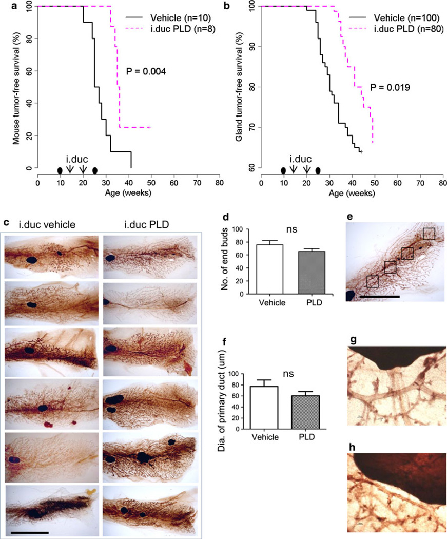
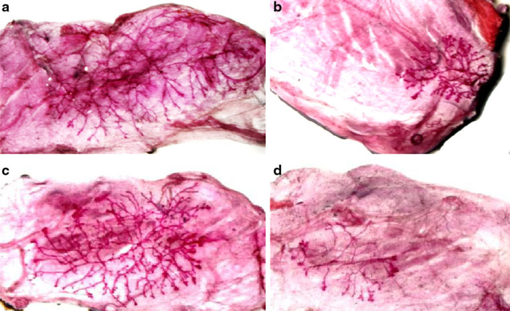
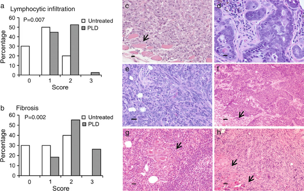
Previously, we have shown that the intraductal (i.duc) administration of pegylated liposomal doxorubicin (PLD) to Her2/neu transgenic mice is associated with mammary tumor regression and prevention. Exploring the mechanism underlying the protection afforded by PLD, we studied: the effects of i.duc PLD-treatment with a subsequent pregnancy on outgrowth of tumors in Her2/neu mice; whether the i.duc PLD antitumor effect was mediated partially through changes in normal mammary stem cells (MaSCs); and the long-term safety of i.duc PLD into the normal mouse mammary gland. Her2/neu mice were treated with two i.duc injections of PLD given four weeks apart; pregnancy was induced and mice were followed up for changes in physiology, and tumor formation. We found that all pups born to i.duc PLD-treated Her2/neu mice died without weight gain within 7 days after birth. Despite an additional pregnancy, compared to vehicle control PLD-treated Her2/neu mice had a significantly longer latency and lower frequency of tumor development. Mammary epithelial cells isolated from untreated and i.duc PLD-treated 6-8 months-old multiparous FVB/N mice were analyzed for their repopulating ability in mammary fat pads of naïve recipients. Mice were also monitored for abnormalities in mammary gland morphology and function, including tumor formation. PLD-treated FVB/N mice displayed histomorphologic changes and a significant reduction in the outgrowth potential of cells from the mammary glands. Thus, i.duc PLD administration altered the mammary gland structurally and functionally by reducing the MaSC population, which could compromise milk production. Followed long term, i.duc PLD-treated FVB/N mice developed malignant mammary tumors, confirming similar published findings on doxorubicin injected into the mammary gland of rats. Unless there are fundamental species differences in PLD metabolism in rodents and humans, this finding seriously limits the consideration of i.duc PLD use in the clinic for treatment or prevention of breast cancer.
Conflict of interest statement
Figures
References
-
- Molofsky AV, Pardal R, Morrison SJ. Diverse mechanisms regulate stem cell self-renewal. Curr Opin Cell Biol. 2004;16:700–707. - PubMed
-
- Reya T, Morrison SJ, Clarke MF, Weissman IL. Stem cells, cancer and cancer stem cells. Nature. 2001;414:105–111. - PubMed
-
- Smith GH. Stem cells and mammary cancer in mice. Stem Cell Rev. 2005;1:215–223. - PubMed
-
- Williams JM, Daniel CW. Mammary ductal elongation: differentiation of myoepithelium and basal lamina during branching morphogenesis. Dev Biol. 1983;97:274–290. - PubMed
Publication types
MeSH terms
Substances
Grants and funding
LinkOut - more resources
Full Text Sources
Medical
Research Materials
Miscellaneous
