

Prevalence of apical periodontitis detected in cone beam CT images of a Brazilian subpopulation
- PMID: 22752318
- PMCID: PMC3729192
- DOI: 10.1259/dmfr/80179163
Prevalence of apical periodontitis detected in cone beam CT images of a Brazilian subpopulation
Abstract
Objectives: The aim of this study was to determine the prevalence of apical periodontitis (AP) detected in cone beam CT (CBCT) images from a database.
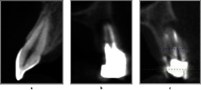
Methods: CBCT images of 300 Brazilian patients were assessed. AP images were measured in three dimensions. Age, gender, number and location of total teeth in each patient were considered. AP location was considered according to tooth groups. The extent of AP was determined by the largest diameter in any of the three dimensions. Percentages and the χ(2) test were used for statistical analysis.
Results: AP was found in 51.4% of the patients and in 3.4% of the teeth. Higher prevalence of AP was found in 60- to 69-year-olds (73.1%) and in mandibular molars (5.9%) (p < 0.05). Inadequate endodontic treatment presented higher prevalence of AP (78.1%).
Conclusions: AP can be frequently found in CBCT examinations. The presence of AP has a significant association with patients' age, and tooth type and condition. CBCT databases are useful for cross-sectional studies about AP prevalence in a population.
Figures
Similar articles
-
Evaluation of technical quality and periapical health of root-filled teeth by using cone-beam CT.J Appl Oral Sci. 2014 Nov-Dec;22(6):502-8. doi: 10.1590/1678-775720140110. J Appl Oral Sci. 2014. PMID: 25591019 Free PMC article.
-
Prevalence of apical periodontitis and root filled teeth in a Belgian subpopulation found on CBCT images.Int Endod J. 2017 Apr;50(4):317-329. doi: 10.1111/iej.12631. Epub 2016 Apr 6. Int Endod J. 2017. PMID: 26992464
-
Assessment of periapical health, quality of root canal filling, and coronal restoration by using cone-beam computed tomography.Niger J Clin Pract. 2016 Sep-Oct;19(5):673-7. doi: 10.4103/1119-3077.188697. Niger J Clin Pract. 2016. PMID: 27538559
-
Influence of length of root canal obturation on apical periodontitis detected by periapical radiography and cone beam computed tomography.J Endod. 2009 Jun;35(6):805-9. doi: 10.1016/j.joen.2009.03.013. J Endod. 2009. PMID: 19482175
-
Prevalence of Apical Periodontitis and Conventional Nonsurgical Root Canal Treatment in General Adult Population: An Updated Systematic Review and Meta-analysis of Cross-sectional Studies Published between 2012 and 2020.J Endod. 2020 Oct;46(10):1371-1386.e8. doi: 10.1016/j.joen.2020.07.007. Epub 2020 Jul 14. J Endod. 2020. PMID: 32673634
Cited by
-
A retrospective three-dimensional assessment of the prevalence of apical periodontitis and quality of root canal treatment in Mid-West Indian population.J Conserv Dent. 2021 Mar-Apr;24(2):184-189. doi: 10.4103/jcd.jcd_44_21. Epub 2021 Oct 9. J Conserv Dent. 2021. PMID: 34759587 Free PMC article.
-
Prevalence of Lateral Radiolucency, Apical Root Resorption and Periapical Lesions in Portuguese Patients: A CBCT Cross-Sectional Study with a Worldwide Overview.Eur Endod J. 2021 Apr;6(1):56-71. doi: 10.14744/eej.2021.29981. Epub 2021 Mar 23. Eur Endod J. 2021. PMID: 33762535 Free PMC article.
-
Machine Learning to Predict Apical Lesions: A Cross-Sectional and Model Development Study.J Clin Med. 2023 Aug 23;12(17):5464. doi: 10.3390/jcm12175464. J Clin Med. 2023. PMID: 37685531 Free PMC article.
-
Prevalence of Periapical Radiolucency and Conventional Root Canal Treatment in Adults: A Systematic Review of Cross-Sectional Studies.Cureus. 2023 Jan 3;15(1):e33302. doi: 10.7759/cureus.33302. eCollection 2023 Jan. Cureus. 2023. PMID: 36741594 Free PMC article. Review.
-
Prevalence of Asymptomatic Apical Periodontitis and its Association with Coronary Artery Disease in a Brazilian Subpopulation.Acta Stomatol Croat. 2017 Jun;51(2):106-112. doi: 10.15644/asc51/2/3. Acta Stomatol Croat. 2017. PMID: 28827847 Free PMC article.
References
-
- Abbott PV. Classification, diagnosis and clinical manifestations of apical periodontitis. Endod Topics 2004;8:36–54.
-
- Nair PNR. Pathogenesis of apical periodontitis and the causes of endodontic failures. Crit Rev Oral Biol Med 2004;15:348–381. - PubMed
-
- Lofthag-Hansen S, Huumonen S, Grondahl K, Grondahl HG. Limited cone-beam CT and intraoral radiography for the diagnosis of periapical pathology. Oral Surg Oral Med Oral Pathol Oral Radiol Endod 2007;103:114–119. - PubMed
-
- Huumonen S, Orstavik D. Radiological aspects of apical periodontitis. Endod Topics 2002;1:3–25.
-
- Tyndall DA, Rathore S. Cone-beam CT diagnostic applications: caries, periodontal bone assessment, and endodontic applications. Dent Clin North Am 2008;52:825–841. - PubMed
Publication types
MeSH terms
LinkOut - more resources
Full Text Sources
Other Literature Sources
