. 2012 Sep;11(9):1968-77.
doi: 10.1158/1535-7163.MCT-12-0248. Epub 2012 Jul 2.

Evidence for the ubiquitin protease UBP43 as an antineoplastic target

Affiliations

Evidence for the ubiquitin protease UBP43 as an antineoplastic target

Yongli Guo et al. Mol Cancer Ther. 2012 Sep.

Abstract

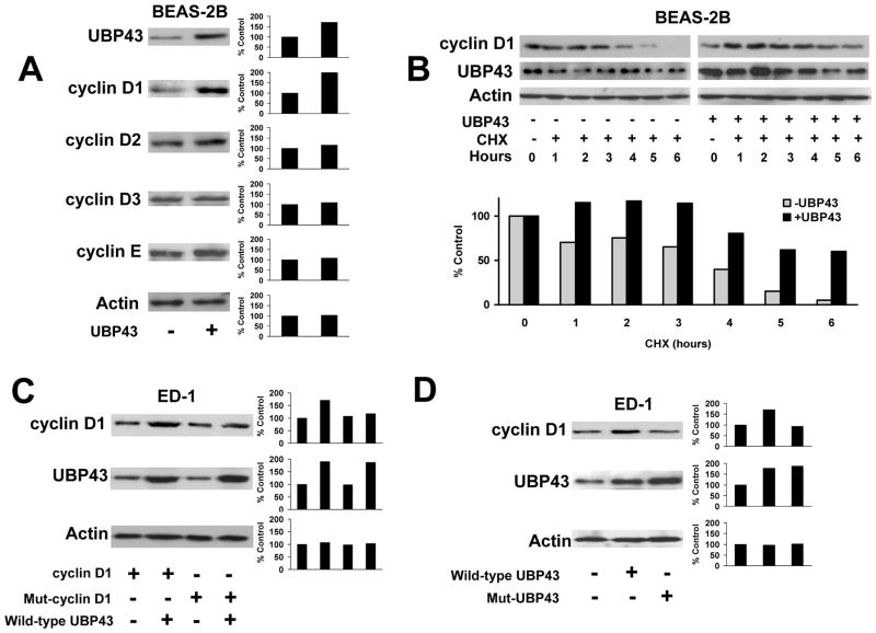
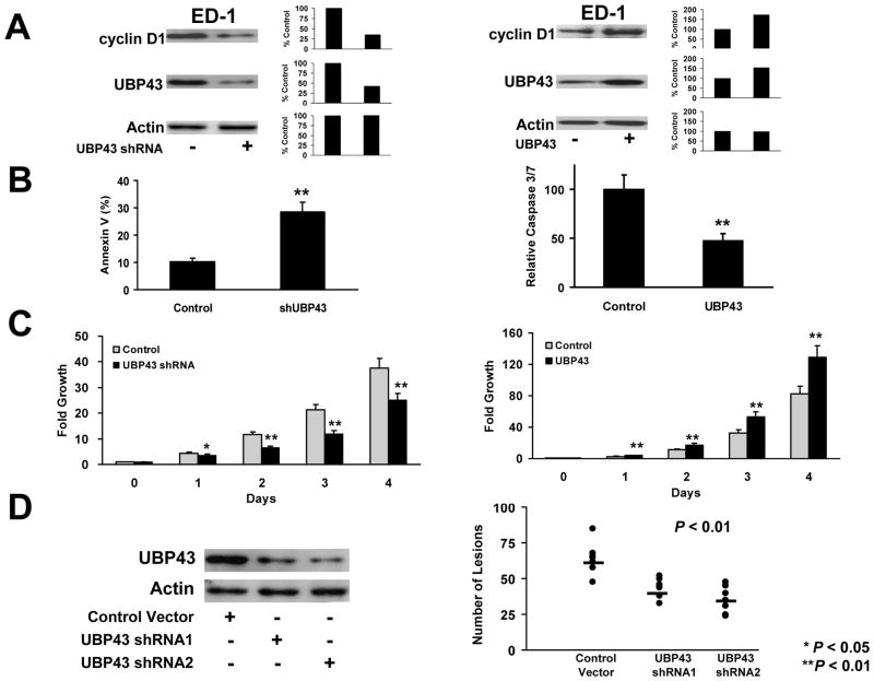
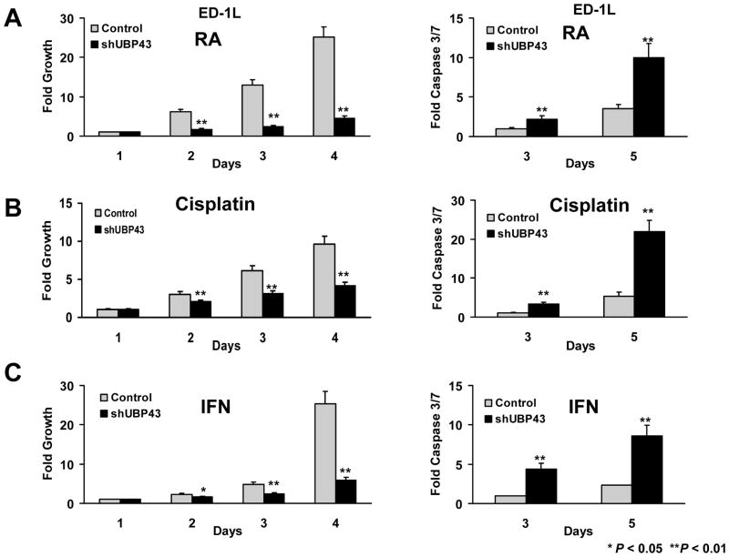
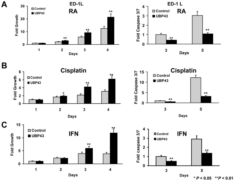
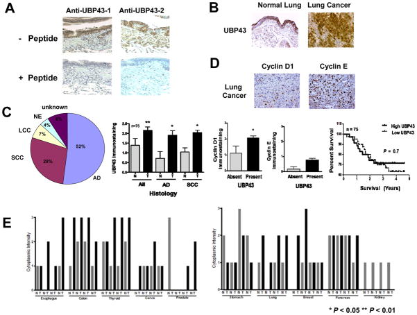
New pharmacologic targets are needed for lung cancer. One candidate pathway to target is composed of the E1-like ubiquitin-activating enzyme (UBE1L) that associates with interferon-stimulated gene 15 (ISG15), which complexes with and destabilizes cyclin D1. Ubiquitin protease 43 (UBP43/USP18) removes ISG15 from conjugated proteins. This study reports that gain of UBP43 stabilized cyclin D1, but not other D-type cyclins or cyclin E. This depended on UBP43 enzymatic activity; an enzymatically inactive UBP43 did not affect cyclin D1 stability. As expected, small interfering RNAs that reduced UBP43 expression also decreased cyclin D1 levels and increased apoptosis in a panel of lung cancer cell lines. Forced cyclin D1 expression rescued UBP43 apoptotic effects, which highlighted the importance of cyclin D1 in conferring this. Short hairpin RNA-mediated reduction of UBP43 significantly increased apoptosis and reduced murine lung cancer growth in vitro and in vivo after transplantation of these cells into syngeneic mice. These cells also exhibited increased response to all-trans-retinoic acid, interferon, or cisplatin treatments. Notably, gain of UBP43 expression antagonized these effects. Normal-malignant human lung tissue arrays were examined independently for UBP43, cyclin D1, and cyclin E immunohistochemical expression. UBP43 was significantly (P < 0.01) increased in the malignant versus normal lung. A direct relationship was found between UBP43 and cyclin D1 (but not cyclin E) expression. Differential UBP43 expression was independently detected in a normal-malignant tissue array with diverse human cancers. Taken together, these findings uncovered UBP43 as a previously unrecognized antineoplastic target.


Conflict of interest statement

Disclosure of Potential Conflicts of Interest

No potential conflicts of interest were disclosed.

Figures

Figure 1
Figure 1
Effects of UBP43 on cyclin D1 protein stability. (A) Effects of UBP43 on individual transiently-transfected D-type and cyclin E species. UBP43 transfection (+) or empty vector transfection (−) was accomplished in BEAS-2B HBE cells with immunoblot analyses subsequently performed. Actin expression served as a loading control. Quantification of each respective signal is displayed in the right panel. The percent change in expression of each of these cyclins relative to respective actin expression is presented. (B) Effects of UBP43 co-transfection on transfected HA-tagged cyclin D1 immunoblot expression in the presence (+) or absence (−) of cycloheximide (CHX) treatment of BEAS-2B cells. UBP43 stabilized (versus actin control) exogenous cyclin D1 protein, which was detected by an anti-HA antibody. Exogenous cyclin D1 expression was enhanced despite CHX treatment. Quantification of signals is provided in the panel below this immunoblot. (C) Consequences of UBP43 transfection on wild-type cyclin D1 and independently on lysine-less cyclin D1 (Mut-cyclin D1) species. These respective species were independently transfected into ED-1 lung cancer cells in the presence (+) or absence (−) of co-transfected UBP43. Compared with effects on wild-type cyclin D1, UBP43 did not appreciably augment lysine-less cyclin D1 expression. Quantification of signals is provided in right panels. (D) Effects of cyclin D1 protein stability on wild-type (wild-type UBP43) or an enzymatically-inactive UBP43 species (Mut-UBP43) after their respective co-transfections. Wild-type and mutant UBP43 species were independently co-transfected into ED-1 cells along with HA-tagged-cyclin D1 species. Compared to wild-type UBP43, this mutant UBP43 species did not stabilize cyclin D1 expression. Quantification of these respective signals is in the right panel.
Figure 2
Figure 2
UBP43 effects on ISG15-conjugation and cellular apoptosis. (A) A549 lung cancer cells were transiently transfected with vector-control or plasmids containing HA-ISG15, UBE1L, or Flag-tagged-UbcH8 species in the absence or the presence of wild-type or the described mutant UBP43 species. The level of overall ISG15 conjugation was assessed with an anti-HA antibody that recognized the HA-tagged ISG15. As compared with wild-type UBP43 transfection, transfected mutant UBP43 (Mut-UBP43) species did not reduce ISG15 conjugates, consistent with its enzymatic inactivity (B) UBP43 knock-down in lung cancer and HBE cells caused apoptosis. Independent transient transfection of two independent UBP43-targeting siRNAs versus a control siRNA for 24 hours reduced UBP43 and cyclin D1 immunoblot expression in ED-1, BEAS-2B, A549, H23 and HOP62 cells (upper panel). UBP43 knock-down by each siRNA-targeting UBP43 significantly induced an apoptosis marker versus transfected control siRNA (lower panel). (C) Cyclin D1 involvement in UBP43-dependent apoptosis in A549 lung cancer cells. Assays confirmed that siRNA-mediated knock-down of UBP43 led to an induced apoptosis marker (gray bars) versus control siRNA. Transient transfection of independent cyclin D1-targeting siRNAs increased apoptosis versus a control siRNA. Co-transfection of cyclin D1 with respective siRNAs-targeting UBP43 or cyclin D1 species significantly reduced apoptosis (black bars). The expected changes in expressed UBP43 and cyclin D1 mRNAs as well as findings from rescue experiments are shown in Supplemental Fig. 2. Standard deviation bars are shown. Symbols refer to *P < 0.05 and ** P < 0.01.
Figure 3
Figure 3
Engineered UBP43 knock-down versus UBP43 over-expression in ED-1 lung cancer cells. (A) Stable UBP43 knock-down by lentiviral-mediated shRNA transduction or engineered UBP43 over-expression via retroviral-mediated transduction of UBP43 in ED-1 cells. UBP43 shRNA transduction (+) destabilized endogenous cyclin D1 protein expression as compared with a transfected empty vector (−) (left panel). Retroviral transduction (+) of UBP43 increased UBP43 expression and augmented cyclin D1 expression in ED-1 cells (right panel). Actin expression confirmed similar protein loading in each line. Quantification of signals is provided. (B) Lentiviral-mediated knock-down of UBP43 triggered significant apoptosis (left panel). UBP43 retroviral-mediated transduction inhibited apoptosis (right panel) in ED-1 cells versus the empty control vector transduction. (C) Lentiviral-mediated knock-down of UBP43 significantly inhibited proliferation (left panel) and engineered gain of UBP43 expression independently augmented proliferation (right panel) of ED-1 cells versus these empty control transduced cells. (D) Engineered UBP43 knock-down in ED-1 cells significantly reduced (P< 0.01) tumorigenicity of these transduced lung cancer cells. The indicated transfected UBP43 shRNA knock-downs of ED-1 cells repressed in vivo lung tumorigenicity after tail vein injections into FVB mice. The horizontal bars represented the respective median numbers of lung cancers for UBP43 shRNA-mediated transduction of ED-1 cells relative to mice injected with control vector transduced ED-1 cells. Each circle in this figure represents an individual mouse. Standard deviation bars are shown. Symbols refer to * P < 0.05 and ** P < 0.01.
Figure 4
Figure 4
Growth and apoptotic effects of drug treatments in lung cancer cells engineered with UBP43 knock-down (designated as shUBP43) versus control vector (Control). (A) Treatment with RA (1μM) inhibited cell growth (left panel) and triggered a significant increase in apoptosis (right panel) in these ED-1L cells having lentiviral-mediated UBP43 knock-down. (B) Cisplatin (2.5μM) treatments of the same UBP43-mediated knock-down cells inhibited proliferation (left panel) and promoted apoptosis (right panel). (C) IFN (1000U) treatments decreased cell growth and increased apoptosis in the same UBP43 knock-down cells. Standard deviation bars are shown. Symbols refer to * P < 0.05 and ** P < 0.01.
Figure 5
Figure 5
Growth and apoptotic effects of drug treatments in lung cancer cells engineered with UBP43 over-expression (designated UBP43) versus control vector (Control). (A) Treatment with RA (1μM) reduced growth inhibition (left panel) and significantly decreased apoptosis (right panel) in engineered ED-1L cells having UBP43 over-expression. (B) Cisplatin (2.5μM) treatments inhibited growth suppression (left panel) and apoptosis (right panel) in ED-1L cells over-expressing UBP43 relative to controls. (C) IFN (1000U) treatments increased cell growth and decreased apoptosis in UBP43 over-expressing ED-1L cells versus controls. Standard deviation bars are shown. Symbols refer to * P < 0.05 and ** P < 0.01.
Figure 6
Figure 6
UBP43 expression profiles in paired normal-malignant tissue arrays. (A) Immunohistochemical assays for UBP43 expression levels were performed using two different anti-UBP43 antibodies. Blocking peptides for each respective antibody were used. UBP43 protein expression was detected in the normal human lung. These peptides blocked UBP43 staining by each respective antibody. (B) UBP43 immunohistochemical expression was enhanced in the malignant versus normal human lung tissues. (C) Differential expression profiles for UBP43, cyclin D1 and cyclin E in a paired normal-malignant lung tissue array. Different histopathologic lung cancers are shown (All = NSCLCs, AD = adenocarcinoma, and SCC = squamous cell carcinoma). Significant associations were found between UBP43 and cyclin D1, but not with cyclin E. The Venn diagram displayed lung cancer histopathologies (NE = neuroendocrine, LCC = large cell carcinoma, AD = adenocarcinoma, and SCC = squamous cell carcinoma). There was no significant survival difference in lung cancer cases with high versus low UBP43 expression (right panel). (D) Representative immunohistochemical expression of cyclin D1 and cyclin E in human lung cancers. (E) Differential UBP43 immunohistochemical expression profiles in a tissue array with diverse normal-malignant human tissues. Symbols refer to * P < 0.05 and ** P < 0.01.

Similar articles

Cited by

References

    1. Siegel R, Ward E, Brawley O, Jemal A. Cancer statistics, 2011: the impact of eliminating socioeconomic and racial disparity on premature deaths. CA Cancer J Clin. 2011;61:212–36. - PubMed
    1. Dragnev KH, Stover D, Dmitrovsky E. Lung cancer prevention: the guidelines. Chest. 2003;123:60S–71S. - PubMed
    1. Freemantle SJ, Guo Y, Dmitrovsky E. Retinoid chemoprevention trials: cyclin D1 in the crosshairs. Cancer Prev Res. 2009;2:3–6. - PMC - PubMed
    1. Petty WJ, Dragnev KH, Dmitrovsky E. Cyclin D1 as a target for chemoprevention. Lung Cancer. 2003;41:S155–61. - PubMed
    1. Lonardo F, Rusch V, Langenfeld J, Dmitrovsky E, Klimstra DS. Overexpression of cyclins D1 and E is frequent in bronchial preneoplasia and precedes squamous cell carcinoma development. Cancer Res. 1999;59:2470–6. - PubMed

Publication types

MeSH terms