Review
. 2012 Jul;20(5):635-56.
doi: 10.1007/s10577-012-9290-3.

Centromeres of filamentous fungi

Affiliations
Review

Centromeres of filamentous fungi

Kristina M Smith et al. Chromosome Res. 2012 Jul.

Abstract

How centromeres are assembled and maintained remains one of the fundamental questions in cell biology. Over the past 20 years, the idea of centromeres as precise genetic loci has been replaced by the realization that it is predominantly the protein complement that defines centromere localization and function. Thus, placement and maintenance of centromeres are excellent examples of epigenetic phenomena in the strict sense. In contrast, the highly derived "point centromeres" of the budding yeast Saccharomyces cerevisiae and its close relatives are counter-examples for this general principle of centromere maintenance. While we have learned much in the past decade, it remains unclear if mechanisms for epigenetic centromere placement and maintenance are shared among various groups of organisms. For that reason, it seems prudent to examine species from many different phylogenetic groups with the aim to extract comparative information that will yield a more complete picture of cell division in all eukaryotes. This review addresses what has been learned by studying the centromeres of filamentous fungi, a large, heterogeneous group of organisms that includes important plant, animal and human pathogens, saprobes, and symbionts that fulfill essential roles in the biosphere, as well as a growing number of taxa that have become indispensable for industrial use.


Conflict of interest statement

The authors have no conflicting interests.

Figures

Figure 1
Figure 1. Different Neurospora strains have vastly different DNA sequence at their centromeres
The centromere region of LG VII is identified by the absence of genes and enrichment of CenH3 by ChIP-seq (“CenH3”). Genomic HTS of strains with diverse genetic backgrounds demonstrates DNA sequence differences in centromere regions. There are regions of missing sequence and high levels of SNPs in the centromere of NMF37, the Mauriceville wild-collected strain. Both FGSC3566 and FGSC7022 have mixed genetic backgrounds, and FGSC2261 was backcrossed into the St. Lawrence lineage two times. Tracks show SNPs per kb (“SNP”) and coverage (“COV”) as read coverage per base (if not specified, tracks show coverage).
Figure 2
Figure 2. Mapping of Fusarium centromeres by ChIP-seq with CenH3
A. Region that contains the presumed centromeric DNA on Chromosome 1 of Fusarium graminearum. Genes flanking the centromere are shown in blue, CenH3 ChIP-seq-derived reads are shown in purple (two replicates), enrichment for the silencing H3K9me3 modification is shown in red, and the activating H3K4me2 modification is shown in green. The region under the purple line above the two CenH3 tracks contains only “N”s. The two arrows indicate position of misassembled DNA that cannot be amplified from the genome by PCR. B. Mapping of F. graminearum CenH3 ChIP-seq reads to the F. asiaticum Cen1 region. Only the edges of the centromeric regions are identified. The core region is so divergent that essentially no reads map to this AT- and repeat-rich region. One silencing (H3K27me3, red) and one activating (H3K4me2, green) histone modification are also shown. In both A. and B. CenH3 enrichment was observed at the edges of the presumed centromeric regions (and even overlapping genes, as in A.), likely because shearing by sonication can result in arrays of several nucleosomes.
Figure 3
Figure 3. Centromere proteins bind centromeric DNA with a non-uniform but stable pattern
A. CenH3 localization at the centromere is stable through many rounds of mitotic and meiotic cell divisions. The entire LG II (top) or 0.5 Mb including Cen II (bottom) shows distribution of CenH3 in four different strains. Localization of CenH3 is similar in each strain and stable even following continued growth of NMF327 in race tubes. E, early time point, and L, late time point. B. The inner kinetochore proteins CEN-C and CEN-T colocalize with CenH3. LG IV and Cen IV coverage by CenH3, CEN-C and two replicates of CEN-T (CEN-T1 and CEN-T2) ChIP-seq. C. Binning CenH3 enrichment as reads per kb reveals periodicity in enrichment for three CenH3 replicates (CenH3 A–C), and to a lesser extent for CEN-C. CEN-T seems to be more evenly distributed.
Figure 4
Figure 4. Neurospora centromeres are heterochromatic
A. The first 4 Mb of LG IV (top) and Cen-IV (bottom) show colocalization of CenH3 and H3K9me3 at the centromere. Together with cytosine DNA methylation (5meC), H3K9me3 is also found in relatively short pericentric regions directly adjacent to the regions enriched for CenH3, and in dispersed regions of heterochromatin (e.g. around 1.1 Mb). H3K4me2 and –me3 are localized in gene-rich regions and excluded from the centromere. B. Binning CenH3 enrichment (reads/kb) reveals periodicity that is essentially absent from H3K9me3 enrichment. C. Phasing observed in WT strains (CenH3, A–C) is altered in dim-5 strains that lack H3K9me3 (CenH3, dim-5) but similar to WT in hpo strains (CenH3, hpo).
Figure 5
Figure 5. Phylogeny of fungal CENPB homologs
Putative CEN-B homologs from S. pombe and filamentous fungi were aligned with ClustalW and phylogenies constructed on Biology Workbench (http://seqtool.sdsc.edu). Phylip alignments were rendered with FigTree (version 1.3.1.; http://tree.bio.ed.ac.uk/software/figtree/). A consensus tree with node ages is shown. This tree suggests the existence of three CEN-B clades in the fungi: (1) Homologs of S. pombe Abp1/Cbh1/Cbh2, the heterogeneous “Cbh group”; (2) homologs of Neurospora CEN-B1 (“CEN-B1 group”); and (3) homologs of Neurospora CEN-B2 (“CEN-B2 group”). On this tree, fungal pogo-like transposases, the Cbh group and human CENP-B reside on a different branch than the CEN-B homologs of filamentous fungi.
Figure 6
Figure 6. Evolution of fungal CENPC homologs
A. Analysis of CEN-C proteins identified from representative fungal genomes suggests that CEN-C has one short region (blue line) that may be under positive selection, while the N- and C-terminal regions are under negative selection (gray), as is the CEN-C motif (red line). Numerous charged amino acid residues (shown in blue) in the putatively adaptive region may be involved in DNA-binding by CEN-C. B. Neurospora CEN-C proteins have two short regions (green lines) that may be under positive selection and that surround a less variable region (orange line). This region mostly coincides with the putative motif under positive selection when comparing representative fungal taxa (see A.). Alignments were done by MEGA (Tamura et al., 2007) or MEME (Bailey et al., 2009) and the graphs show Pi(a)/Pi(s), the ratio of non-synonymous to synonymous mutations calculated by analyzing sequences in DNAsp (Rozas and Rozas, 1995).
Figure 7
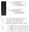
Figure 7. Phylogeny of fungal CENP-T and CENP-W
A. Neurospora CEN-T-GFP localizes to chromocenters, here shown in a cluster of asexual spores. Spores typically contain 2 – 3 nuclei and each nucleus has a single bright focus. Phylogenies for CEN-T (B) and CEN-W (C) were constructed from predicted protein sequences of representative filamentous fungi as well as S. pombe, zebrafish and human. Phylogenetic relationships between the taxa are maintained in these single-protein trees. Alignments of the conserved histone-like domain of CEN-T homologs (D) or the whole CEN-W proteins (E) from selected filamentous fungi as well as S. pombe, zebrafish and human. Residues to be predicted as necessary for tetramer formation (Nishino et al., 2012) are underlined and appear conserved between animal and fungal CEN-T. Residues that may be involved in DNA-binding are shown in bold.
Figure 8
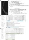
Figure 8. Phylogeny of fungal CENP-S and CENP-X
A. Neurospora CEN-S-GFP localizes to chromocenters, here shown in a germinating asexual spore (lower right). Chromocenters are oriented towards the growing tip (top center), and each nucleus has one single bright CEN-S-GFP focus. Phylogenies for CEN-S (B) and CEN-X (C) were constructed from predicted protein sequences of representative filamentous fungi as well as A. gossypii, S. pombe, zebrafish and human. Phylogenetic relationships between the taxa are largely maintained in these single-protein trees. Alignments of CEN-S homologs (D) or the histone-like domain from CEN-X proteins (E) from selected filamentous fungi as well as A. gossypii,S. pombe, zebrafish and human. Residues to be predicted as necessary for tetramer formation (Nishino et al., 2012) are underlined and appear conserved between animal and fungal CEN-T. Residues that may be involved in DNA-binding are shown in bold.

Similar articles

Cited by

References

    1. Allshire RC. Centromeres, checkpoints and chromatid cohesion. Curr Opin Genet Dev. 1997;7:264–273. - PubMed
    1. Bailey TL, Boden M, Buske FA, et al. MEME SUITE: tools for motif discovery and searching. Nucleic Acids Res. 2009;37:W202–208. - PMC - PubMed
    1. Baker RE, Rogers K. Phylogenetic analysis of fungal centromere H3 proteins. Genetics. 2006;174:1481–1492. - PMC - PubMed
    1. Baum M, Clarke L. Fission yeast homologs of human CENP-B have redundant functions affecting cell growth and chromosome segregation. Mol Cell Biol. 2000;20:2852–2864. - PMC - PubMed
    1. Baum M, Sanyal K, Mishra PK, Thaler N, Carbon J. Formation of functional centromeric chromatin is specified epigenetically in Candida albicans. Proc Natl Acad Sci U S A. 2006;103:14877–14882. - PMC - PubMed

Publication types

MeSH terms