. 2012 Aug;91(2):139-48.
doi: 10.1007/s00223-012-9623-4. Epub 2012 Jul 1.

Rosiglitazone inhibits bone regeneration and causes significant accumulation of fat at sites of new bone formation

Affiliations

Rosiglitazone inhibits bone regeneration and causes significant accumulation of fat at sites of new bone formation

Lichu Liu et al. Calcif Tissue Int. 2012 Aug.

Abstract

Thiazolidinediones (TZDs), peroxisome proliferator-activated receptor gamma activators, and insulin sensitizers represent drugs used to treat hyperglycemia in diabetic patients. Type 2 diabetes mellitus (T2DM) is associated with a twofold increase in fracture risk, and TZDs use increases this risk by an additional twofold. In this study, we analyzed the effect of systemic administration of the TZD rosiglitazone on new bone formation in two in vivo models of bone repair, a model of drilled bone defect regeneration (BDR) and distraction osteogenesis (DO) and a model of extended bone formation. Rosiglitazone significantly inhibited new endosteal bone formation in both models. This effect was correlated with a significant accumulation of fat cells, specifically at sites of bone regeneration. The diminished bone regeneration in the DO model in rosiglitazone-treated animals was associated with a significant decrease in cell proliferation measured by the number of cells expressing proliferating cell nuclear antigen and neovascularization measured by both the number of vascular sinusoids and the number of cells producing proangiogenic vascular endothelial growth factor at the DO site. In summary, rosiglitazone decreased new bone formation in both BDR and DO models of bone repair by mechanisms which include both intrinsic changes in mesenchymal stem cell proliferation and differentiation and changes in the local environment supporting angiogenesis and new bone formation. These studies suggest that bone regeneration may be significantly compromised in T2DM patients on TZD therapy.


Conflict of interest statement

The authors have stated that they have no conflict of interest.

Figures

Fig. 1
Fig. 1
Sagittal μCT renderings of murine tibia model of drilled bone defect regeneration (BDR). The skeletal injury was generated in the tibia of a C57BL/6 mouse as described in “Materials and Methods” section. Mice were killed at different time points after surgery, representing the inflammatory (1 week), hard callus (2 weeks), and remodeling (3 and 4 weeks) phases of healing [21]. New bone at the injury site with a poorly organized trabecular structure forms approximately 2 weeks postsurgery. Three weeks postsurgery the injury site is covered by a layer of bone on the periosteal side and the process of remodeling of newly formed woven bone in the endosteum is initiated. Almost complete cortical bone restoration and woven bone remodeling are observed at 4 weeks postsurgery
Fig. 2
Fig. 2
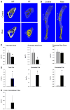
The effect of rosiglitazone on bone regeneration and fat accumulation in the BDR model. Two weeks after surgery both tibiae, with a defect and contralateral, were harvested for μCT assessment of mineralized tissue mass and fat mass at the site of the defect or at the corresponding site in the contralateral tibia. a Representative μCT renderings of horizontal cross sections of the tibia with a drilled cortical defect harvested from control and rosiglitazone-treated animals. MT renderings of mineralized tissues, FT renderings of fat stained with osmium tetroxide after bone demineralization, as described in “Materials and Methods” section. b μCT renderings of the whole tibia harvested 2 weeks postsurgery, demineralized, and stained for fat with osmium tetroxide. White brackets mark the area of the drilled bone defect. c μCT measurements of a new bone formed either at the entire defect site (total new bone) or at the endosteal (endosteal new bone) or periosteal (periosteal new bone) location of the bone defect. d μCT measurements of fat content at the entire defect site (total fat) or at the endosteal or periosteal location of the defect site. e Measurement of fat content in the contralateral nonoperated tibia in the region corresponding to the drilled defect in the operated tibia. Gray bars, control; black bars, rosiglitazone-treated animals. *p < 0.05 vs. control (Color figure online)
Fig. 3
Fig. 3
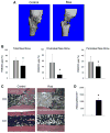
The effect of rosiglitazone on bone regeneration and fat accumulation in the DO model. a μCT renderings of newly formed bone in the DO gap after 2 weeks of distraction. b μCT measurements of new bone occupying either the entire DO gap (total new bone [TNB]) or the endosteal (endosteal new bone [ENB]) or periosteal (periosteal new bone [PNB]) location of the DO gap. c Histological visualization of adipocytes at the DO site and in the corresponding region of contralateral, nonoperated tibia (CO) from mice treated or not with rosiglitazone. Specimens were stained with hematoxylin and eosin. Note decreased new bone formation at the DO site and large quantities of fat (white oval spaces) accumulated in the DO zone of microcolumn formation and in the marrow adjacent to the DO site in rosiglitazone-treated animals (magnification ×10). d Quantification of adipocytes at the DO site of nontreated and rosiglitazone-treated mice. Gray bars, control; black bars, rosiglitazone. *p < 0.05 vs. control (Color figure online)
Fig. 4
Fig. 4
Immunohistochemical analyses of PCNA-positive and hematopoietic VEGF-producing cells and the number of hematopoietic sinusoids at the DO gap after 2 weeks of distraction. a Visualization and quantification of cells stained for PCNA (brown) on the surface and in close proximity to the newly formed bone at the DO site. Magnification ×40. b Quantification of VEGF-positive cells in the DO gap of control and rosiglitazone-treated animals. Microphotograph illustrates a typical location of VEGF-positive cells on the surface of new bone formed in the DO gap (brown). c Number of hematopoietic sinusoids in the DO gap area. Sinusoids were identified by the presence of CD34-positive cells in the surrounding peripheral area (brown). Red arrows indicate cells stained positively for the tested antigen. Magnification ×40. Gray bars, control; black bars, rosiglitazone. *p < 0.05 vs. control (Color figure online)

References

    1. Lecka-Czernik B. Bone loss in diabetes: use of antidiabetic thiazolidinediones and secondary osteoporosis. Curr Osteoporos Rep. 2010;8:178–184. - PMC - PubMed
    1. Vestergaard P, Rejnmark L, Mosekilde L. Diabetes and its complications and their relationship with risk of fractures in type 1 and 2 diabetes. Calcif Tissue Int. 2009;84:45–55. - PubMed
    1. Janghorbani M, Van Dam RM, Willett WC, Hu FB. Systematic review of type 1 and type 2 diabetes mellitus and risk of fracture. Am J Epidemiol. 2007;166:495–505. - PubMed
    1. Kahn SE, Zinman B, Lachin JM, Haffner SM, Herman WH, Holman RR, Kravitz BG, Yu D, Heise MA, Aftring RP, Viberti G. Rosiglitazone-associated fractures in type 2 diabetes: an analysis from a diabetes outcome progression trial (ADOPT) Diabetes Care. 2008;31:845–851. - PubMed
    1. Loke YK, Singh S, Furberg CD. Long-term use of thiazolidinediones and fractures in type 2 diabetes: a meta-analysis. CMAJ. 2009;180:32–39. - PMC - PubMed

Publication types

MeSH terms