. 2012 Sep 1;303(5):H605-18.
doi: 10.1152/ajpheart.00366.2012. Epub 2012 Jun 29.

Apelin-13 increases myocardial progenitor cells and improves repair postmyocardial infarction

Affiliations

Apelin-13 increases myocardial progenitor cells and improves repair postmyocardial infarction

Lanfang Li et al. Am J Physiol Heart Circ Physiol. .

Abstract

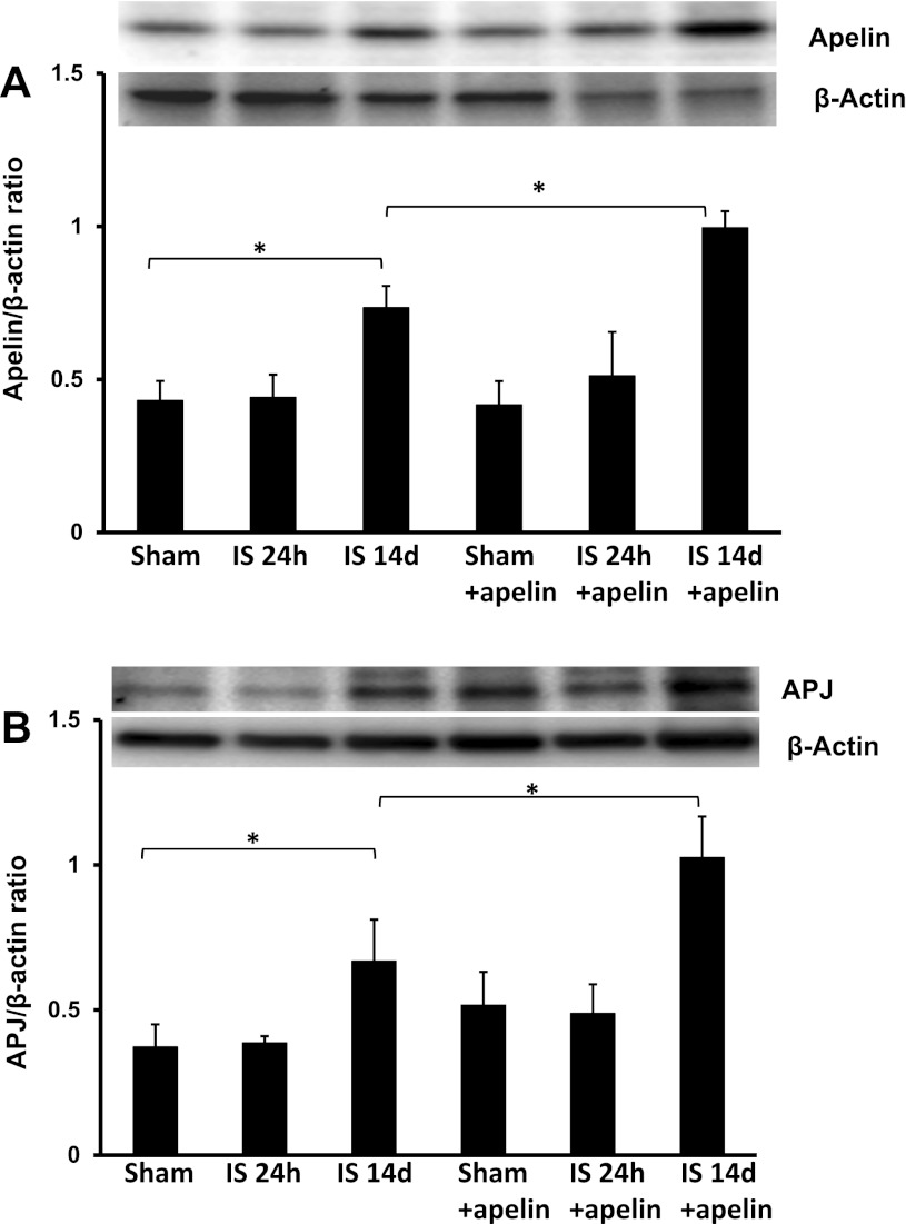
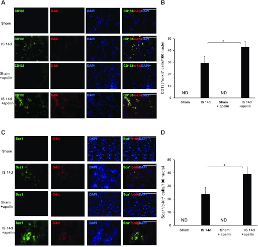
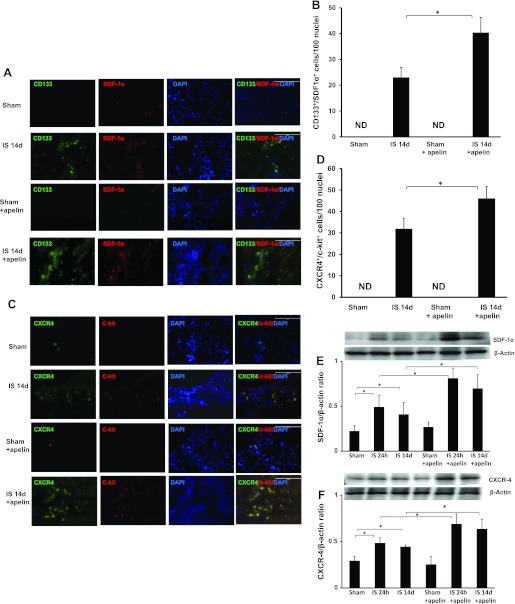
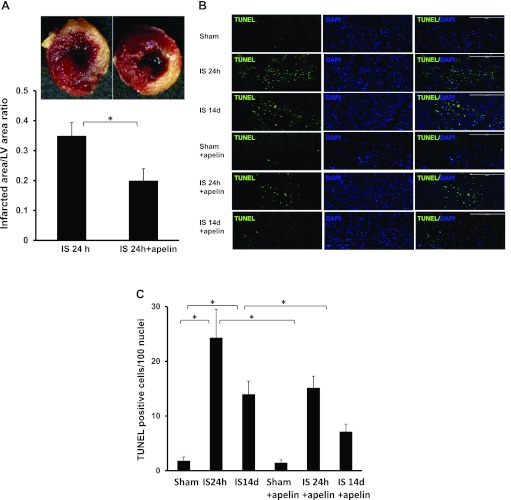
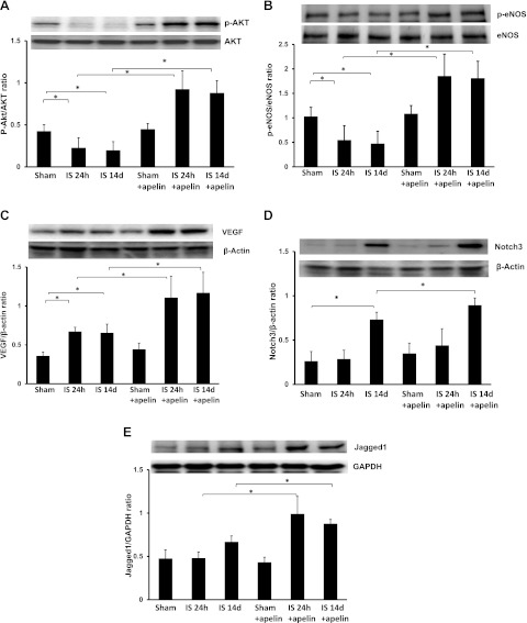
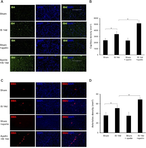
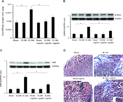
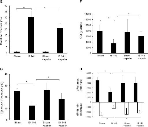
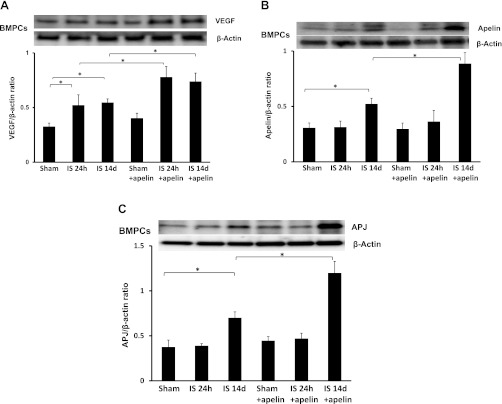
Apelin is an endogenous ligand for the angiotensin-like 1 receptor (APJ) and has beneficial effects against myocardial ischemia-reperfusion injury. Little is known about the role of apelin in the homing of vascular progenitor cells (PCs) and cardiac functional recovery postmyocardial infarction (post-MI). The present study investigated whether apelin affects PC homing to the infarcted myocardium, thereby mediating repair and functional recovery post-MI. Mice were infarcted by coronary artery ligation, and apelin-13 (1 mg·kg(-1)·day(-1)) was injected for 3 days before MI and for 14 days post-MI. Homing of vascular PCs [CD133(+)/c-Kit(+)/Sca1(+), CD133(+)/stromal cell-derived factor (SDF)-1α(+), and CD133(+)/CXC chemokine receptor (CXCR)-4(+)] into the ischemic area was examined. Myocardial Akt, endothelial nitric oxide synthase (eNOS), VEGF, jagged1, notch3, SDF-1α, and CXCR-4 expression were assessed at 24 h and 14 days post-MI. Functional analyses were performed on day 14 post-MI. Mice that received apelin-13 treatment demonstrated upregulation of SDF-1α/CXCR-4 expression and dramatically increased the number of CD133(+)/c-Kit(+)/Sca1(+), CD133(+)/SDF-1α(+), and c-Kit(+)/CXCR-4(+) cells in infarcted hearts. Apelin-13 also significantly increased Akt and eNOS phosphorylation and upregulated VEGF, jagged1, and notch3 expression in ischemic hearts. This was accompanied by a significant reduction of myocardial apoptosis. Furthermore, treatment with apelin-13 promoted myocardial angiogenesis and attenuated cardiac fibrosis and hypertrophy together with a significant improvement of cardiac function at 14 days post-MI. Apelin-13 increases angiogenesis and improves cardiac repair post-MI by a mechanism involving the upregulation of SDF-1α/CXCR-4 and homing of vascular PCs.


Figures

Fig. 1.
Fig. 1.
Apelin-13 increases apelin and angiotensin-like 1 receptor (APJ) expression in ischemic hearts. A: results from Western blot analysis demonstrating that exposure to ischemia (IS) for 14 days leads to a significant increase in apelin expression compared with sham-operated (sham) control mice. Treatment with apelin-13 further upregulated apelin expression in ischemic mouse hearts at 14 days. These changes were not seen at 24 h (n = 4 mice/group). *P < 0.05. B: results from Western blot analysis demonstrating that exposure to ischemia for 14 days leads to a significant increase in APJ expression compared with sham control mice. Treatment with apelin-13 further increased APJ expression in ischemic mouse hearts at 14 days. These changes were not apparent at 24 h (n = 4 mice/group). *P < 0.05.
Fig. 2.
Fig. 2.
Apelin-13 promotes vascular progenitor cell (PC) homing into ischemic hearts. A: representative images showing the colocalization of CD133 with c-Kit in the border zone of infarcted mouse hearts at 14 days postmyocardial infaction (post-MI). CD133+ cells (green), c-Kit+ cells (red), and nuclei stained by 4′,6-diamidino-2-phenylindole (DAPI; blue) are shown. Magnification: ×40. CD133+ and c-Kit+ cells were recruited into the ischemic area of mouse hearts at day 14 post-MI. Merged images show that CD133+ cells colocalized with c-Kit+ cells (yellow) in post-MI animals and that the increase was more striking in post-MI animals that received apelin-13. No specific staining was observed in the sham control groups. B: results from quantitative analysis of CD133+/c-Kit+ cells demonstrating that the number of CD133+/c-Kit+ cells was significantly increased in apelin-13-treated mice subjected to MI compared with control ischemic mice. CD133+/c-Kit+ cells are expressed as total numbers per 100 nuclei. ND, not detected. All data are means ± SD; n = 4 mice/group. *P < 0.05. C: immunofluorescence images showing the colocalization of Sca1 and c-Kit in the border zone of ischemic mouse hearts. Sca1 was stained with mouse Sca1 antibody (green), c-Kit was stained with c-Kit antibody (red), and nuclei were stained with DAPI (blue). Magnification: ×40. Merged images showed that Sca1 colocalized with c-Kit (yellow) in hearts from post-MI mice and that the increase was more dramatic in post-MI animals that received apelin-13. No specific staining was observed in the sham control groups. D: results from quantitative analysis of Sca1+/c-Kit+ cells demonstrating that the number of Sca1+/c-Kit+ cells was significantly increased in apelin-13-treated mice subjected to MI compared with control ischemic mice. Sca1+/c-Kit+ cells are expressed as total numbers per 100 nuclei. All data are means ± SD; n = 4 mice/group. *P < 0.05.
Fig. 3.
Fig. 3.
Apelin-13 upregulates stromal cell-derived factor (SDF)-1α and C-X-C chemokine receptor (CXCR)-4 expression in ischemic hearts. A: immunofluorescence images showing the colocalization of CD133 with SDF-1α in the border zone of infarcted mouse hearts. CD133+ cells (green), SDF-1α+ cells (red), and nuclei stained by DAPI (blue) are shown. Magnification: ×40. Merged images show that CD133+ cells colocalized with SDF-1α+ in ischemic control and apelin-13-treated ischemic mice. No specific staining was observed in the sham control groups. B: results from quantitative analysis of CD133+/SDF-1α+ cells demonstrating that the number of CD133+/SDF-1α+ cells was significantly increased in apelin-13-treated mice subjected to MI compared with control ischemic mice. CD133+/SDF-1α+ cells are expressed as total numbers per 100 nuclei. All data are means ± SD; n = 4 mice/group. *P < 0.05. C: immunofluorescence images showing colocalization of c-Kit with CXCR-4 in the border zone of infarcted mouse hearts. CXCR-4+ cells (green), c-Kit+ cells (red), and nuclei stained by DAPI (blue) are shown. Magnification: ×40. Merged images show that CXCR-4+ cells colocalized with c-Kit+ in ischemic control and apelin-13-treated ischemic mice. No specific staining was observed in the sham control groups. D: results from quantitative analysis of CXCR-4+/c-Kit+ cells demonstrating that the number of CXCR-4+/c-Kit+ cells was significantly increased in apelin-13-treated mice subjected to MI compared with control ischemic mice. CXCR-4+/c-Kit+ cells are expressed as total numbers per 100 nuclei. All data are means ± SD; n = 4 mice/group. *P < 0.05. E: results from Western blot and densitometric analyses demonstrating that SDF-1α expression was significantly increased in mouse hearts subjected to ischemia for 24 h and 14 days compared with sham control hearts. Treatment with apelin-13 significantly increased SDF-1α expression in ischemic hearts compared with ischemic control harts. All data are means ± SD; n = 4 mice/group. *P < 0.05. F: results from Western blot and densitometric analyses of myocardial CXCR-4 expression showing that expression was significantly increased in mouse hearts subjected to ischemia for 24 h and 14 days compared with sham control hearts. Treatment with apelin-13 significantly increased CXCR-4 expression in ischemic hearts compared with ischemic control hearts. All data are means ± SD; n = 4 mice/group. *P < 0.05.
Fig. 4.
Fig. 4.
Apelin-13 attenuates myocardial apoptosis and reduces infarct size. A: representative images and quantification of infarct size in the left ventricle of control post-MI and apelin-13-treated mice after 24 h of ischemia. The noninfarcted area appears red and the infarct area appears white after 2,3,5-triphenyltetrazolium chloride (TTC) staining. The myocardial infarct area was significantly reduced in apelin-13-treated mice compared with control post-MI mice (n = 3 mice/group). *P < 0.05. B: representative images of TUNEL-stained heart sections of the infarcted area from sham control and apelin-13-treated mice 24 h and 14 days post-MI. Apoptotic cells in the infarcted area of the left ventricle were identified by TUNEL staining (green) and total nuclei by DAPI counterstaining (blue). Magnification: ×10. TUNEL staining was seen post-MI at 24 h and 14 days, but in apelin-treated mice, fewer TUNEL+ cells were found at each time point. C: results from quantitative analysis of apoptotic cells in the infarcted area of control and apelin-13-treated mouse hearts 24 h and 14 days after sham surgery or ischemia. TUNEL+ cells are expressed as total numbers per 100 nuclei in the infracted area. Apoptotic cells were significantly increased post-MI at 24 and 14 days. Significantly fewer TUNEL+ cells were seen in apelin-13-treated mice at both time points (n = 4 mice/group). *P < 0.05.
Fig. 5.
Fig. 5.
Apelin-13 increases survival and angiogenic growth factor expression in ischemic hearts. A: representative results from Western blot and densitometric analyses of myocardial Akt phosphorylation. Phosphorylated (p-)Akt was significantly reduced in mouse hearts subjected to ischemia for 24 h and 14 days compared with sham control hearts. Treatment with apelin-13 significantly prevented ischemia-induced downregulation of Akt phosphorylation and increased levels over sham control values at both time points (n = 4 mice/group). *P < 0.05. B: representative results from Western blot and densitometric analyses of myocardial endothelial nitric oxide synthase (eNOS) phosphorylation. p-eNOS was significantly reduced in mouse hearts subjected to ischemia for 24 h and 14 days compared with sham control hearts. Treatment with apelin-13 significantly prevented and increased levels over sham control levels of ischemia-induced downregulation of eNOS phosphorylation. (n = 4 mice/group). *P < 0.05. C: representative results from Western blot and densitometry analyses of myocardial VEGF protein expression at 24 h and 14 days after myocardial ischemia. VEGF expression was significantly increased in ischemic control mice compared with sham control mice at 24 h and 14 days. Treatment with apelin-13 resulted in a significant increase in VEGF expression compared with control mice subjected to myocardial ischemia for 24 h and 14 days and increased levels over sham controls (n = 4 mice/group). *P < 0.05. D: representative results from Western blot and densitometric analyses of Notch3 expression. Notch3 expression was significantly increased in ischemic control mice compared with sham control mice at 14 days. Treatment with apelin-13 resulted in significant increases in Notch3 expression compared with ischemic control mice at 14 days (n = 4 mice/group). *P < 0.05. E: representative results from Western blot and densitometric analyses of jagged1 expression. Jagged1 expression was similar in sham control mice at 24 h and 14 days post-MI. Treatment with apelin-13 resulted in a significant increase in jagged1 expression compared with ischemic control mice at 24 h and 14 days post-MI (n = 4 mice/group). *P < 0.05.
Fig. 6.
Fig. 6.
Apelin-13 enhances myocardial neovascularization in ischemic hearts. A and B: representative images (A) and quantitative analysis (B) showing that myocardial ischemia significantly increased myocardial capillary density by isolectin B4 (IB4) staining at 14 days. Treatment of ischemic mice with apelin-13 significantly increased capillary formation compared with ischemic control mice (n = 5 mice/group). *P < 0.05. C and D: representative images (C) and quantitative analysis (D) of α-smooth muscle actin (SMA) staining showing that myocardial ischemia significantly increased myocardial arteriole density in ischemic control mice compared with sham control mice at 14 days. Treatment of ischemic mice with apelin-13 caused a significant increase in arteriole formation compared with ischemic control mice (n = 6 mice/group). *P < 0.05.
Fig. 7.
Fig. 7.
Apelin-13 reduces cardiac hypertrophy/fibrosis and improves cardiac functional recovery in post-MI mice. A: heart weight-to-body weight ratio in sham control, ischemic, and apelin-13-treated mouse hearts at 24 h and 14 days post-MI. A significant increase in cardiac hypertrophy was observed in mouse hearts after 14 days of ischemia. Apelin-13 treatment significantly suppressed cardiac hypertrophy compared with mice treated with saline (n = 4–6 mice/group). *P < 0.05. B: results from Western blot and densitometric analyses demonstrating that β-myosin heavy chain (β-MHC) expression was significantly increased in 14-day post-MI mice compared with sham control mice. Treatment with apelin-13 significantly decreased β-MHC expression in 14-day post-MI mice compared with ischemic control mice. All data are means ± SD; n = 4 mice/group. *P < 0.05. C: results from Western blot and densitometric analyses demonstrating that atrial natriuretic peptide (ANP) expression was significantly increased in 14-day post-MI mice compared with sham control mice. Treatment with apelin-13 significantly attenuated ANP expression in 14-day post-MI mice compared with ischemic control mice. All data are means ± SD; n = 4 mice/group. *P < 0.05. D and E: representative images of cardiac fibrosis in the infarction zone (D) and quantitative analysis of the fibrotic area (E) in mice treated with saline or apelin-13 (Masson's trichrome stain). Apelin-13 significantly reduced the area of cardiac fibrosis (blue) in ischemic mice compared with saline-treated ischemic mice. All data are means ± SD; n = 5 mice/group. *P < 0.05. F: cardiac output (CO) was significantly reduced in 14-day post-MI mice (n = 6) compared with sham control mice (n = 5). Apelin-13 treatment (n = 6) significantly increased CO compared with ischemic control mice. All data are means ± SD; *P < 0.05. G: myocardial ejection fraction (EF; in %) was significantly decreased in 14-day post-MI mice (n = 6) compared with sham control mice (n = 5). Apelin-13 treatment (n = 6) significantly increased EF compared with ischemic control mice. All data are means ± SD. *P < 0.05. H: left ventricular function was significantly improved in ischemic mice treated with apelin-13 compared with saline-treated ischemic mice, as shown by the higher +dP/dtmax and lower −dP/dtmin at 14 days of ischemia. All data are means ± SD; n = 6 apelin-13-treated ischemic mice, 6 saline-treated ischemic mice, and 5 sham control mice. *P < 0.05.
Fig. 7.
Fig. 7.
Apelin-13 reduces cardiac hypertrophy/fibrosis and improves cardiac functional recovery in post-MI mice. A: heart weight-to-body weight ratio in sham control, ischemic, and apelin-13-treated mouse hearts at 24 h and 14 days post-MI. A significant increase in cardiac hypertrophy was observed in mouse hearts after 14 days of ischemia. Apelin-13 treatment significantly suppressed cardiac hypertrophy compared with mice treated with saline (n = 4–6 mice/group). *P < 0.05. B: results from Western blot and densitometric analyses demonstrating that β-myosin heavy chain (β-MHC) expression was significantly increased in 14-day post-MI mice compared with sham control mice. Treatment with apelin-13 significantly decreased β-MHC expression in 14-day post-MI mice compared with ischemic control mice. All data are means ± SD; n = 4 mice/group. *P < 0.05. C: results from Western blot and densitometric analyses demonstrating that atrial natriuretic peptide (ANP) expression was significantly increased in 14-day post-MI mice compared with sham control mice. Treatment with apelin-13 significantly attenuated ANP expression in 14-day post-MI mice compared with ischemic control mice. All data are means ± SD; n = 4 mice/group. *P < 0.05. D and E: representative images of cardiac fibrosis in the infarction zone (D) and quantitative analysis of the fibrotic area (E) in mice treated with saline or apelin-13 (Masson's trichrome stain). Apelin-13 significantly reduced the area of cardiac fibrosis (blue) in ischemic mice compared with saline-treated ischemic mice. All data are means ± SD; n = 5 mice/group. *P < 0.05. F: cardiac output (CO) was significantly reduced in 14-day post-MI mice (n = 6) compared with sham control mice (n = 5). Apelin-13 treatment (n = 6) significantly increased CO compared with ischemic control mice. All data are means ± SD; *P < 0.05. G: myocardial ejection fraction (EF; in %) was significantly decreased in 14-day post-MI mice (n = 6) compared with sham control mice (n = 5). Apelin-13 treatment (n = 6) significantly increased EF compared with ischemic control mice. All data are means ± SD. *P < 0.05. H: left ventricular function was significantly improved in ischemic mice treated with apelin-13 compared with saline-treated ischemic mice, as shown by the higher +dP/dtmax and lower −dP/dtmin at 14 days of ischemia. All data are means ± SD; n = 6 apelin-13-treated ischemic mice, 6 saline-treated ischemic mice, and 5 sham control mice. *P < 0.05.
Fig. 8.
Fig. 8.
Treatment with apelin-13 increases VEGF and apelin/APJ expression in bone marrow (BM)-derived PCs (BMPCs). A: results from Western blot analyses of VEGF expression in BMPCs. VEGF expression was significantly upregulated in BMPCs of apelin-13-treated ischemic mice compared with saline-treated mice at 24 h and 14 days post-MI. B and C: results from Western blot analyses of apelin (B) and APJ (C) expression in BMPCs. Apelin and APJ expression were upregulated in BMPCs of 14-day post-MI mice compared with sham control mice. Treatment with apelin-13 for 14 days further increased apelin and APJ expression in BMPCs of post-MI mice.

Similar articles

Cited by

References

    1. Arboleda-Velasquez JF, Zhou Z, Shin HK, Louvi A, Kim HH, Savitz SI, Liao JK, Salomone S, Ayata C, Moskowitz MA, Artavanis-Tsakonas S. Linking notch signaling to ischemic stroke. Proc Natl Acad Sci USA 105: 4856–4861, 2008 - PMC - PubMed
    1. Askari AT, Unzek S, Popovic ZB, Goldman CK, Forudi F, Kiedrowski M, Rovner A, Ellis SG, Thomas JD, DiCorleto PE, Topol EJ, Penn MS. Effect of stromal-cell-derived factor 1 on stem-cell homing and tissue regeneration in ischaemic cardiomyopathy. Lancet 362: 697–703, 2003 - PubMed
    1. Barcelos LS, Duplaa C, Krankel N, Graiani G, Invernici G, Katare R, Siragusa M, Meloni M, Campesi I, Monica M, Simm A, Campagnolo P, Mangialardi G, Stevanato L, Alessandri G, Emanueli C, Madeddu P. Human CD133+ progenitor cells promote the healing of diabetic ischemic ulcers by paracrine stimulation of angiogenesis and activation of Wnt signaling. Circ Res 104: 1095–1102, 2009 - PMC - PubMed
    1. Britten MB, Abolmaali ND, Assmus B, Lehmann R, Honold J, Schmitt J, Vogl TJ, Martin H, Schachinger V, Dimmeler S, Zeiher AM. Infarct remodeling after intracoronary progenitor cell treatment in patients with acute myocardial infarction (TOPCARE-AMI): mechanistic insights from serial contrast-enhanced magnetic resonance imaging. Circulation 108: 2212–2218, 2003 - PubMed
    1. Chen JX, Stinnett A. Ang-1 gene therapy inhibits hypoxia-inducible factor-1α (HIF-1α)-prolyl-4-hydroxylase-2, stabilizes HIF-1α expression, and normalizes immature vasculature in db/db mice. Diabetes 57: 3335–3343, 2008 - PMC - PubMed

Publication types

MeSH terms

Substances