. 2012 Nov;11(8):1219-25.
doi: 10.1007/s10237-012-0413-8. Epub 2012 Jul 3.

Cardiomyocytes from late embryos and neonates do optimal work and striate best on substrates with tissue-level elasticity: metrics and mathematics

Affiliations

Cardiomyocytes from late embryos and neonates do optimal work and striate best on substrates with tissue-level elasticity: metrics and mathematics

Stephanie F Majkut et al. Biomech Model Mechanobiol. 2012 Nov.

Abstract

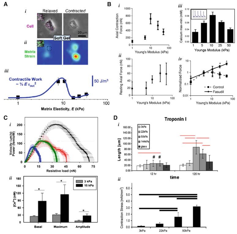
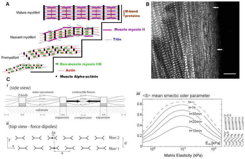
In this review, we discuss recent studies on the mechanosensitive morphology and function of cardiomyocytes derived from embryos and neonates. For early cardiomyocytes cultured on substrates of various stiffnesses, contractile function as measured by force production, work output and calcium handling is optimized when the culture substrate stiffness mimics that of the tissue from which the cells were obtained. This optimal contractile function corresponds to changes in sarcomeric protein conformation and organization that promote contractile ability. In light of current models for myofibillogenesis, a recent mathematical model of striation and alignment on elastic substrates helps to illuminate how substrate stiffness modulates early myofibril formation and organization. During embryonic heart formation and maturation, cardiac tissue mechanics change dynamically. Experiments and models highlighted here have important implications for understanding cardiomyocyte differentiation and function in development and perhaps in regeneration processes.


Figures

Fig. 1
Fig. 1
Isolated cardiomyocytes plated on gels of various stiffnesses. a Engler et al. (2008) characterized the morphological and functional effects of substrate stiffness on embryonic cardiomyocytes isolated from E7 chick embryos cultured on collagen-I-coated polyacrylamide (PA) gels. They found that the cells put out the most work on substrates of ~10 kPa. This optimal substrate stiffness matches that of E7 myocardium as they measured by AFM. b Jacot et al. (2008) cultured at NRVM on PA gels of 1, 10, and 50 kPa. They found that 10 kPa optimized NRVM function measured by contractile force-generation and calcium activity (Jacot et al. 2010). Furthermore, inhibition of ROCK and RhoA pathways eliminates the decreased force production of NRVM on stiff gels. c Rodriguez et al. (2011) cultured NRVM on fibronectin-coated microposts with an narrow range of effective moduli ranging from 3 to 20 kPa and found that twitch force, work, and power increased with substrate stiffness. In addition, calcium activity increased in NRVM on stiffer substrates. d Bhana et al. (2009) co-cultured both NRVM and fibroblasts isolated from the same tissue on PA gels for 5 days and monitored not just NRVM morphology and function, but the relative population changes in fibroblasts and cardiomyocytes. They found that cardiomyocyte function and population relative to fibroblasts were optimal at substrate stiffness of 22–50 kPa relative to soft (3 kPa) and stiff (144 kPa)
Fig. 2
Fig. 2
Myofibril formation and registration modulation by substrate stiffness. a Premyofibril model for myofibril formation (Sanger et al. 2005). Striated premyofibrils comprised of alpha-actinin-enriched z-bodies, short actin thin filaments, and nonmuscle myosin IIB filaments mature into mature myofibrils by replacement of nonmuscle myosin II with muscle myosin II and incorporation of other sarcomeric proteins. b Premyofibril formation, registration, and maturation into mature myofibrils have been visualized in live spreading cardiomyocytes, as shown Sanger et al. (2005), in precardiac explants, and in whole embryonic hearts (Sanger et al. 2010). White arrows indicate premoyfibrils deposited near the edge of the spreading cell. c Theoretical model proposed by Friedrich et al. (2011) showing how aligned striated fibers on elastic substrates may come into registry in a substrate stiffness–dependent manner. i Striated fibers apply stresses at the cell–substrate interface that can be modeled as periodic line of force dipoles. ii Adjacent fibers interact through the laterally propagating strain fields they produce. iii These interactions lead fibers to come into registry with each other in a nonmonotonic substrate-elasticity-dependent way. The smectic order S of the resulting arrays of striations is a measure of the level of registration

Similar articles

Cited by

References

    1. Bajaj P, Tang X, Saif TA, Bashir R. Stiffness of the substrate influences the phenotype of embryonic chicken cardiac myocytes. J Biomed Mater Res Part A. 2010;95(4):1261–1269. - PubMed
    1. Bers DM. Excitation-contraction coupling and cardiac contractile force. 2. Kluwer; Dordrecht: 2001.
    1. Bhana B, Iyer RK, Chen WLK, Zhao R, Sider KL, Likhitpanichkul M, Simmons CA, Radisic M. Influence of substrate stiffness on the phenotype of heart cells. Biotechnol Bioeng. 2009;105(6):2151–2162. - PubMed
    1. Cadete VJJ, Sawicka J, Polewicz D, Doroszko A, Wozniak M, Sawicki G. Effects of the Rho kinase inhibitor Y-27632 on the proteome of hearts with ischemia-reperfusion injury. Proteomics. 2010;10(24):4377–4385. - PubMed
    1. Engler AJ, Carag-Krieger C, Johnson CP, Raab M, Tang H, Spelcher DW, Sanger JW, Sanger JM, Discher DE. Embryonic cardiomyocytes beat best on a matrix with heart-like elasticity: scar-like rigidity inhibits beating. J Cell Sci. 2008;121:3794–3802. - PMC - PubMed

Publication types

LinkOut - more resources