. 2013 Jan;26(1):98-109.
doi: 10.1007/s10548-012-0235-0. Epub 2012 Jun 30.

Electrophysiological correlation patterns of resting state networks in single subjects: a combined EEG-fMRI study

Affiliations

Electrophysiological correlation patterns of resting state networks in single subjects: a combined EEG-fMRI study

Matthias C Meyer et al. Brain Topogr. 2013 Jan.

Abstract

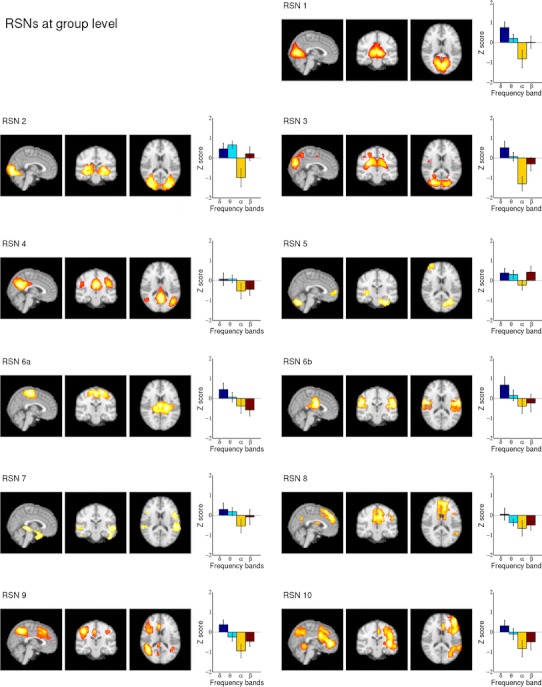
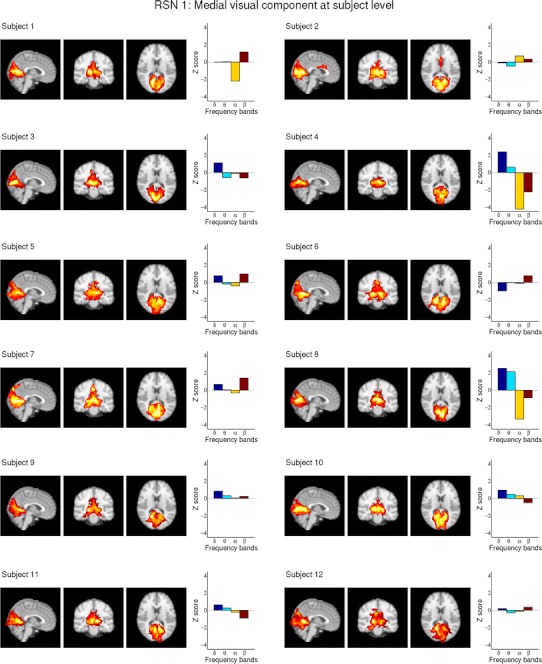
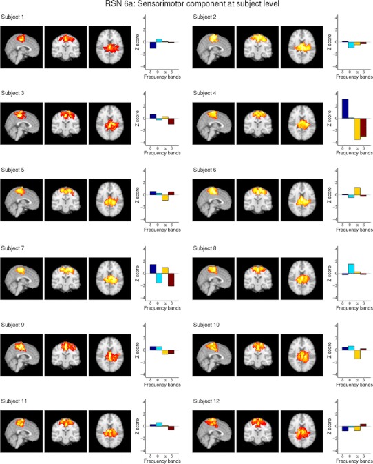
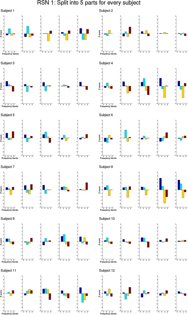
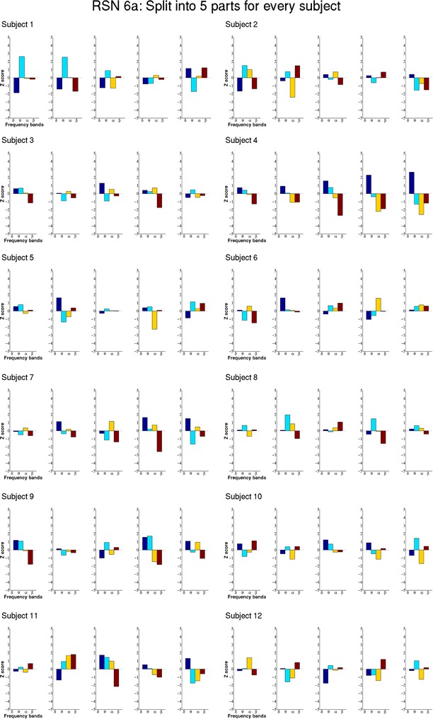
With combined EEG-fMRI a powerful combination of methods was developed in the last decade that seems promising for answering fundamental neuroscientific questions by measuring functional processes of the human brain simultaneously with two complementary modalities. Recently, resting state networks (RSNs), representing brain regions of coherent BOLD fluctuations, raised major interest in the neuroscience community. Since RSNs are reliably found across subjects and reflect task related networks, changes in their characteristics might give insight to neuronal changes or damage, promising a broad range of scientific and clinical applications. The question of how RSNs are linked to electrophysiological signal characteristics becomes relevant in this context. In this combined EEG-fMRI study we investigated the relationship of RSNs and their correlated electrophysiological signals [electrophysiological correlation patterns (ECPs)] using a long (34 min) resting state scan per subject. This allowed us to study ECPs on group as well as on single subject level, and to examine the temporal stability of ECPs within each subject. We found that the correlation patterns obtained on group level show a large inter-subject variability. During the long scan the ECPs within a subject show temporal fluctuations, which we interpret as a result of the complex temporal dynamic of the RSNs.


Figures

Fig. 1
Fig. 1
All 11 group fMRI RSNs as maximum intensity projection on the central slices and their group ECPs, representing the average Z scores (12 subjects) for the four EEG frequency bands. Only clusters larger than 15 voxels were plotted. The large standard errors indicate the large variability of the subject-specific ECPs
Fig. 2
Fig. 2
Depicts RSN 1 (medial visual component) after dual regression on single subject level as maximum intensity projection on the central slices and the subject specific ECPs for all 12 subjects, showing the high inter-subject variability of the ECPs but also significant negative alpha correlation in subject 4 and subject 8. For visualization purposes a cluster threshold of 100 voxels and a minimum intensity threshold of 25 % was used
Fig. 3
Fig. 3
Depicts RSN6a (sensorimotor component) after dual regression on single subject level as maximum intensity projection on the central slices and the subject specific ECPs for all 12 subjects, showing the high inter-subject variability of the ECPs. Subject 4 shows significant positive delta correlation and negative alpha and beta correlation. For visualization purposes a cluster threshold of 100 voxels and a minimum intensity threshold of 25 % was used
Fig. 4
Fig. 4
Shows the ECPs of RSN 1 (medial visual component) for all five parts of the split datasets for all 12 subjects. The ECPs show higher Z scores at these shorter time intervals and the patterns change over time
Fig. 5
Fig. 5
Shows the ECPs of RSN 6a (sensorimotor component) for all five parts of the split datasets for all 12 subjects. The ECPs show higher Z scores at these shorter time intervals and the patterns change over time

References

    1. Acar Z, Makeig S. Neuroelectromagnetic forward head modeling toolbox. J Neurosci Methods. 2010;190(2):258–270. doi: 10.1016/j.jneumeth.2010.04.031. - DOI - PMC - PubMed
    1. Aguirre GK, Zarahn E, D’esposito M. The variability of human. BOLD hemodynamic responses. NeuroImage. 1998;8(4):360–369. doi: 10.1006/nimg.1998.0369. - DOI - PubMed
    1. Allen PJ, Polizzi G, Krakow K, Fish DR, Lemieux L. Identification of EEG events in the MR scanner: the problem of pulse artifact and a method for its subtraction. NeuroImage. 1998;8(3):229–239. doi: 10.1006/nimg.1998.0361. - DOI - PubMed
    1. Allen PJ, Josephs O, Turner R. A method for removing imaging artifact from continuous EEG recorded during functional MRI. NeuroImage. 2000;12(2):230–239. doi: 10.1006/nimg.2000.0599. - DOI - PubMed
    1. Biswal B, Yetkin FZ, Haughton VM, Hyde JS. Functional connectivity in the motor cortex of resting human brain using echo-planar MRI. Magn Reson Med. 1995;34(4):537–541. doi: 10.1002/mrm.1910340409. - DOI - PubMed

Publication types

LinkOut - more resources