. 2012 Aug 17;111(5):521-31.
doi: 10.1161/CIRCRESAHA.112.265736. Epub 2012 Jun 29.

Direct and indirect involvement of microRNA-499 in clinical and experimental cardiomyopathy

Affiliations

Direct and indirect involvement of microRNA-499 in clinical and experimental cardiomyopathy

Scot J Matkovich et al. Circ Res. .

Abstract

Rationale: MicroRNA-499 and other members of the myomiR family regulate myosin isoforms in pressure-overload hypertrophy. miR-499 expression varies in human disease, but results of mouse cardiac miR-499 overexpression are inconsistent, either protecting against ischemic damage or aggravating cardiomyopathy after pressure overload. Likewise, there is disagreement over direct and indirect cardiac mRNAs targeted in vivo by miR-499.

Objective: To define the associations between regulated miR-499 level in clinical and experimental heart disease and modulation of its predicted mRNA targets and to determine the consequences of increased cardiac miR-499 on direct mRNA targeting, indirect mRNA modulation, and on myocardial protein content and posttranslational modification.

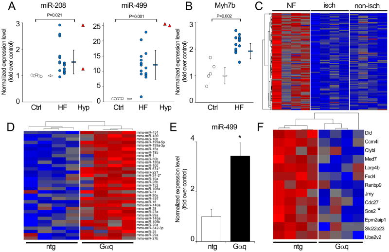
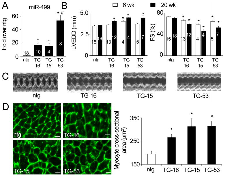
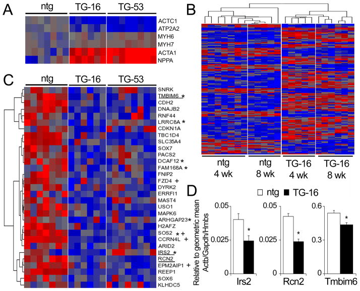
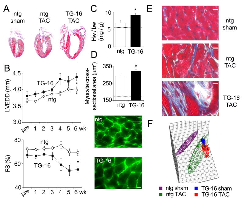
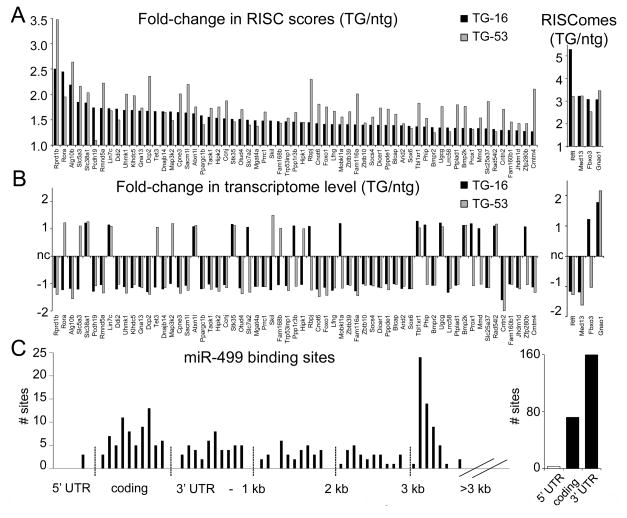
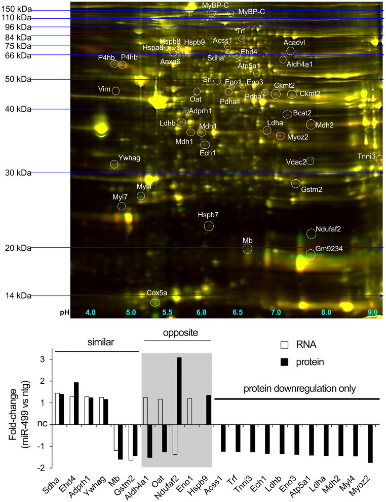
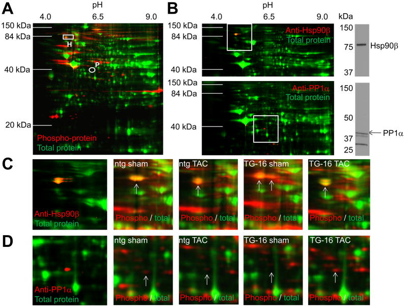
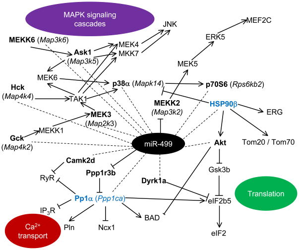
Methods and results: miR-499 levels were increased in failing and hypertrophied human hearts and associated with decreased levels of predicted target mRNAs. Likewise, miR-499 is increased in Gq-mediated murine cardiomyopathy. Forced cardiomyocyte expression of miR-499 at levels comparable to human cardiomyopathy induced progressive murine heart failure and exacerbated cardiac remodeling after pressure overloading. Genome-wide RNA-induced silencing complex and RNA sequencing identified 67 direct, and numerous indirect, cardiac mRNA targets, including Akt and MAPKs. Myocardial proteomics identified alterations in protein phosphorylation linked to the miR-499 cardiomyopathy phenotype, including of heat shock protein 90 and protein serine/threonine phosphatase 1-α.

Conclusions: miR-499 is increased in human and murine cardiac hypertrophy and cardiomyopathy, is sufficient to cause murine heart failure, and accelerates maladaptation to pressure overloading. The deleterious effects of miR-499 reflect the cumulative consequences of direct and indirect mRNA regulation, modulation of cardiac kinase and phosphatase pathways, and higher-order effects on posttranslational modification of myocardial proteins.


Conflict of interest statement

The authors declare that they have no conflicts of interest relating to this manuscript.

Figures

Figure 1
Figure 1. miR-499 upregulation and target suppression in human heart failure and murine genetic cardiomyopathy
(A) Increased miR-208, miR-499 levels in human heart failure; white = non-failing (Ctrl), blue = failing hearts (HF), red = hypertrophied non-failing hearts (Hyp). The miR expression arrays used did not distinguish between miRs-208a and -208b. Individual data are shown together with mean ± s.d. (B) Increased Myh7b levels in human heart failure; white = non-failing (Ctrl) and blue = failing hearts (HF). (C) Downregulation of 98 miR-499 target mRNAs, predicted by TargetScan, in human heart failure (greater than 1.2-fold downregulation, P<0.05, Online Table I). Four representative hearts each are shown for non-failing (NF), and failing hearts with cardiomyopathy of ischemic origin (isch) or non-ischemic origin (non-isch). Red = higher expression, blue = lower expression. (D) miR microarray analysis of Gαq transgenic hearts , displaying miRs according to degree of upregulation (1.2-fold or greater, P<0.05; miR-451, 4.4 fold-upregulated in Gq, is at the top). (E) miR-499 analyzed by RT-qPCR relative to combined reference of 5S rRNA, U6 snRNA and Gapdh mRNA in nontransgenic (n=3) and Gαq hearts (n=6). Data shown are mean +/minus; s.e.m., arbitrary units (nontransgenic = 1); * denotes P=0.0006, 2-tailed unpaired t-test. (F) Downregulation of 13 miR-499 target mRNAs, predicted by TargetScan (greater than 1.2-fold downregulation, P<0.05) in Gαq transcriptomes analyzed by RNA-sequencing . Red, higher expression; blue, lower expression. * denotes downregulation of the same mRNA in human heart failure.
Figure 2
Figure 2. Cardiac miR-499 overexpression causes heart failure in mice
(A) Cardiac-directed overexpression of miR-499 in two transgenic mouse lines at levels similar to human heart failure (TG-16 and TG-15) and in a higher-overexpressing line (TG-53). * denotes P<0.0001 relative to nontransgenic (1-way ANOVA); # denotes significant difference to all other groups (Tukey’s pairwise comparisons). (B) Progressive dilated cardiomyopathy in miR-499 transgenic hearts shown by increases in left-ventricular end-diastolic dimension (LVEDD) and decreases in fractional shortening (FS%) at 20 weeks of age. Two-way ANOVA was used to assess effects of timepoint and genotype (full details in Online Table II); * denotes significant difference vs age-matched nontransgenic. For both (A) and (B), data shown are mean ± s.e.m.; numbers of hearts are shown in columns. (C) Representative M-mode echocardiograms from 20 week-old mouse hearts. (D) Myocyte cross-sectional area shown by fluorescent wheat germ agglutinin staining of 20 week-old mouse hearts. White scale bar represents 10 μm. Quantitation represents mean values from ~600 myocytes, from each of two hearts. * denotes P=0.0004 relative to nontransgenic (1-way ANOVA).
Figure 3
Figure 3. Transcriptome regulation in miR-499 transgenic mice
(A) Markers of the hypertrophic / fetal recapitulation gene program; α-cardiac actin (Actc1), SERCA2a (Atp2a2), α-MHC (Myh6), β-MHC (Myh7), α-skeletal actin (Acta1) and ANP (Nppa) in 4 week-old hearts. (B) Heatmap of 136 mRNAs indirectly regulated by miR-499 in 4 week-old and 8 week-old transgenic hearts (details in Online Table III). (C) Downregulation of 31 miR-499 target mRNAs predicted by TargetScan in miR-499 transgenic hearts (greater than 1.2-fold downregulation, P<0.05, Online Table IV). * denotes downregulation of the same mRNA in human heart failure; + denotes downregulation of the same mRNA in Gαq mice (Figure 1). Underlined mRNAs are those confirmed by RT-qPCR in panel D. Red = higher expression, blue = lower expression. (D) TaqMan RT-qPCR validation of 3 predicted miR-499 target mRNAs. Expression level is normalized to a combined reference of Actb, Gapdh and Hmbs. Relative abundances of Irs2, Rcn2 and Tmbim6 correlate to RNA-sequencing determinations (Online Table IV). * denotes P<0.05, unpaired 2-tailed t-test.
Figure 4
Figure 4. miR-499 overexpression worsens the response to pressure overload in mice
(A) Representative sections of nontransgenic sham, nontransgenic pressure-overloaded (TAC) and miR-499 TG-16 pressure-overloaded hearts 6 weeks after surgery. (B) Development of hypertrophy shown by echocardiography of nontransgenic (n=5) and miR-499 TG-16 (n=3) animals. Data shown are mean ± s.e.m. ; * denotes significant difference at 6 weeks (P=0.03 for LVEDD and P=0.007 for FS%, 2-tailed unpaired t-test). (C) Heart weight / body weight ratios after 6 weeks TAC. * denotes significant difference (P=0.036, 1-tailed unpaired t-test). (D) Myocyte cross-sectional area shown by fluorescent wheat germ agglutinin staining. White scale bar represents 10 μm. Quantitation represents mean values from ~600 myocytes, from each of 2 hearts. * denotes P=0.04. Dotted lines, C and D: typical levels in age-matched, untreated nontransgenic mice. (E) Fibrosis after 6 weeks of TAC shown by Masson’s trichrome staining. White scale bar represents 10 μm. (F, left) Principal components analysis of 6,500 mRNAs (those expressed at >=3 copies/cell) in nontransgenic and miR-499 TG-16 hearts, sham-treated and after 1 week of TAC (5 hearts per treatment). Individual hearts are shown as filled spheres; wireframes enclose all hearts of a treatment group. Reduction of variance to 3 principal components (the 3 dimensions shown in the plot) accounts for 49.8% of the total variance in the data set.
Figure 5
Figure 5. Direct targets of miR-499 in mouse hearts
(A, left) Fold-change in RISC score (RISCome expression/transcriptome expression) between transgenic and nontransgenic mice for 63 direct targets of miR-499 identified by RISC-sequencing. (A, right) Change in RISCome expression for a further 4 miR-499 targets. (B) Fold-change in transcriptome expression for the miR-499 targets shown in (A). nc = no change. Black bars, miR-499 transgenic TG-16; gray bars, miR-499 transgenic TG-53. (C) Schematic depiction of miR-499 recognition sites in miR-499 targets defined by RISC-sequencing. Graph on the left shows number of sites according to mRNA position; graph on the right shows aggregate counts. The majority (68%) of putative binding sites are in the 3′ untranslated region (UTR), with 31% in coding regions and 1% in the 5′ UTR.
Figure 6
Figure 6. Similar and differential regulation of mRNA and protein for indirect targets of miR-499
Upper panel: Representative 2D DiGE gel image. Green = miR-499, red = nontransgenic cardiac protein. Lower panel: Comparison of mRNA and protein regulation for 22 gene products. Mybpc3 (MyBP-C), Ckmt2, Mdh1, P4hb and Pdha1 migrated as multiple species and were not analyzed. Also identified, but with neither mRNA nor protein regulation: Acadvl, Anxa6, Cox5a, Hspa8, Hspb8, Myl7, Vdac2, Vim. White bars, mRNA regulation; black bars, protein regulation. nc = no change. Regulated mRNAs had changes of ≥20%, P<0.05. Proteins were classed as regulated if changes of ≥20% in the same direction were identified on at least 3 of 5 gels.
Figure 7
Figure 7. Differential phosphorylation in nontransgenic and miR-499 transgenic mice at 1 week of pressure overload
(A) Representative phosphoproteome 2D gel image of nontransgenic sham-treated cardiac protein samples. Green is total protein stain; red is ProQ-diamond phosphoprotein; rectangle, HSP90β (H); circle, PP1α (P). (B) 2D immunoblots of HSP90β and PP1α; 1D SDS-PAGE of a nontransgenic, sham-treated cardiac protein sample is shown to at right. White squares denote regions shown in panels C and D. (C) Left: magnified view of 2D immunoblot for HSP90β. Right: same region in representative phosphoproteome 2D gels for nontransgenic and TG-16, sham- and TAC-treated hearts. (D) as for (C) but for PP1α. A total of 5 hearts were used in each treatment group (Online Figure VI).
Figure 8
Figure 8. Signaling networks influenced by miR-499
MetaCore was used to explore signaling pathways involving direct and indirect targets of miR-499. Dotted lines, indirect action; solid lines, direct action (arrowheads denote activation, while bars denote inhibition). miR-499 targets are in bold, and those with altered phosphorylation (HSP90β and PP1α) are in blue.

Comment in

  • Targeting microRNA targets.
    Da Costa Martins PA, De Windt LJ. Da Costa Martins PA, et al. Circ Res. 2012 Aug 17;111(5):506-8. doi: 10.1161/CIRCRESAHA.112.276717. Circ Res. 2012. PMID: 22904037 No abstract available.

Similar articles

Cited by

References

    1. van Rooij E, Quiat D, Johnson BA, Sutherland LB, Qi X, Richardson JA, Kelm RJ, Jr, Olson EN. A family of microRNAs encoded by myosin genes governs myosin expression and muscle performance. Dev Cell. 2009;17:662–673. - PMC - PubMed
    1. Wang JX, Jiao JQ, Li Q, Long B, Wang K, Liu JP, Li YR, Li PF. miR-499 regulates mitochondrial dynamics by targeting calcineurin and dynamin-related protein-1. Nat Med. 2011;17:71–78. - PubMed
    1. Shieh JT, Huang Y, Gilmore J, Srivastava D. Elevated miR-499 levels blunt the cardiac stress response. PLoS One. 2011;6:e19481. - PMC - PubMed
    1. Matkovich SJ, Van Booven DJ, Youker KA, Torre-Amione G, Diwan A, Eschenbacher WH, Dorn LE, Watson MA, Margulies KB, Dorn GW., 2nd Reciprocal regulation of myocardial microRNAs and messenger RNA in human cardiomyopathy and reversal of the microRNA signature by biomechanical support. Circulation. 2009;119:1263–1271. - PMC - PubMed
    1. Bell ML, Buvoli M, Leinwand LA. Uncoupling of expression of an intronic microRNA and its myosin host gene by exon skipping. Mol Cell Biol. 2010;30:1937–1945. - PMC - PubMed

Publication types

MeSH terms