Transferring isolated mitochondria into tissue culture cells
- PMID: 22753025
- PMCID: PMC3479163
- DOI: 10.1093/nar/gks639
Transferring isolated mitochondria into tissue culture cells
Abstract
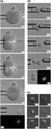
We have developed a new method for introducing large numbers of isolated mitochondria into tissue culture cells. Direct microinjection of mitochondria into typical mammalian cells has been found to be impractical due to the large size of mitochondria relative to microinjection needles. To circumvent this problem, we inject isolated mitochondria through appropriately sized microinjection needles into rodent oocytes or single-cell embryos, which are much larger than tissue culture cells, and then withdraw a 'mitocytoplast' cell fragment containing the injected mitochondria using a modified holding needle. These mitocytoplasts are then fused to recipient cells through viral-mediated membrane fusion and the injected mitochondria are transferred into the cytoplasm of the tissue culture cell. Since mouse oocytes contain large numbers of mouse mitochondria that repopulate recipient mouse cells along with the injected mitochondria, we used either gerbil single-cell embryos or rat oocytes to package injected mouse mitochondria. We found that the gerbil mitochondrial DNA (mtDNA) is not maintained in recipient rho0 mouse cells and that rat mtDNA initially replicated but was soon completely replaced by the injected mouse mtDNA, and so with both procedures mouse cells homoplasmic for the mouse mtDNA in the injected mitochondria were obtained.
Figures


Similar articles
-
Microinjection of cytoplasm or mitochondria derived from somatic cells affects parthenogenetic development of murine oocytes.Biol Reprod. 2005 Jun;72(6):1397-404. doi: 10.1095/biolreprod.104.036129. Epub 2005 Feb 16. Biol Reprod. 2005. PMID: 15716395
-
Mitochondrial transfer between oocytes: potential applications of mitochondrial donation and the issue of heteroplasmy.Hum Reprod. 1998 Oct;13(1O):2857-68. doi: 10.1093/humrep/13.10.2857. Hum Reprod. 1998. PMID: 9804246
-
Microinjection of serum-starved mitochondria derived from somatic cells affects parthenogenetic development of bovine and murine oocytes.Mitochondrion. 2010 Mar;10(2):137-42. doi: 10.1016/j.mito.2009.12.144. Epub 2009 Dec 11. Mitochondrion. 2010. PMID: 20005304
-
Functional consequences of mitochondrial mismatch in reconstituted embryos and offspring.J Reprod Dev. 2019 Dec 18;65(6):485-489. doi: 10.1262/jrd.2019-089. Epub 2019 Aug 29. J Reprod Dev. 2019. PMID: 31462597 Free PMC article. Review.
-
Transmission of Dysfunctional Mitochondrial DNA and Its Implications for Mammalian Reproduction.Adv Anat Embryol Cell Biol. 2019;231:75-103. doi: 10.1007/102_2018_3. Adv Anat Embryol Cell Biol. 2019. PMID: 30617719 Review.
Cited by
-
Mitochondrial Transfer by Photothermal Nanoblade Restores Metabolite Profile in Mammalian Cells.Cell Metab. 2016 May 10;23(5):921-9. doi: 10.1016/j.cmet.2016.04.007. Cell Metab. 2016. PMID: 27166949 Free PMC article.
-
Mitochondria transplantation between living cells.PLoS Biol. 2022 Mar 23;20(3):e3001576. doi: 10.1371/journal.pbio.3001576. eCollection 2022 Mar. PLoS Biol. 2022. PMID: 35320264 Free PMC article.
-
Optimization of mitochondrial isolation techniques for intraspinal transplantation procedures.J Neurosci Methods. 2017 Aug 1;287:1-12. doi: 10.1016/j.jneumeth.2017.05.023. Epub 2017 May 26. J Neurosci Methods. 2017. PMID: 28554833 Free PMC article.
-
mtDNA Heteroplasmy at the Core of Aging-Associated Heart Failure. An Integrative View of OXPHOS and Mitochondrial Life Cycle in Cardiac Mitochondrial Physiology.Front Cell Dev Biol. 2021 Feb 22;9:625020. doi: 10.3389/fcell.2021.625020. eCollection 2021. Front Cell Dev Biol. 2021. PMID: 33692999 Free PMC article. Review.
-
Modifying the Mitochondrial Genome.Cell Metab. 2016 May 10;23(5):785-96. doi: 10.1016/j.cmet.2016.04.004. Cell Metab. 2016. PMID: 27166943 Free PMC article. Review.
References
-
- King MP, Attardi G. Human cells lacking mtDNA repopulation with exogenous mitochondria by complementation. Science. 1989;246:500–503. - PubMed
-
- Trounce I, Wallace DC. Production of transmitochondrial mouse cell lines by cybrid rescue of rhodamine-6G pre-treated L-cells. Somat. Cell Mol. Genet. 1996;22:81–85. - PubMed
-
- Maniura-Weber K, Helm M, Engemann K, Eckertz S, Mollers M, Schauen M, Hayrapetyan A, von Kleist-Retzow JC, Lightowlers RN, Bindoff LA, et al. Molecular dysfunction associated with the human mitochondrial 3302A>G mutation in the MTTL1 (mt-tRNALeu(UUR)) gene. Nucleic Acids Res. 2006;34:6404–6415. - PMC - PubMed

