. 2012 Sep 1;40(17):8425-39.
doi: 10.1093/nar/gks638. Epub 2012 Jun 29.

Fanconi anemia proteins FANCD2 and FANCI exhibit different DNA damage responses during S-phase

Affiliations

Fanconi anemia proteins FANCD2 and FANCI exhibit different DNA damage responses during S-phase

Archana Sareen et al. Nucleic Acids Res. .

Abstract

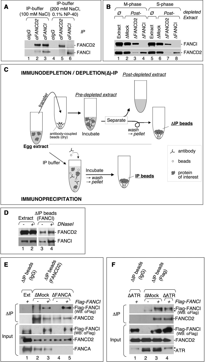
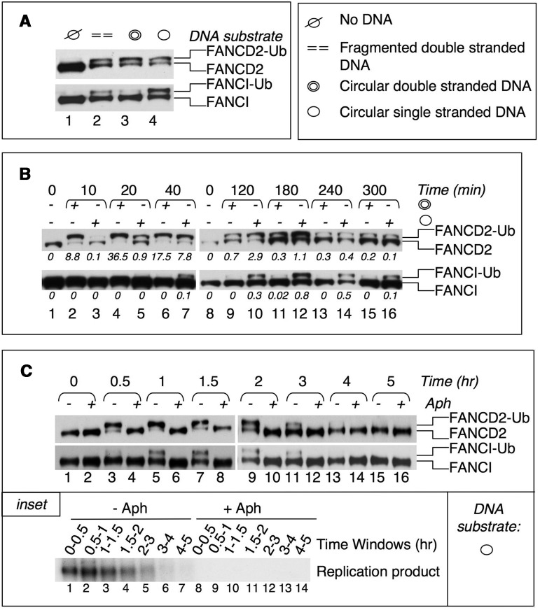
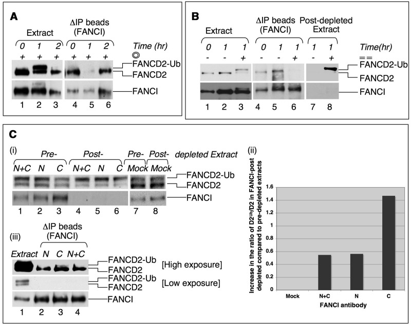
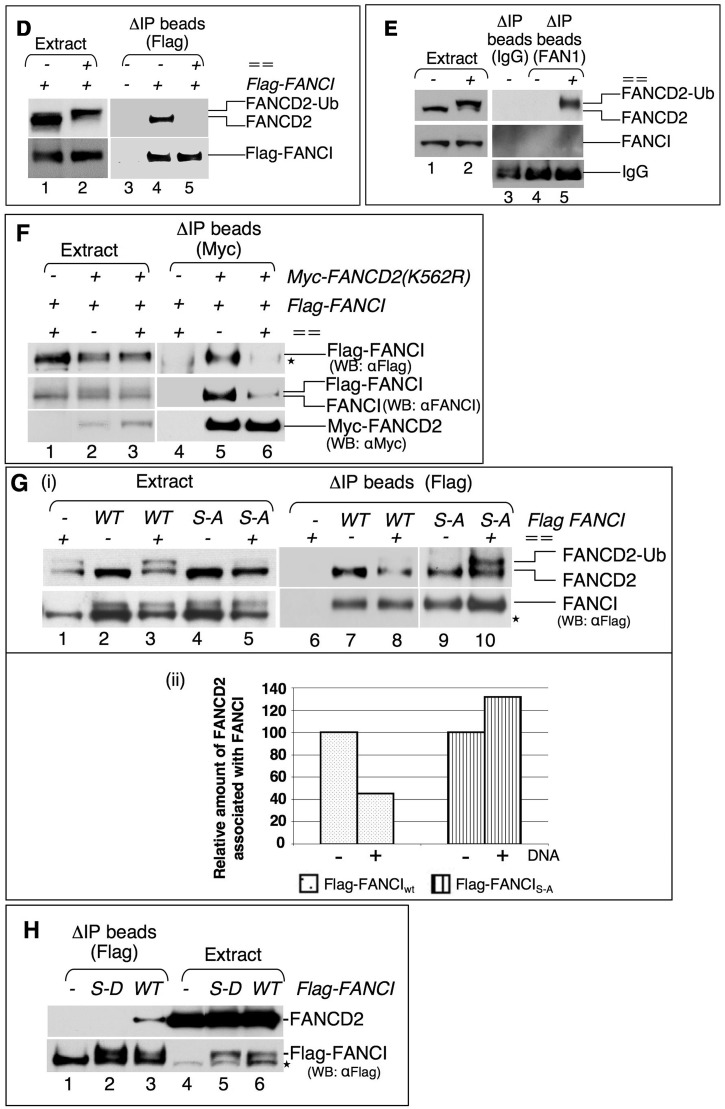
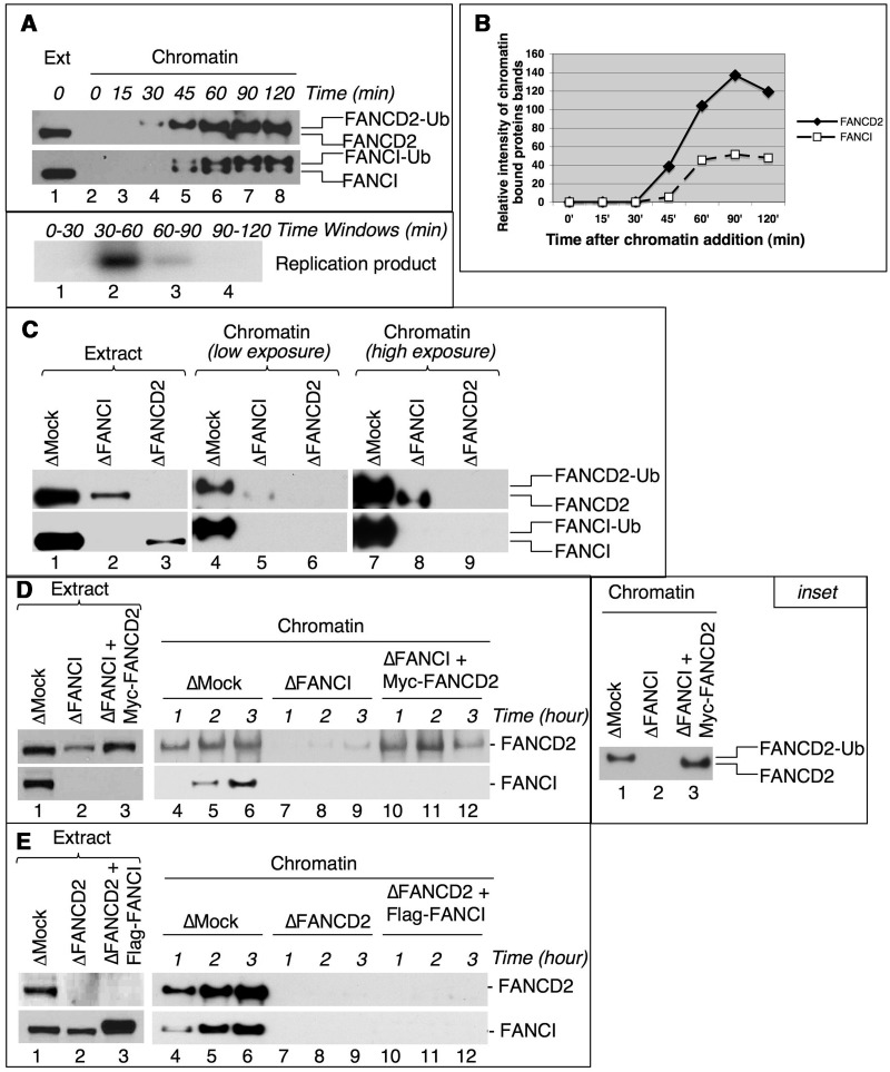
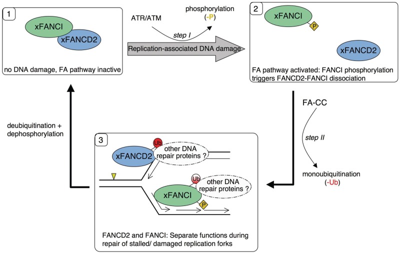
Fanconi anemia (FA) pathway members, FANCD2 and FANCI, contribute to the repair of replication-stalling DNA lesions. FA pathway activation relies on phosphorylation of FANCI by the ataxia telangiectasia and Rad3-related (ATR) kinase, followed by monoubiquitination of FANCD2 and FANCI by the FA core complex. FANCD2 and FANCI are thought to form a functional heterodimer during DNA repair, but it is unclear how dimer formation is regulated or what the functions of the FANCD2-FANCI complex versus the monomeric proteins are. We show that the FANCD2-FANCI complex forms independently of ATR and FA core complex, and represents the inactive form of both proteins. DNA damage-induced FA pathway activation triggers dissociation of FANCD2 from FANCI. Dissociation coincides with FANCD2 monoubiquitination, which significantly precedes monoubiquitination of FANCI; moreover, monoubiquitination responses of FANCD2 and FANCI exhibit distinct DNA substrate specificities. A phosphodead FANCI mutant fails to dissociate from FANCD2, whereas phosphomimetic FANCI cannot interact with FANCD2, indicating that FANCI phosphorylation is the molecular trigger for FANCD2-FANCI dissociation. Following dissociation, FANCD2 binds replicating chromatin prior to-and independently of-FANCI. Moreover, the concentration of chromatin-bound FANCD2 exceeds that of FANCI throughout replication. Our results suggest that FANCD2 and FANCI function separately at consecutive steps during DNA repair in S-phase.


Figures

Figure 1.
Figure 1.
FANCD2 and FANCI form a weak complex throughout the cell cycle in a manner independently of the FA core complex or ATR. (A) FANCD2 and FANCI co-immunoprecipitate (co-IP) from S-phase extracts under low-stringency conditions. S-phase extracts were diluted in low-stringency buffer (lanes 1–3) or high-stringency buffer (lanes 4–6) (see also ‘Materials and Methods’ section), and incubated with rabbit IgG (lanes 1 and 4), FANCD2 antibody (lanes 2 and 5) or FANCI antibody (lanes 3 and 6), followed by incubation with sepharose beads. Beads were washed in the respective IP buffer and analyzed for bound FANCD2 and FANCI by SDS–PAGE and western blot. (B) FANCD2 and FANCI interact in M- and S-phase. Xenopus M-phase (lanes 1–4) or S-phase (lanes 5–8) egg extracts were mock- (lanes 2 and 6), FANCD2- (lanes 3 and 7), or FANCI-depleted (lanes 4 and 8). Depleted extracts were assayed for the presence of FANCD2 and FANCI by SDS–PAGE and western blot. One microliter of untreated M-phase extract (lane 1) or S-phase extract (lane 5) was used as a loading control. (C) Overview of Immunodepletion/Depletion-IP and Immunoprecipitation strategies. Immunodepletion/Depletion (Δ)-IP: Extracts were added to dry antibody-coupled beads and incubated on ice. Following incubation, beads were separated from the extract, washed in low stringency IP buffer and analyzed for bound protein (ΔIP beads). Aliquots were taken from the bead–extract mix just before separation of beads and extract (Pre-depleted extract) and from extracts after removal of beads (Post-depleted extract). Immunoprecipitation: Extracts were diluted in (low or high stringency) IP buffer first, followed by incubation with antibody and then beads (rotation at 4°C). Following incubation, beads were washed in the respective IP buffer and analyzed for bound protein (IP beads). (D) The FANCD2–FANCI complex persists in DNaseI-treated S-phase extracts. S-phase extracts were untreated (lane 1) or DNaseI-treated (lane 2), followed by ΔIP with FANCI antibody-beads. ΔIP beads were analyzed for the presence of FANCD2 and FANCI by western blot (lanes 3 and 4). (E) Recombinant Flag–FANCI and extract-endogenous FANCD2 interact in absence of FANCA. Input panels: S-phase extracts were undepleted (lane 1), mock- (lanes 2 and 3) or FANCA-depleted (lanes 4 and 5), and either untreated (lanes 2 and 4) or incubated with recombinant Flag–FANCI protein (lanes 1, 3 and 5) for 30 min. ΔIP panels: the different extracts were then subjected to ΔIP by incubation with beads coupled to rabbit IgG (lane 1) or FANCD2 antibody (lanes 2–5). ΔIP beads were assayed for bound FANCD2 and FANCI by SDS–PAGE and western blot. (F) Recombinant Flag–FANCI and extract-endogenous FANCD2 interact in absence of ATR. Input panels: S-phase extracts were mock- (lanes 2 and 3) or ATR depleted (lanes 1 and 4). Depleted extracts were untreated (lane 2) or incubated with recombinant Flag–FANCI (lanes 1, 3 and 4) for 30 min. ΔIP panels: the different extracts were then subjected to ΔIP by incubation with beads coupled to rabbit IgG (lane 1) or Flag antibody (lanes 2–4). ΔIP beads were assayed for bound FANCD2 and FANCI by SDS–PAGE and western blot [ATR depleted, Flag–FANCI-treated extracts instead of undepleted, Flag–FANCI-treated extracts were used for mock-depletion (lane 1) in Figure 1F]. The asterisk marks a non-specific band that is sometimes recognized by the Flag antibody (upper panel) and a non-specific band typically recognized by the ATR antibody (bottom panel).
Figure 2.
Figure 2.
FANCD2 and FANCI exhibit different monoubiquitination responses. (A) FANCD2 and FANCI are not monoubiquitinated in response to the same DNA substrates. S-phase extracts were either untreated (lane 1) or treated with 50 ng/µl of fragmented dsDNA (lane 2), circular dsDNA (lane 3) or circular ssDNA (lane 4) for 1 h. Following incubation, 1 µl of extract was assayed for FANCD2 and FANCI by western blot. (B) Monoubiquitination responses of FANCD2 and FANCI follow a different time course. Extracts were either untreated (lanes 1 and 8) or treated with 50 ng/µl of circular dsDNA (lanes 2, 4, 6, 9, 11, 13 and 15) or circular ssDNA (lanes 3, 5, 7, 10, 12, 14 and 16) for the indicated time points. Following incubation, 1 µl of extract was assayed for FANCD2 and FANCI by western blot. The ratio of monoubiquitinated to non-ubiquitinated FANCD2 and FANCI is shown below each lane. A graphical presentation of FANCD2Ub/FANCD2 and FANCIUb/FANCI ratios is provided in Supplementary Figure S2A. (C) Monoubiquitination of FANCD2 and FANCI is replication dependent in presence of circular ssDNA. S-phase extracts were supplemented with 50 ng/µl of circular ssDNA and either untreated (lanes 1, 3, 5, 7, 9, 11, 13 and 15), or treated with the replicative DNA polymerase inhibitor, aphidicolin (lanes 2, 4, 6, 8, 10, 12, 14 and 16) A graphical presentation of FANCD2Ub/FANCD2 and FANCIUb/FANCI ratios is provided in Supplementary Figure S2B. Inset: the efficiency of replication inhibition in presence of aphidicolin was monitored in a parallel replication assay using aliquots of egg extracts that were supplemented with circular ssDNA and either untreated (lanes 1–7) or treated with aphidicolin (lanes 8–14).
Figure 3.
Figure 3.
The FANCD2–FANCI complex dissociates in response to FA pathway activation. (A) DNA damage-induced dissociation and subsequent reassociation of FANCD2 and FANCI. S-phase extracts were incubated with 50 ng/µl of circular dsDNA for 0, 1 or 2 h (lanes 1–3, respectively). At the indicated time points, extracts were transferred to ice and subjected to ΔIP by incubation with beads coupled to FANCI antibody. ΔIP beads were assayed for bound FANCD2 and FANCI by SDS–PAGE and western blot (lanes 4–6). (B) the FANCI–FANCD2 complex dissociates in response to dsDNA fragments. S-phase extracts were either untreated, or treated with 50 ng/µl of fragmented dsDNA (lanes 1–3) for 1 h. At the indicated time points, extracts were transferred onto ice and subjected to ΔIP by incubation with beads coupled to FANCI antibody. ΔIP beads (lanes 4–6) and ‘post-depleted’ extracts (lanes 7 and 8) were assayed for the presence of FANCD2 and FANCI. (C) N- and C-terminal FANCI antibodies do not interrupt the FANCD2–FANCI complex. (i) S-phase egg extracts were incubated with 50 ng/µl of circular dsDNA for 60 min, transferred to ice, and subjected to ΔIP by incubation with beads coupled to antibodies against the FANCI N-terminus (N, lane 2), C-terminus (C, lane 3) or both (N+C, lane 1), or with beads coupled to rabbit IgG (lane 7). One microliter aliquots were taken at the end of the 90 min incubation time (‘pre-depleted extract’, lanes 1–3 and 7), followed by separation of extract (‘post-depleted extract’, lanes 4–6 and 8) and ΔIP beads [see (iii)]. Aliquots of pre- and post-depleted extracts were assayed for FANCD2 and FANCI by SDS–PAGE and immunoblot. The ratio of monoubiquitinated to non-ubiquitinated FANCD2 (FANCD2Ub/FANCD2) was determined in pre- and post-depleted extracts and the ratio increase in post-depleted extracts compared with pre-depleted extracts was plotted and is shown in (ii). The ΔIP beads were assayed for the presence of FANCD2 and FANCI (iii). One microliter of undepleted, dsDNA-treated extract was used as a size control in (iii) (lane 1). (D) A de novo formed complex of Flag–FANCI and endogenous FANCD2 dissociates in response to fragmented dsDNA. Egg extracts were incubated with Flag–FANCI for 30 min, then untreated (lane 1) or treated with dsDNA fragments (lane 2) for an additional 30 min. DNA-free or DNA-containing extracts were subjected to ΔIP by incubation with Flag antibody-coupled beads. ΔIP beads were analyzed for bound FANCD2 and FANCI (lanes 4 and 5). ΔIP with Flag antibody-beads from untreated extracts (no Flag–FANCI, no DNA) was used as negative control (lane 3). (E) FANCI is not associated with the FAN1–FANCD2Ub complex. S-phase extracts were untreated (lane 1) or treated with 50 ng/µl of fragmented dsDNA (lane 2) for 1 h, and then subjected to ΔIP by incubation with FAN1 antibody-beads. ΔIP beads were assayed for bound FANCD2 and FANCI (lanes 4 and 5). Lane 3 shows a control ΔIP with mouse IgG. (F) A monoubiquitination-dead FANCD2 mutant dissociates from FANCI in response to dsDNA fragments. Egg extracts were incubated with Flag–FANCIwt alone (lane 1) or with Flag–FANCIwt and Myc–FANCD2K562R (lanes 2 and 3) for 30 min, then untreated (lane 2) or treated with dsDNA fragments (lanes 1 and 3) for an additional 30 min. DNA-free or DNA-containing extracts were subjected to ΔIP by incubation with Myc antibody-coupled beads. ΔIP with Myc antibody-beads from extracts lacking Myc–FANCD2K562R was used as negative control (lane 4). ΔIP beads were analyzed for bound FANCI and FANCD2 (lanes 4–6). (G) A phospho-dead FANCI mutant is unable to dissociate from FANCD2 in response to dsDNA fragments. (i) Egg extracts were incubated with Flag–FANCIwt (lanes 2 and 3) or Flag–FANCI6S→A (lanes 4 and 5) for 30 min, then untreated (lanes 2 and 4) or treated with dsDNA fragments (lanes 1, 3 and 5) for an additional 60 min. DNA-free or DNA-containing extracts were subjected to ΔIP by incubation with Flag antibody-coupled beads. ΔIP beads were analyzed for bound FANCD2 and FANCI (lanes 6–10). ΔIP with Flag antibody-beads from DNA-treated extracts lacking recombinant FANCI was used as negative control (lane 6). (ii) The intensity of co-immunoprecipitated FANCD2 and FANCI protein bands shown in lanes 6–10 of Figure 3G (i) was determined by densitometry using Image J software. The relative intensity of each FANCD2 protein band compared with the corresponding FANCI protein band in the same lane was calculated. The relative amount of FANCD2 associated with Flag–FANCIwt or Flag–FANCIS-A in DNA-free extracts was set at a value of 100 and compared with the relative amount of FANCD2 associated with Flag–FANCIwt or with Flag–FANCIS-A in dsDNA fragment-treated extracts. [Densitometry of FANCD2 protein bands did include both FANCD2 isoforms (FANCD2 and FANCD2Ub) where present (Figure 3G (i), lane 10)]. (H) A phosphomimetic FANCI mutant is unable to interact with FANCD2. DNA-free S-phase extracts were untreated (lane 4), or incubated for 30 min with Flag–FANCI6S-D (lane 5) or Flag–FANCIwt (lane 6). Extracts were subjected to ΔIP by incubation with Flag antibody-beads, and ΔIP beads were assayed for bound FANCD2 and FANCI (lanes 1–3). In all figures, the asterisk marks a non-specific band that is sometimes recognized by the Flag antibody.
Figure 3.
Figure 3.
The FANCD2–FANCI complex dissociates in response to FA pathway activation. (A) DNA damage-induced dissociation and subsequent reassociation of FANCD2 and FANCI. S-phase extracts were incubated with 50 ng/µl of circular dsDNA for 0, 1 or 2 h (lanes 1–3, respectively). At the indicated time points, extracts were transferred to ice and subjected to ΔIP by incubation with beads coupled to FANCI antibody. ΔIP beads were assayed for bound FANCD2 and FANCI by SDS–PAGE and western blot (lanes 4–6). (B) the FANCI–FANCD2 complex dissociates in response to dsDNA fragments. S-phase extracts were either untreated, or treated with 50 ng/µl of fragmented dsDNA (lanes 1–3) for 1 h. At the indicated time points, extracts were transferred onto ice and subjected to ΔIP by incubation with beads coupled to FANCI antibody. ΔIP beads (lanes 4–6) and ‘post-depleted’ extracts (lanes 7 and 8) were assayed for the presence of FANCD2 and FANCI. (C) N- and C-terminal FANCI antibodies do not interrupt the FANCD2–FANCI complex. (i) S-phase egg extracts were incubated with 50 ng/µl of circular dsDNA for 60 min, transferred to ice, and subjected to ΔIP by incubation with beads coupled to antibodies against the FANCI N-terminus (N, lane 2), C-terminus (C, lane 3) or both (N+C, lane 1), or with beads coupled to rabbit IgG (lane 7). One microliter aliquots were taken at the end of the 90 min incubation time (‘pre-depleted extract’, lanes 1–3 and 7), followed by separation of extract (‘post-depleted extract’, lanes 4–6 and 8) and ΔIP beads [see (iii)]. Aliquots of pre- and post-depleted extracts were assayed for FANCD2 and FANCI by SDS–PAGE and immunoblot. The ratio of monoubiquitinated to non-ubiquitinated FANCD2 (FANCD2Ub/FANCD2) was determined in pre- and post-depleted extracts and the ratio increase in post-depleted extracts compared with pre-depleted extracts was plotted and is shown in (ii). The ΔIP beads were assayed for the presence of FANCD2 and FANCI (iii). One microliter of undepleted, dsDNA-treated extract was used as a size control in (iii) (lane 1). (D) A de novo formed complex of Flag–FANCI and endogenous FANCD2 dissociates in response to fragmented dsDNA. Egg extracts were incubated with Flag–FANCI for 30 min, then untreated (lane 1) or treated with dsDNA fragments (lane 2) for an additional 30 min. DNA-free or DNA-containing extracts were subjected to ΔIP by incubation with Flag antibody-coupled beads. ΔIP beads were analyzed for bound FANCD2 and FANCI (lanes 4 and 5). ΔIP with Flag antibody-beads from untreated extracts (no Flag–FANCI, no DNA) was used as negative control (lane 3). (E) FANCI is not associated with the FAN1–FANCD2Ub complex. S-phase extracts were untreated (lane 1) or treated with 50 ng/µl of fragmented dsDNA (lane 2) for 1 h, and then subjected to ΔIP by incubation with FAN1 antibody-beads. ΔIP beads were assayed for bound FANCD2 and FANCI (lanes 4 and 5). Lane 3 shows a control ΔIP with mouse IgG. (F) A monoubiquitination-dead FANCD2 mutant dissociates from FANCI in response to dsDNA fragments. Egg extracts were incubated with Flag–FANCIwt alone (lane 1) or with Flag–FANCIwt and Myc–FANCD2K562R (lanes 2 and 3) for 30 min, then untreated (lane 2) or treated with dsDNA fragments (lanes 1 and 3) for an additional 30 min. DNA-free or DNA-containing extracts were subjected to ΔIP by incubation with Myc antibody-coupled beads. ΔIP with Myc antibody-beads from extracts lacking Myc–FANCD2K562R was used as negative control (lane 4). ΔIP beads were analyzed for bound FANCI and FANCD2 (lanes 4–6). (G) A phospho-dead FANCI mutant is unable to dissociate from FANCD2 in response to dsDNA fragments. (i) Egg extracts were incubated with Flag–FANCIwt (lanes 2 and 3) or Flag–FANCI6S→A (lanes 4 and 5) for 30 min, then untreated (lanes 2 and 4) or treated with dsDNA fragments (lanes 1, 3 and 5) for an additional 60 min. DNA-free or DNA-containing extracts were subjected to ΔIP by incubation with Flag antibody-coupled beads. ΔIP beads were analyzed for bound FANCD2 and FANCI (lanes 6–10). ΔIP with Flag antibody-beads from DNA-treated extracts lacking recombinant FANCI was used as negative control (lane 6). (ii) The intensity of co-immunoprecipitated FANCD2 and FANCI protein bands shown in lanes 6–10 of Figure 3G (i) was determined by densitometry using Image J software. The relative intensity of each FANCD2 protein band compared with the corresponding FANCI protein band in the same lane was calculated. The relative amount of FANCD2 associated with Flag–FANCIwt or Flag–FANCIS-A in DNA-free extracts was set at a value of 100 and compared with the relative amount of FANCD2 associated with Flag–FANCIwt or with Flag–FANCIS-A in dsDNA fragment-treated extracts. [Densitometry of FANCD2 protein bands did include both FANCD2 isoforms (FANCD2 and FANCD2Ub) where present (Figure 3G (i), lane 10)]. (H) A phosphomimetic FANCI mutant is unable to interact with FANCD2. DNA-free S-phase extracts were untreated (lane 4), or incubated for 30 min with Flag–FANCI6S-D (lane 5) or Flag–FANCIwt (lane 6). Extracts were subjected to ΔIP by incubation with Flag antibody-beads, and ΔIP beads were assayed for bound FANCD2 and FANCI (lanes 1–3). In all figures, the asterisk marks a non-specific band that is sometimes recognized by the Flag antibody.
Figure 4.
Figure 4.
FANCD2 binds replicating chromatin prior to and independently of FANCI. (A) FANCD2 is recruited to replicating chromatin prior to FANCI. Sperm chromatin was added to S-phase extracts and reisolated at the indicated time points during replication. Chromatin fractions (lanes 2–8) were analyzed for bound FANCD2 and FANCI by SDS–PAGE and western blot. Lane 1: 1 µl extract (loading control). Inset: replication assay. Throughout the experimental procedure described in the legend to panel A, replication was monitored by pulsing replicating extract aliquots with [α-32P]GTP at time windows as indicated. (B) The concentration of chromatin-bound FANCD2 is higher than that of FANCI. The intensity of chromatin-bound FANCD2 and FANCI protein bands shown in Figure 4A was determined by densitometry using Image J software. The relative intensity of each chromatin-bound protein band (lanes 2–8) compared with the protein band in the extract lane (lane 1) was plotted for each time point. (An independent repeat of this experiment is shown in Supplementary Figure S6A and B). (C) Residual FANCD2 binds chromatin in FANCI-depleted extracts. S-phase extracts were mock depleted (lane 1), FANCI depleted (lane 2) or FANCD2 depleted (lane 3). Sperm chromatin was added to the extracts and allowed to replicate. Chromatin was reisolated at 150 min (post-replication) and chromatin fractions were analyzed for bound FANCD2 and FANCI (lanes 4–6, short exposure; lanes 7–9, long exposure) by SDS–PAGE and western blotting. (D) Recombinant Myc–FANCD2wt binds replicating chromatin in FANCI-depleted extracts. S-phase extracts were mock depleted (lane 1), FANCI depleted (lane 2) or FANCI depleted (thus partially co-depleting FANCD2) and reconstituted with Myc–FANCD2wt (lane 3). Sperm chromatin was added to the extracts and allowed to replicate. Chromatin was reisolated at the indicated time points and chromatin fractions were analyzed for bound FANCD2 and FANCI (lanes 4–6: mock depleted; lanes 7–9: FANCI depleted; lanes 10–12: FANCI depleted and reconstituted with Myc–FANCD2wt) by SDS–PAGE and western blotting. Inset: FANCI-depleted extracts (deficient in FANCD2Ub formation) support chromatin recruitment of non-ubiquitinated Myc–FANCD2. The same chromatin fractions isolated from mock depleted (Figure 4D, lane 4), FANCI depleted (Figure 4D, lane 7) and FANCI depleted + Myc–FANCD2 (Figure 4D, lane 10) extracts were run on a lower percentage gel to allow separation of non-ubiquitinated and monoubiquitinated FANCD2 isoforms. (E) Recombinant Flag–FANCIwt is unable to bind replicating chromatin in FANCD2-depleted extracts. S-phase extracts were mock depleted (lane 1), FANCD2 depleted (lane 2) or FANCD2 depleted (thus partially co-depleting FANCI) and reconstituted with Flag–FANCIwt (lane 3). Sperm chromatin was added to the extracts and allowed to replicate. Chromatin was reisolated at the indicated time points and chromatin fractions were analyzed for bound FANCD2 and FANCI (lanes 4–6: mock depleted; lanes 7–9: FANCD2 depleted; lanes 10–12: FANCD2 depleted and reconstituted with Flag–FANCIwt) by SDS–PAGE and western blotting.
Figure 5.
Figure 5.
Dynamic FA pathway model. The FANCD2–FANCI dimer represents the inactive state of FANCD2 and FANCI (1). When DNA damage (yellow triangle) is encountered during DNA replication, the FA pathway is activated via ATR/ATM-mediated phosphorylation of FANCI. FANCI phosphorylation triggers dissociation of the FANCD2–FANCI dimer (2), immediately followed by monoubiquitination of FANCD2 by the FA core complex (FA-CC). Once dissociated, the monomeric FANCD2 and FANCI proteins are recruited separately to stalled replication forks where they have distinct functions during repair of stalled/collapsed replication forks. (3) One function of dissociated activated FANCD2 and FANCI may be to recruit different DNA repair protein complexes during their activated state. Once the DNA lesion is repaired, FANCD2 and FANCI modifications are removed and unmodified FANCD2 and FANCI return to their inactive, heterodimeric state (1).

Similar articles

Cited by

References

    1. Alter BP, Kupfer G. Fanconi anemia Gene Reviews [Internet] In: Dynan W Dr, editor. Seattle, WA: University of Washington; 1993.
    1. Joenje H, Patel KJ. The emerging genetic and molecular basis of Fanconi anaemia. Nat. Rev. Genet. 2001;2:446–457. - PubMed
    1. Kee Y, D’Andrea AD. Expanded roles of the Fanconi anemia pathway in preserving genomic stability. Genes Dev. 2010;24:1680–1694. - PMC - PubMed
    1. Kitao H, Takata M. Fanconi anemia: a disorder defective in the DNA damage response. Int. J. Hematol. 2011;93:417–424. - PubMed
    1. Garner E, Smogorzewska A. Ubiquitylation and the Fanconi anemia pathway. FEBS Lett. 2011;585:2853–2860. - PMC - PubMed

Publication types

Substances