. 2012 Sep 1;40(17):8519-35.
doi: 10.1093/nar/gks630. Epub 2012 Jun 29.

RORγ directly regulates the circadian expression of clock genes and downstream targets in vivo

Affiliations

RORγ directly regulates the circadian expression of clock genes and downstream targets in vivo

Yukimasa Takeda et al. Nucleic Acids Res. .

Abstract

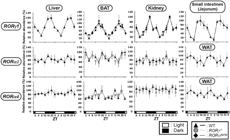
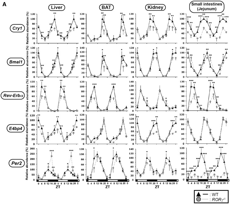
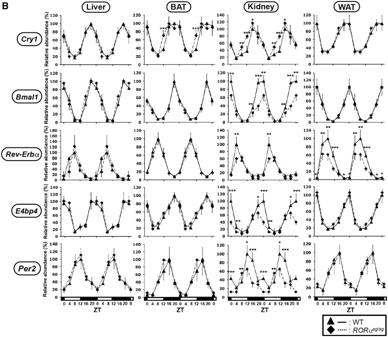
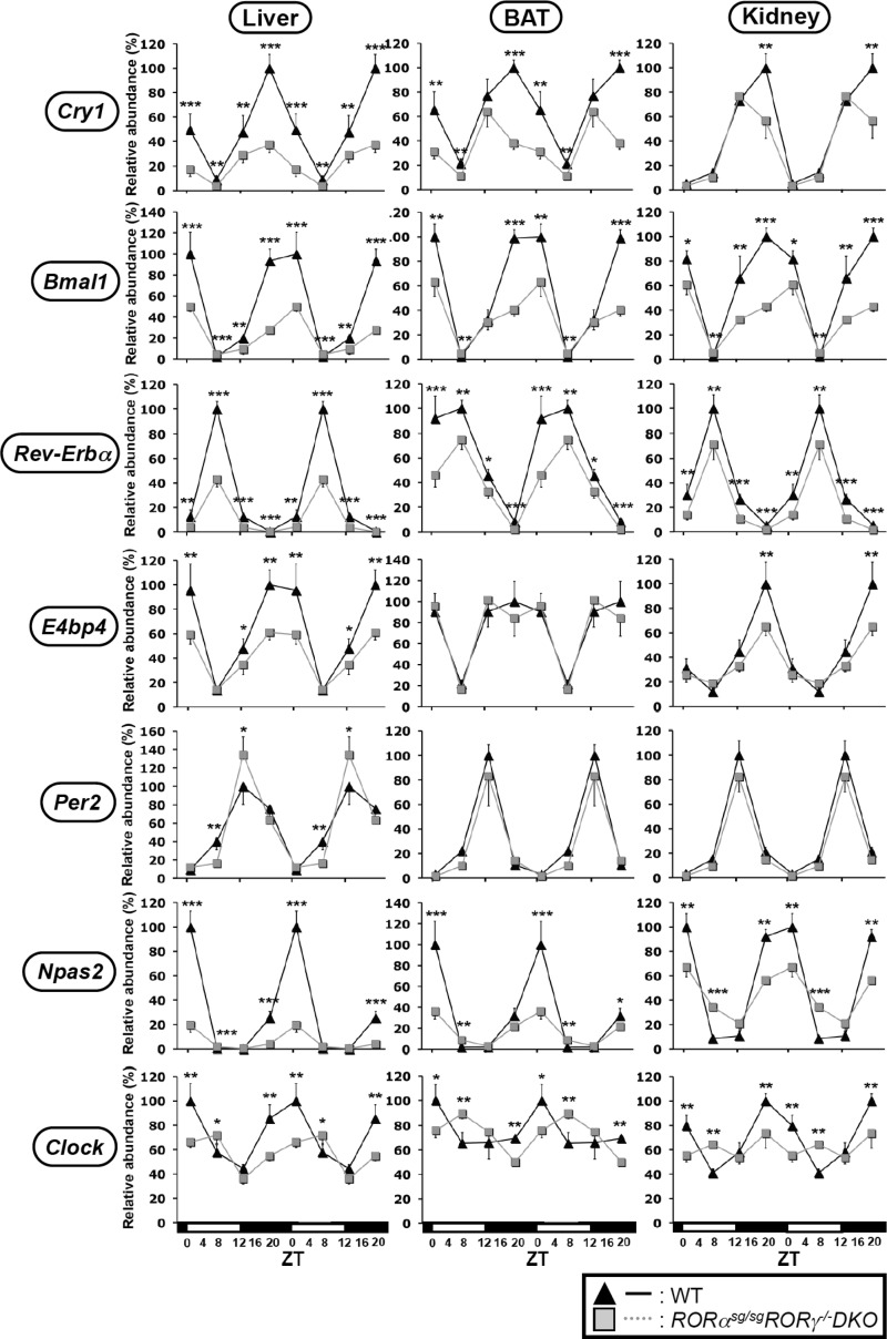
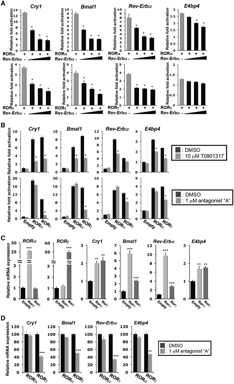
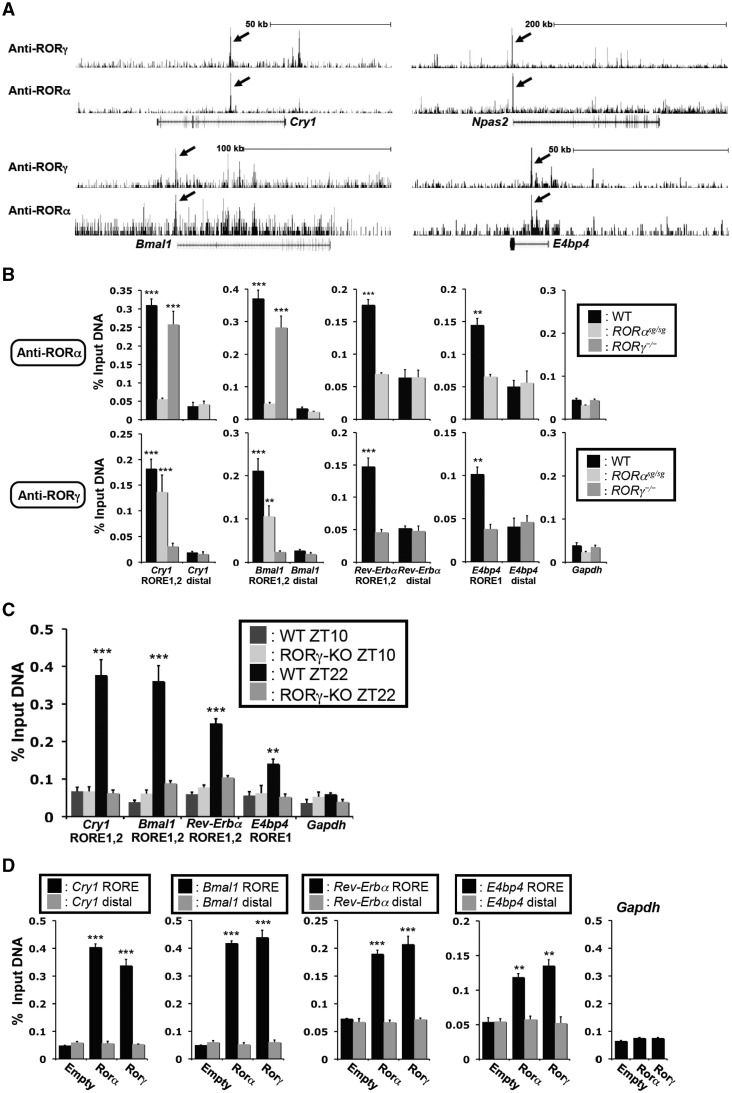
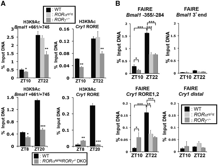
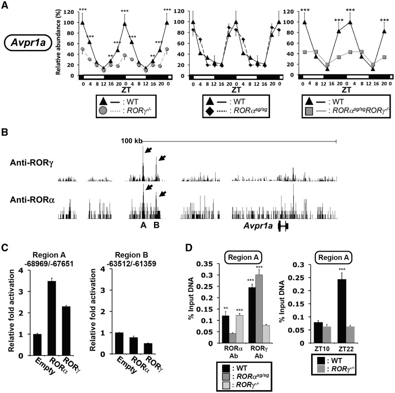
In this study, we demonstrate that the lack of retinoic acid-related orphan receptor (ROR) γ or α expression in mice significantly reduced the peak expression level of Cry1, Bmal1, E4bp4, Rev-Erbα and Per2 in an ROR isotype- and tissue-selective manner without affecting the phase of their rhythmic expression. Analysis of RORγ/RORα double knockout mice indicated that in certain tissues RORγ and RORα exhibited a certain degree of redundancy in regulating clock gene expression. Reporter gene analysis showed that RORγ was able to induce reporter gene activity through the RORE-containing regulatory regions of Cry1, Bmal1, Rev-Erbα and E4bp4. Co-expression of Rev-Erbα or addition of a novel ROR antagonist repressed this activation. ChIP-Seq and ChIP-Quantitative real-time polymerase chain reaction (QPCR) analysis demonstrated that in vivo RORγ regulate these genes directly and in a Zeitgeber time (ZT)-dependent manner through these ROREs. This transcriptional activation by RORs was associated with changes in histone acetylation and chromatin accessibility. The rhythmic expression of RORγ1 by clock proteins may lead to the rhythmic expression of RORγ1 target genes. The presence of RORγ binding sites and its down-regulation in RORγ-/- liver suggest that the rhythmic expression of Avpr1a depends on RORγ consistent with the concept that RORγ1 provides a link between the clock machinery and its regulation of metabolic genes.


Figures

Figure 1.
Figure 1.
Oscillatory pattern of expression of RORγ1, RORα1 and RORα4 in mouse BAT, kidney, WAT and small intestines. Liver, BAT, kidney, WAT and small intestines (jejunum) from WT, RORαsg/sg and RORγ/ mice (n = 4) were isolated every 4 h over a period of 24 h. Subsequently, the expression of RORγ1, RORα1 and RORα4 was analyzed by QRT-PCR. The oscillatory expression patterns in liver were similar to those previously reported (25) using samples from different WT mice. The 24 h expression pattern was double-plotted. The open and solid boxes indicate the 12 h light and dark periods, respectively. Data represent mean ±SD; *P < 0.05, **P < 0.01, ***P < 0.001 by ANOVA.
Figure 2.
Figure 2.
Comparison of the circadian expression pattern of Cry1, Bmal1, Rev-Erbα, E4bp4 and Per2 in several peripheral tissues from WT, RORγ/ and RORαsg/sg mice. Liver, BAT, kidney and small intestines from WT, RORγ/ (A) and RORαsg/sg (B) mice (n = 4) were isolated every 4 h over a period of 24 h and expression of was analyzed by QRT-PCR as indicated. The 24 h expression pattern was double-plotted. Data represent mean ±SD; *P < 0.05, **P < 0.01, ***P < 0.001 by ANOVA.
Figure 2.
Figure 2.
Comparison of the circadian expression pattern of Cry1, Bmal1, Rev-Erbα, E4bp4 and Per2 in several peripheral tissues from WT, RORγ/ and RORαsg/sg mice. Liver, BAT, kidney and small intestines from WT, RORγ/ (A) and RORαsg/sg (B) mice (n = 4) were isolated every 4 h over a period of 24 h and expression of was analyzed by QRT-PCR as indicated. The 24 h expression pattern was double-plotted. Data represent mean ±SD; *P < 0.05, **P < 0.01, ***P < 0.001 by ANOVA.
Figure 3.
Figure 3.
Analysis of the circadian expression pattern of Cry1, Bmal1, Rev-Erbα, E4bp4, Per2, Npas2 and Clock in DKO mice. Liver, BAT and kidney were isolated at ZT2, ZT8, ZT14 and ZT20 from WT and RORαsg/sg RORγ/ DKO mice (n = 4) and the expression of clock genes examined by QRT-PCR. The level of expression was normalized to the expression peak of littermate WT mice controls. Data represent mean ±SD; *P < 0.05, **P < 0.01, ***P < 0.001 by ANOVA.
Figure 4.
Figure 4.
Rev-Erbα and ROR antagonists inhibited the activation of the RORE-containing regulatory regions by RORs. (A) Rev-Erbα co-expression inhibited the activation of the Cry1, Bmal1, Rev-Erbα and E4bp4 regulatory regions by RORs. Huh-7 cells were transfected with p3xFlag-CMV10-RORγ or p3xFlag-CMV10-RORα, pGL4.27 reporter plasmid driven by the RORE-containing regulatory region of Cry1, Bmal1, Rev-Erbα or E4bp4, and increasing concentrations (ROR:Rev-Erbα 1:0.3; 1:1; 1:2) of p3xFlag-CMV10-Rev-Erbα. Twenty-four hours later cells were assayed for reporter gene activity. (B) Inhibition of transcriptional activation by the ROR antagonists, T0901317 and compound A. Huh-7 cells transfected with p3xFlag-CMV10-RORγ or p3xFlag-CMV10-RORα, and the pGL4.27 reporter plasmid driven by the RORE-containing regulatory region of Cry1, Bmal1, Rev-Erbα or E4bp4, were treated with the indicated antagonist. Data represent mean ±SEM; *P < 0.01 by ANOVA. (C) Exogenous expression of RORγ1 or RORα4 in Hepa1-6 cells increased the expression of Cry1, Bmal1, Rev-Erbα and E4bp4. The expression of clock genes in Hepa1-6 cells (n = 5) stably expressing the empty vector, Flag-RORα4 or Flag-RORγ1 was examined by QRT-PCR analysis. The level of clock gene expression in Hepa1-6(Empty) was normalized to 1. (D) Treatment of Hepa1-6 cells with the RORγ-selective antagonist compound A (1 µM) repressed the induction of clock gene expression by RORγ, but not that by RORα. Data represent mean ±SEM; **P < 0.01, ***P < 0.001 by ANOVA.
Figure 5.
Figure 5.
RORα and RORγ were associated with the RORE-containing regulatory regions of Cry1, Bmal1, Rev-Erbα and E4bp4 in vivo. (A) Representative view of ChIP-Seq results using either anti-RORγ or -RORα antibody in mouse liver tissue in Cry1, Npas2, Bmal1 and E4bp4 genes. Arrows indicate the peaks corresponding to ROREs studied in this article. Gene tracks were taken from the UCSC Genome Browser using mouse mm9 reference genome. (B) Recruitment of RORα and RORγ to the ROREs in Cry1, Bmal1, Rev-Erbα and E4bp4 in vivo. ChIP-QPCR analysis was performed with chromatin isolated from livers of WT, RORαsg/sg and RORγ/ mice (n = 4) collected at ZT22 and anti-RORα or -RORγ antibodies. QPCR amplification of distal sites and Gapdh were used as negative controls. (C) The recruitment of RORγ to the ROREs in Cry1, Bmal1, Rev-Erbα and E4bp4 was ZT-dependent. ChIP-QPCR was performed using an anti-RORγ antibody and chromatin from WT livers collected at ZT10 (low expression of RORγ) or ZT22 (high expression of RORγ). (D) Recruitment of RORα and RORγ to the RORE-containing regulatory regions of Cry1, Bmal1, Rev-Erbα and E4bp4 in Hepa1-6 cells. ChIP analysis was performed with chromatin isolated from Hepa1-6(Empty), Hepa1-6(RORα4) and Hepa1-6(RORγ1) cells and anti-Flag M2 antibody. QRT-PCR amplification in samples from Hepa1-6(Empty) cells and of the Gapdh gene served as negative controls. Data represent mean ±SEM; **P < 0.01, ***P < 0.001 by ANOVA.
Figure 6.
Figure 6.
The recruitment of RORα and RORγ to the RORE-containing regulatory regions of Cry1 and Bmal1 was associated with chromatin modifications. (A) The association of H3K9Ac with the RORE-containing regulatory region of Cry1 and Bmal1 was analyzed by ChIP analysis using chromatin samples prepared from liver of WT, RORαsg/sg, RORγ/ and RORαsg/sgRORγ/ DKO mice (n = 4) and an anti-H3K9Ac antibody. (B) DNA accessibility at the RORE sites was assessed by FAIRE-QPCR analysis. FAIRE-QPCR at the RORE-containing region indicated and at a downstream distal site was performed using chromatin prepared from WT, RORαsg/sg and RORγ/ livers collected at ZT10 or ZT22. Data represent mean ±SEM; *P < 0.05, ***P < 0.001 by ANOVA.
Figure 7.
Figure 7.
The activation of the RORγ1 promoter by Bmal1/Clock was repressed by Cry1, and associated with recruitment of Clock and increased accessibility to the RORγ1(E-box1, 2) site. (A, B) Activation of the RORγ1 promoter by Bmal1/Clock. Huh-7 cells were co-transfected with pGL4 reporter plasmids containing the wild type or mutant RORγ1(–1338/–1) (A) or RORγ1(–1338/–968) (B) promoter region, pCMV-β-Gal, empty vector, and pCMV-Sport6 expression plasmids for Clock, Bmal1 and Cry1 as indicated. The E-box 1 and 2 (solid boxes) were mutated from CACGTG to CAATTG (crossed box) as indicated. (C) Recruitment of Clock protein to the RORγ1(E-box1,2) promoter site in vivo. ChIP-QPCR at the RORγ1(–1179-/–1042) site was performed using an anti-Clock antibody and chromatin isolated from mouse livers collected at ZT10 or ZT22. ChIP with an anti-IgG and QPCR amplification of a distal site (–6.2 kb) of RORγ1 were used as negative controls. ChIP analysis targeting the E-box in Cry1 was used as a positive control. The experiment was repeated twice independently. (D) Chromatin accessibility at the RORγ1(E-box1, 2) region was assessed by FAIRE-QPCR analysis using chromatin samples prepared from WT livers isolated at ZT10 or ZT22. FAIRE-QPCR at a distal RORγ1 site was used as a negative control. (E) Activation of the RORγ promoter correlated with an increase in the association of H3K9Ac. ChIP-QPCR analysis was performed using an anti-H3K9Ac antibody and chromatin isolated from mouse livers collected at ZT8 or ZT20. Data represent mean ±SEM; ***P < 0.001 by ANOVA.
Figure 8.
Figure 8.
Regulation of the circadian expression of Avpr1a by RORγ. Liver from WT, RORγ/, RORαsg/sg and RORαsg/sgRORγ/ DKO mice (n = 4) were isolated every 4 h over a period of 24 h and Avpr1a mRNA was quantified by QRT-PCR. The 24 h expression pattern was double-plotted. Data represent mean ±SD; **P < 0.01 and ***P < 0.001 by ANOVA. (B) Analysis of our genome-wide map of ROR binding sites showed strong association of RORγ with two regions, A and B, upstream from the Avpr1a transcription start and weak association of RORα. Data were derived from ChIP-Seq analysis using ChIPed chromatin from liver and an anti-RORγ or -RORα antibody. (C) Exogenous expression of RORα/γ was able to induce region A-dependent trans-activation, but not region B-mediated trans-activation in Huh-7 cells. (D) ChIP-QPCR analysis was performed with chromatin isolated from livers of WT, RORαsg/sg and RORγ/ mice (n = 4) collected at ZT22 and an anti-RORα or -RORγ antibody (left panel). The recruitment of RORγ to region A was ZT-dependent. ChIP-QPCR was performed using an anti-RORγ antibody and chromatin from WT livers collected at ZT10 (low expression of RORγ) or ZT22 (high expression of RORγ) (right panel). Data represent mean ±SEM; **P < 0.01, ***P < 0.001 by ANOVA.

Similar articles

Cited by

References

    1. Jetten AM. Retinoid-related orphan receptors (RORs): critical roles in development, immunity, circadian rhythm, and cellular metabolism. Nucleic Recept. Signal. 2009;7:e003. - PMC - PubMed
    1. Gold DA, Baek SH, Schork NJ, Rose DW, Larsen DD, Sachs BD, Rosenfeld MG, Hamilton BA. RORalpha coordinates reciprocal signaling in cerebellar development through sonic hedgehog and calcium-dependent pathways. Neuron. 2003;40:1119–1131. - PMC - PubMed
    1. Steinmayr M, Andre E, Conquet F, Rondi-Reig L, Delhaye-Bouchaud N, Auclair N, Daniel H, Crepel F, Mariani J, Sotelo C, et al. Staggerer phenotype in retinoid-related orphan receptor alpha-deficient mice. Proc. Natl Acad. Sci. USA. 1998;95:3960–3965. - PMC - PubMed
    1. Kang HS, Angers M, Beak JY, Wu X, Gimble JM, Wada T, Xie W, Collins JB, Grissom SF, Jetten AM. Gene expression profiling reveals a regulatory role for ROR alpha and ROR gamma in phase I and phase II metabolism. Physiol. Genomics. 2007;31:281–294. - PubMed
    1. Lau P, Fitzsimmons RL, Raichur S, Wang SC, Lechtken A, Muscat GE. The orphan nuclear receptor, RORalpha, regulates gene expression that controls lipid metabolism: staggerer (SG/SG) mice are resistant to diet-induced obesity. J. Biol. Chem. 2008;283:18411–18421. - PubMed

Publication types

MeSH terms

Substances