. 2012 Sep;194(17):4630-41.
doi: 10.1128/JB.00668-12. Epub 2012 Jun 29.

Transcriptional repression mediated by a TetR family protein, PfmR, from Thermus thermophilus HB8

Affiliations

Transcriptional repression mediated by a TetR family protein, PfmR, from Thermus thermophilus HB8

Yoshihiro Agari et al. J Bacteriol. 2012 Sep.

Abstract

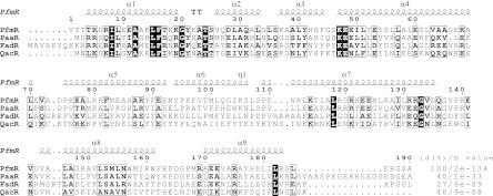
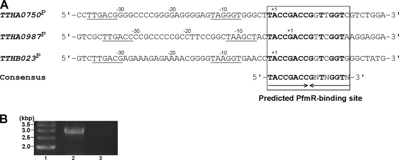
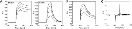
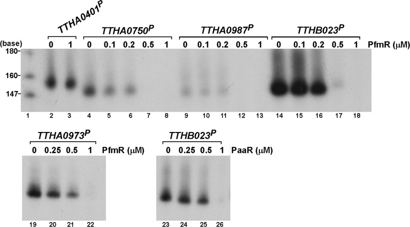
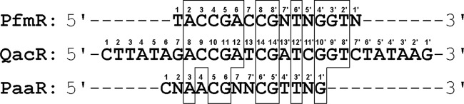
PfmR is one of four TetR family transcriptional regulators found in the extremely thermophilic bacterium, Thermus thermophilus HB8. We identified three promoters with strong negative regulation by PfmR, both in vivo and in vitro. PfmR binds pseudopalindromic sequences, with the consensus sequence of 5'-TACCGACCGNTNGGTN-3' surrounding the promoters. According to the amino acid sequence and three-dimensional structure analyses of the PfmR-regulated gene products, they are predicted to be involved in phenylacetic acid and fatty acid metabolism. In vitro analyses revealed that PfmR weakly cross-regulated with the TetR family repressor T. thermophilus PaaR, which controls the expression of the paa gene cluster putatively involved in phenylacetic acid degradation but not with another functionally identified TetR family repressor, T. thermophilus FadR, which is involved in fatty acid degradation. The X-ray crystal structure of the N-terminal DNA-binding domain of PfmR and the nucleotide sequence of the predicted PfmR-binding site are quite similar to those of the TetR family repressor QacR from Staphylococcus aureus. Similar to QacR, two PfmR dimers bound per target DNA. The bases recognized by QacR within the QacR-binding site are conserved in the predicted PfmR-binding site, and they were important for PfmR to recognize the binding site and properly assemble on it. The center of the PfmR molecule contains a tunnel-like pocket, which may be the ligand-binding site of this regulator.


Figures

Fig 1
Fig 1
Sequence alignment of T. thermophilus PfmR with representative homologous proteins. Strictly conserved residues are represented by white letters on a black background, and similar residues are depicted by boxed bold letters. PaaR, T. thermophilus PaaR (33); FadR, T. thermophilus FadR (1); QacR, S. aureus QacR (9). The sequences were aligned using Clustal W2 (16). The secondary structure of PfmR (chain A) was predicted with the DSSP program (13), and the figure was generated with ESPript, version 2.2 (8). α, η, and T represent the α-helix, 310-helix, and turn, respectively. The percent identities [id(%)] and the E values relative to PfmR, determined by BLAST, are indicated on the right.
Fig 2
Fig 2
(A) Nucleotide sequence alignment of the promoters of the TTHA0750, TTHA0987, and TTHB023 genes regulated by T. thermophilus PfmR. The predicted PfmR-binding sites and the consensus sequence are indicated. The conserved bases in the PfmR binding sites are indicated by bold letters. Possible −10 and −35 hexamer sequences of the promoters are underlined. The transcription start site of each gene (+1), as determined by a 5′ RACE experiment, is indicated. (B) RT-PCR analysis to confirm the operon composed of the genes TTHB023 to TTHB018 (lane 2). As a control, PCR was also performed with no RT, using the same primers (lane 3). The samples were fractionated on a 1% agarose gel, which was stained with ethidium bromide and photographed. Lane 1, 500-bp DNA ladder markers.
Fig 3
Fig 3
BIAcore biosensor analyses of the interactions between the T. thermophilus TetR family transcriptional regulators and DNA. (A) A dsDNA fragment corresponding to the upstream region of the TTHB023 gene (see the text), which contains the predicted PfmR-binding site, was immobilized on the sensor chip, and then the PfmR or PaaR protein was injected over the DNA surface at concentrations of 0.4, 0.3, 0.2, and 0.1 μM dimer in buffer A. (B) A dsDNA fragment corresponding to the upstream region of the TTHA0963 gene (33), which contains the PaaR-binding site, was immobilized on the sensor chip, and then the PfmR protein was injected over the DNA surface, in the same manner as described for panel A. (C) A dsDNA fragment corresponding to the upstream region of the TTHA0890 gene (1), which contains the FadR-binding site, was immobilized on the sensor chip, and then the PfmR protein was injected over the DNA surface, in the same manner as described for panel A.
Fig 4
Fig 4
Effects of T. thermophilus TetR family transcriptional regulators on transcription in vitro. Runoff transcription assays were performed with templates containing the upstream sequences of the genes regulated by PfmR (PTTHA0750, PTTHA0987, and PTTHB023), FadR (PTTHA0401) (1), and PaaR (PTTHA0973) (33) in the absence or presence of PfmR or PaaR. After the reaction, the samples were fractionated on the polyacrylamide gel, followed by autoradiography. Lane 1, [α-32P]dCTP-labeled MspI fragments of pBR322.
Fig 5
Fig 5
Sequence alignment of the predicted binding site of T. thermophilus PfmR with binding sites of other TetR family regulators. QacR, S. aureus QacR (34); PaaR, predicted T. thermophilus PaaR (33). One half-site is numbered 1 to 8, 1 to 14, or 1 to 7 (reading from the left) and the other is 1′ to 8′, 1′ to 14′, or 1′ to 7′ (reading from the right). Identical bases are boxed. N represents G, A, T, or C.
Fig 6
Fig 6
X-ray crystal structure of T. thermophilus PfmR. (A) Ribbon diagram of the PfmR dimer chains A (red) and B (gray). (B) Molecular surface representation of the PfmR dimer. Red and blue surfaces represent negative and positive electrostatic potentials (−5 kBT and +5 kBT, where kB is the Boltzmann constant and T is the temperature), respectively. The electrostatic potentials were calculated using the Adaptive Poisson-Boltzmann Solver (APBS) (3) with the PyMol APBS tools. (C) The N-terminal HTH DNA-binding domain of the S. aureus QacR proximal monomer (chain A) in complex with DNA (PDB code 1JT0) (34). DNA strands are shown in yellow and orange. Oxygen, nitrogen, and phosphate atoms are shown in red, blue, and light blue, respectively. The protein domain is gray, and the DNA-binding residues are indicated by stick models. (D) The N-terminal HTH domain of the PfmR monomer (chain A) superimposed on the corresponding domain of the QacR monomer in complex with DNA is shown, as described for panel C. The putative DNA-binding residues are indicated by stick models. (E) Stereo view around the center of the PfmR molecule. Chains A and B are shown in red and gray, respectively. The putative ligand-binding tunnel-like pocket is indicated by a mesh. The residues comprising the pocket are depicted by stick models. (F) Schematic model structures of the residues comprising the tunnel-like pocket of PfmR. The residues in parentheses are from chain A. The other residues are from chain B. These figures were drawn using the Pymol program (http://www.pymol.org/).
Fig 7
Fig 7
Gel filtration analyses of T. thermophilus PfmR and the PfmR-DNA complex. (A) Elution profiles of the samples (26 μl) comprising 50 μM PfmR dimer (a), 50 μM PfmR dimer plus a 25 μM DNA fragment derived from the upstream region of the TTHB023 gene (see the text) (b), 50 μM PfmR dimer plus a 12.5 μM DNA fragment derived from the upstream region of the TTHB023 gene (c), 50 μM PfmR dimer plus a 25 μM DNA fragment derived from the upstream region of the TTHA0890 gene (see the text) (d), 50 μM PfmR dimer plus a 25 μM DNA fragment derived from the upstream region of the TTHB023 gene containing the A6C/T6′G mutations (see the text) (e), and 50 μM PfmR dimer plus a 25 μM DNA fragment derived from the upstream region of the TTHB023 gene containing the C8A/G8′T mutations (see the text) (f). The elution buffer was 10 mM sodium phosphate (pH 7.0) and 0.3 M NaCl. Relative absorbances at 220 nm (solid line) and 260 nm (dashed line) are indicated. The elution volume at the top of each peak is indicated. (B) Molecular masses of PfmR (open triangle) and PfmR with the DNA fragment derived from the upstream region of the TTHB023 gene (closed triangle). The partition coefficient (Kav) value was calculated as Kav = (elution volume − void volume)/(geometric column volume − void volume). As standards, the Kav values of conalbumin (75 kDa), ovalbumin (44 kDa), carbonic anhydrase (29 kDa), RNase A (13.7 kDa), and aprotinin (6.5 kDa) (open circles) were plotted.

References

    1. Agari Y, Agari K, Sakamoto K, Kuramitsu S, Shinkai A. 2011. TetR family transcriptional repressor Thermus thermophilus FadR controls fatty acid degradation. Microbiology 157:1589–1601 - PubMed
    1. Agari Y, Kashihara A, Yokoyama S, Kuramitsu S, Shinkai A. 2008. Global gene expression mediated by Thermus thermophilus SdrP, a CRP/FNR family transcriptional regulator. Mol. Microbiol. 70:60–75 - PubMed
    1. Baker NA, Sept D, Joseph S, Holst MJ, McCammon JA. 2001. Electrostatics of nanosystems: application to microtubules and the ribosome. Proc. Natl. Acad. Sci. U. S. A. 98:10037–10041 - PMC - PubMed
    1. Brünger AT, et al. 1998. Crystallography and NMR system: A new software suite for macromolecular structure determination. Acta Crystallogr. D Biol. Crystallogr. 54:905–921 - PubMed
    1. Collaborative Computational Project Number 4 1994. The CCP4 suite: programs for protein crystallography. Acta Crystallogr. D Biol. Crystallogr. 50:760–763 - PubMed

Publication types

MeSH terms

Associated data