. 2012 Sep;194(17):4669-76.
doi: 10.1128/JB.00957-12. Epub 2012 Jun 29.

The Escherichia coli SMC complex, MukBEF, shapes nucleoid organization independently of DNA replication

Affiliations

The Escherichia coli SMC complex, MukBEF, shapes nucleoid organization independently of DNA replication

Anjana Badrinarayanan et al. J Bacteriol. 2012 Sep.

Abstract

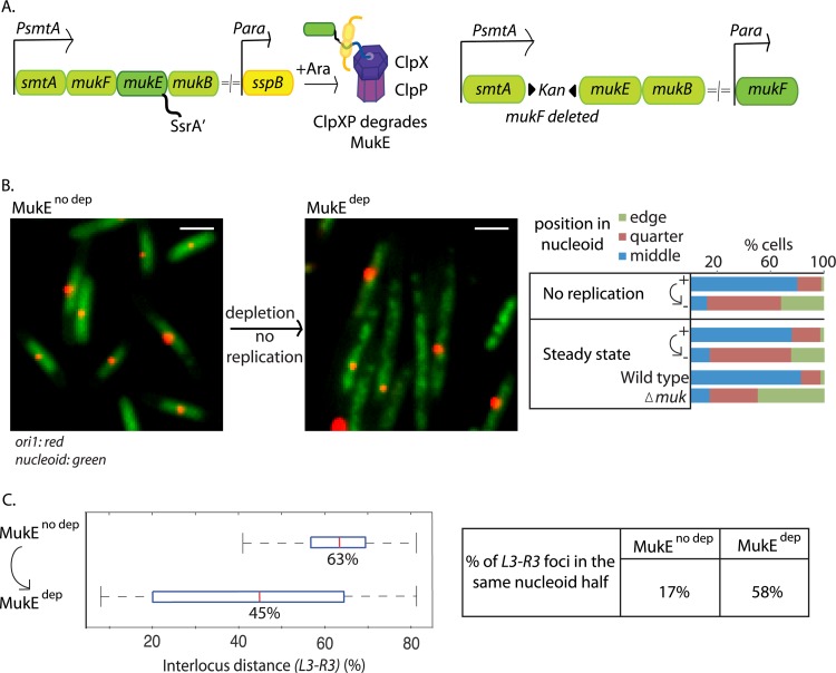
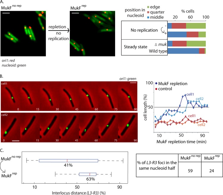
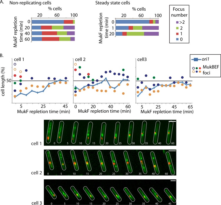
SMC (structural maintenance of chromosomes) complexes function ubiquitously in organizing and maintaining chromosomes. Functional fluorescent derivatives of the Escherichia coli SMC complex, MukBEF, form foci that associate with the replication origin region (ori). MukBEF impairment results in mispositioning of ori and other loci in steady-state cells. These observations led to an earlier proposal that MukBEF positions new replicated sister oris. We show here that MukBEF generates and maintains the cellular positioning of chromosome loci independently of DNA replication. Rapid impairment of MukBEF function by depleting a Muk component in the absence of DNA replication leads to loss of MukBEF foci as well as mispositioning of ori and other loci, while rapid Muk synthesis leads to rapid MukBEF focus formation but slow restoration of normal chromosomal locus positioning.


Figures

Fig 1
Fig 1
MukE depletion leads to mispositioning of ori1 and L3-R3 independently of DNA replication. (A) Depletion and repletion. With depletion, arabinose-induced SspB directs MukE-SsrA′ to ClpXP for degradation (left). With repletion, MukF is expressed from an arabinose-inducible promoter at an ectopic chromosomal location in cells with wild-type MukF deleted (right). (B) MukE depletion results in ori mispositioning independently of replication. ori1 positions before (MukEno dep) and after (MukEdep) MukE depletion (2 h) in steady-state or nonreplicating [dnaC(Ts)] cells (strain Ab64) with a single ori1 focus are estimated as percentage of nucleoid length. Positions are binned into three categories: middle (40 to 60%), quarter (60 to 80%), or edge (80 to 100%) of nucleoid, and the percentage of cells with ori1 in each category is plotted. Wild-type and ΔmukF controls (strains RRL48 and Ab188, respectively) in steady-state cells with a single ori1 focus are also shown. Snapshot images of cells with labeled ori1 (red) and DAPI nucleoid staining (green) are shown before and after MukE depletion (in nonreplicating cells) on the left. n ≥ 300. Bars, 2 μm. (C) MukE depletion results in mispositioning of L3-R3 independently of replication. Interlocus distance (ILD; percentage of nucleoid length) between L3 and R3 before and after MukE depletion (2 h) in nonreplicating dnaC(Ts) cells (snapshot analysis) (strain Ab82). Red lines represent the median ILD. Boxes, interquartile ranges; whiskers, minima and maxima. Median values are shown below the red lines. The percentage of cells with L3 and R3 in the same nucleoid half is also shown (right). n ≥ 200.
Fig 2
Fig 2
MukF repletion repositions ori1 and L3-R3 in the absence of replication. (A) MukF repletion repositions ori1 independently of replication. ori1 position before (MukFno rep) and 1 h after (MukFrep) induction of MukF expression in nonreplicating dnaC(Ts) cells at 37°C (strain Ab174). Snapshot images of cells with labeled ori1 (red) and DAPI nucleoid staining (green) are shown before and after MukF repletion (in nonreplicating cells). n ≥ 300. Protocols, snapshot analysis, and controls were as described for Fig. 1. Bars, 2 μm. (B) ori1 repositioning during MukF repletion in the absence of replication. MukF repletion as a function of time [nonreplicating dnaC(Ts) cells]. Time-lapse analysis (strain Ab174) images were taken every 5 min for 100 min. The data for two representative cells are shown (blue lines), along with a subset of total images. Time-lapse traces of ori1 position for two representative nonreplicating ΔmukF cells are shown in red (strain Ab188). n ≥ 15. Bars, 2 μm. (C) MukF repletion repositions L3-R3 in the absence of replication. Interlocus distance (as percentage of nucleoid length) between L3 and R3 during MukF repletion (1 h) in nonreplicating [dnaA(Ts)] cells is shown (strain Ab229). The percentage of cells with L3 and R3 in the same nucleoid half is also shown (right). n ≥ 300.
Fig 3
Fig 3
Formation of MukB foci is independent of replication. (A) MukB focus appearance during MukF repletion is independent of replication or cell stage. The appearance and number of MukB foci were assessed before (0 min) and 20, 40, and 60 min after induction of MukF repletion (strain Ab174) in nonreplicating cells (left) and at 1 and 20 min after induction of MukF repletion in steady-state cells (strain Ab234). In the steady-state cells, transcription initiation was blocked at each time point by the addition of rifampin. The percentage of cells with 0, 1, 2, or >2 foci per cell are shown. n > 200. (B) MukB focus dynamics with respect to ori1 repositioning during MukF repletion in the absence of replication. Time-lapse analysis of MukB focus formation and ori1 positioning during MukF repletion in nonreplicating cells (strain Ab174). Time-lapse traces of ori1 (blue lines) as well as the position of MukB foci (circles) for three representative cells are shown. ori1, red; MukB, green. Cell outlines are in white. Images were taken every 5 min. Note that the 0-min time point in panel B is not equivalent to that in panel A, since there is a delay of ∼2 min between induction and imaging during the time-lapse experiments. Bars, 2 μm.

Similar articles

Cited by

References

    1. Bachmann BJ. 1972. Pedigrees of some mutant strains of Escherichia coli K-12. Bacteriol. Rev. 36:525–557 - PMC - PubMed
    1. Ben-Yehuda S, Rudner DZ, Losick R. 2003. RacA, a bacterial protein that anchors chromosomes to the cell poles. Science 299:532–536 - PubMed
    1. Carter SD, Sjogren C. 2012. The SMC complexes, DNA and chromosome topology: right or knot? Crit. Rev. Biochem. Mol. Biol. 47:1–16 - PubMed
    1. Cuylen S, Haering CH. 2011. Deciphering condensin action during chromosome segregation. Trends Cell Biol. 21:552–559 - PubMed
    1. D'Ambrosio C, et al. 2008. Identification of cis-acting sites for condensin loading onto budding yeast chromosomes. Genes Dev. 22:2215–2227 - PMC - PubMed

Publication types

MeSH terms

LinkOut - more resources