

Mitochondrial division in rat cardiomyocytes: an electron microscope study
- PMID: 22753088
- PMCID: PMC3744323
- DOI: 10.1002/ar.22523
Mitochondrial division in rat cardiomyocytes: an electron microscope study
Abstract
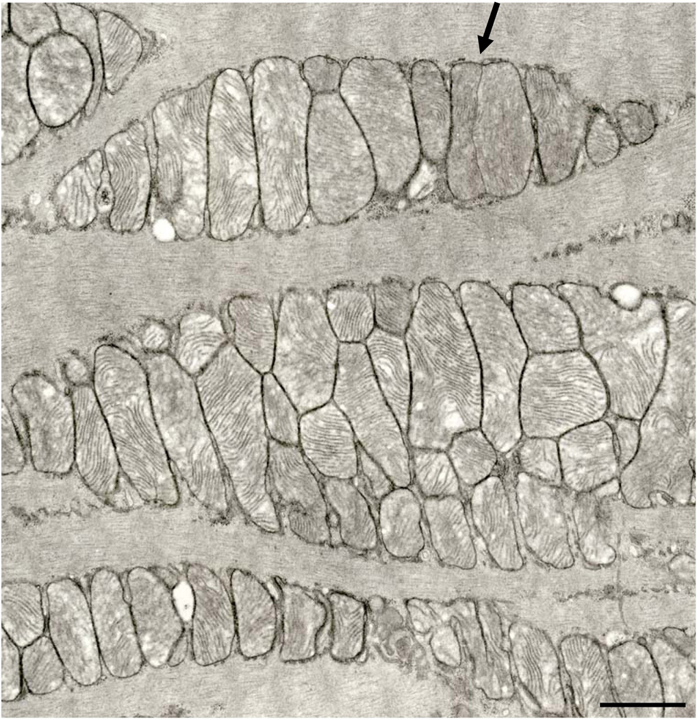
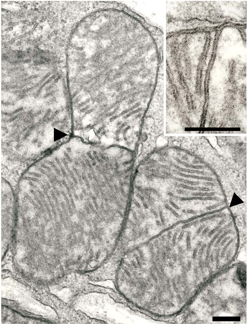
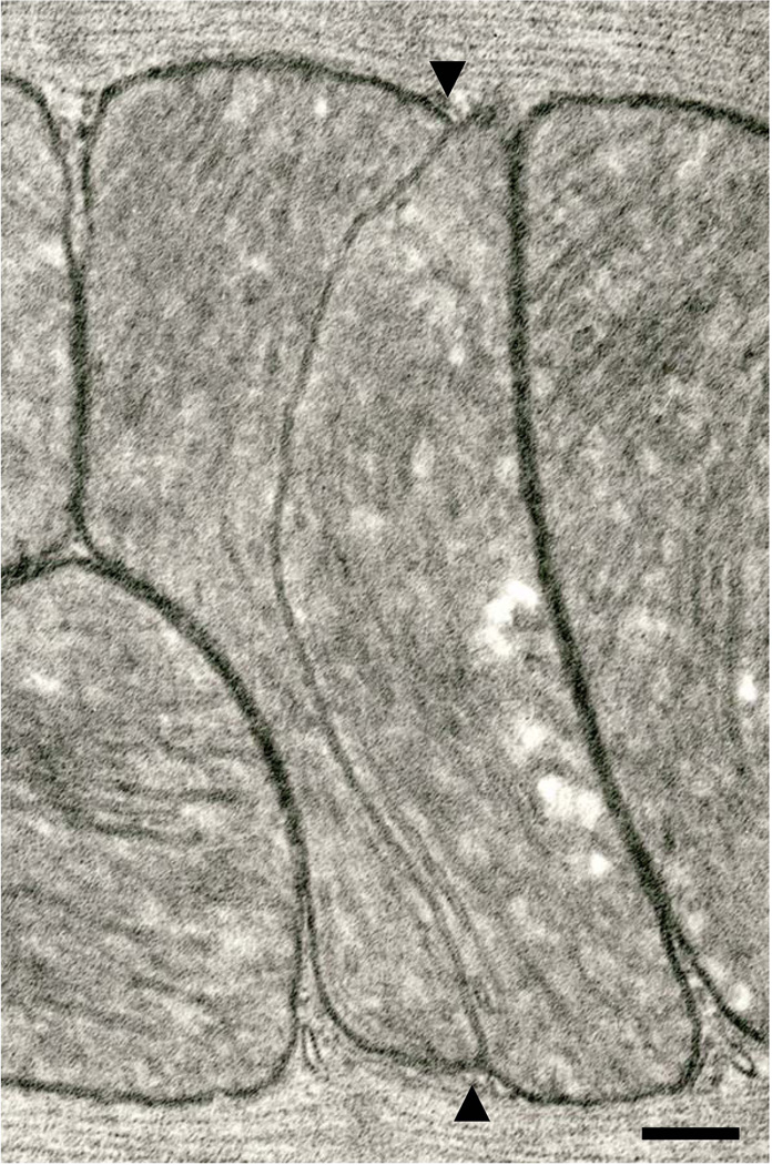
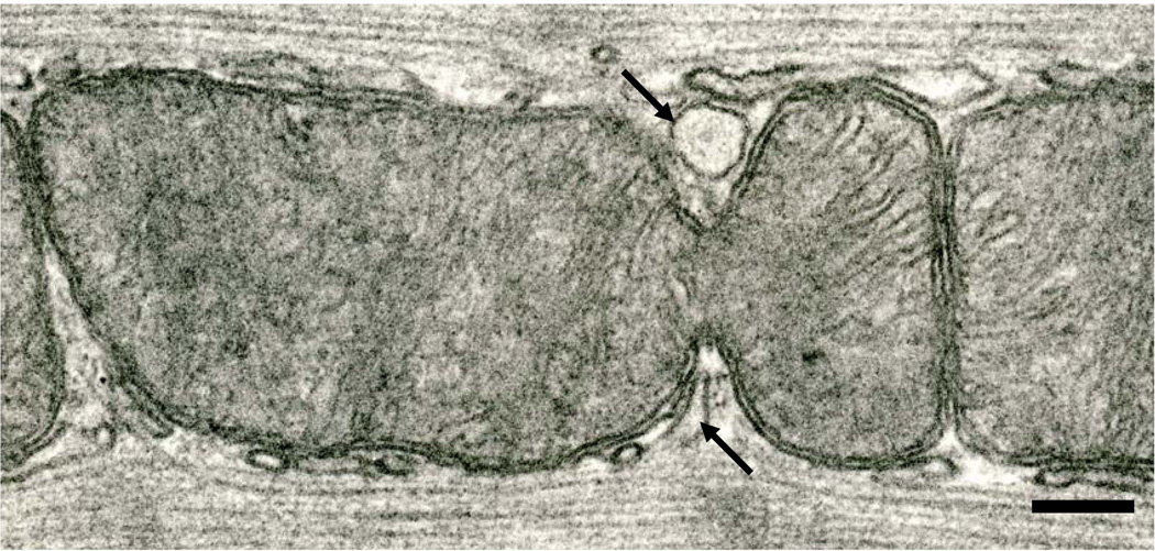
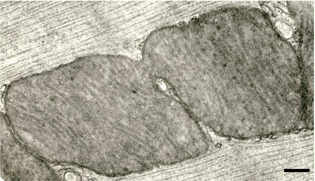
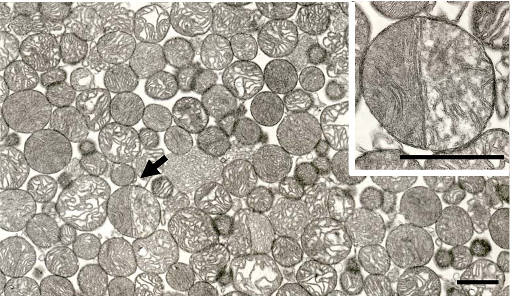
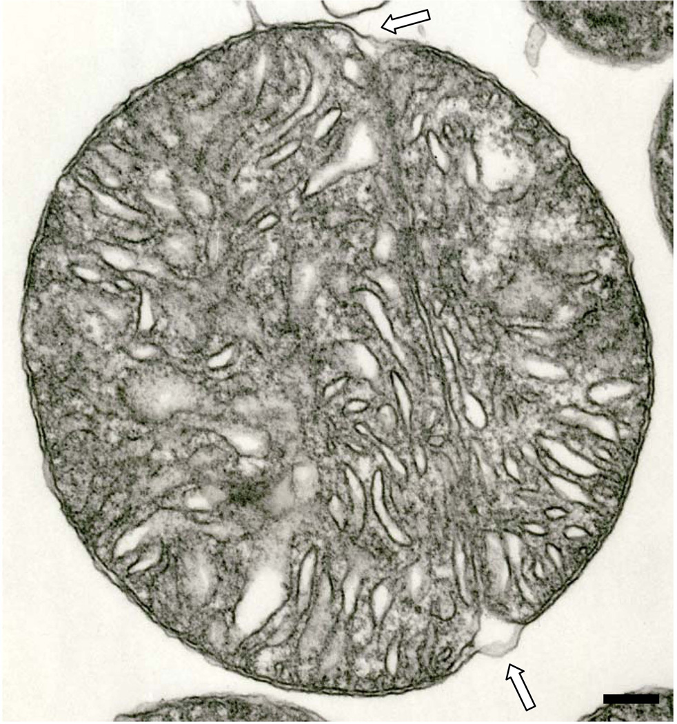
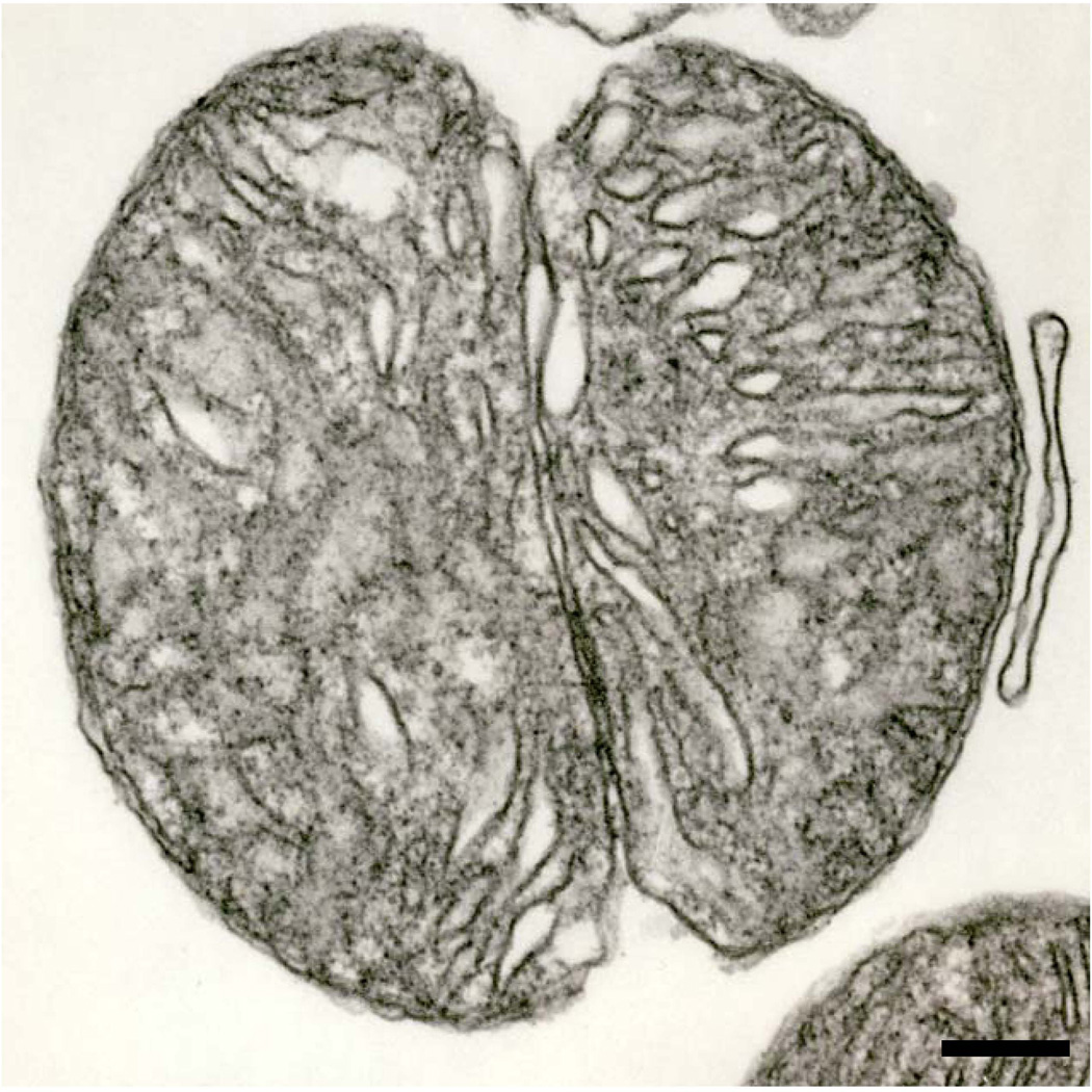
In cardiomyocytes of rats, two distinct mitochondrial division processes are in operation. The predominant process involves extension of a single crista until it spans the full width of a mitochondrion. Ingrowth of the outer membrane ultimately results in scission. The second division process involves "pinching," in which narrowing of the organelle at specific surface locations leads to its attenuation. When limiting membranes from opposite sides meet, mitochondrial fission ensues. When pinching is the operative mode, elements of sarcoplasmic reticulum always are associated with the membrane constrictions. The nuclear control mechanisms that determine which modality of mitochondrial division will prevail are unknown.
Copyright © 2012 Wiley Periodicals, Inc.
Figures
Similar articles
-
[Ultrastructure of mitochondria apparatus of cardiomyocytes in apoptosis induced by long-term anoxia in rats].Tsitologiia. 2003;45(11):1073-82. Tsitologiia. 2003. PMID: 14989146 Russian.
-
[The cytochrome c oxidase activity in mitochondria of cardiomyocytes of isolated cardiac tissue under long-term hypoxic incubation].Tsitologiia. 2008;50(3):268-74. Tsitologiia. 2008. PMID: 18664130 Russian.
-
Mechanical stretch induces mitochondria-dependent apoptosis in neonatal rat cardiomyocytes and G2/M accumulation in cardiac fibroblasts.Cell Res. 2004 Feb;14(1):16-26. doi: 10.1038/sj.cr.7290198. Cell Res. 2004. PMID: 15040886
-
Morphological Pathways of Mitochondrial Division.Antioxidants (Basel). 2018 Feb 15;7(2):30. doi: 10.3390/antiox7020030. Antioxidants (Basel). 2018. PMID: 29462856 Free PMC article. Review.
-
Sarcoplasmic reticulum-mitochondria communication in cardiovascular pathophysiology.Nat Rev Cardiol. 2017 Jun;14(6):342-360. doi: 10.1038/nrcardio.2017.23. Epub 2017 Mar 9. Nat Rev Cardiol. 2017. PMID: 28275246 Review.
Cited by
-
Mitochondrial fission/fusion and cardiomyopathy.Curr Opin Genet Dev. 2016 Jun;38:38-44. doi: 10.1016/j.gde.2016.03.001. Epub 2016 Apr 7. Curr Opin Genet Dev. 2016. PMID: 27061490 Free PMC article. Review.
-
Megamitochondria in Cardiomyocytes of a Knockout (Klf15-/-) Mouse.Ultrastruct Pathol. 2015;39(5):336-9. doi: 10.3109/01913123.2015.1042610. Epub 2015 Jun 25. Ultrastruct Pathol. 2015. PMID: 26111268 Free PMC article.
-
Division of mitochondria in cultured human fibroblasts.Microsc Res Tech. 2013 Dec;76(12):1213-6. doi: 10.1002/jemt.22287. Epub 2013 Sep 5. Microsc Res Tech. 2013. PMID: 24009193 Free PMC article.
-
Lipids activate skeletal muscle mitochondrial fission and quality control networks to induce insulin resistance in humans.Metabolism. 2021 Aug;121:154803. doi: 10.1016/j.metabol.2021.154803. Epub 2021 Jun 4. Metabolism. 2021. PMID: 34090870 Free PMC article. Clinical Trial.
-
Kruppel-like factor 4 is critical for transcriptional control of cardiac mitochondrial homeostasis.J Clin Invest. 2015 Sep;125(9):3461-76. doi: 10.1172/JCI79964. Epub 2015 Aug 4. J Clin Invest. 2015. PMID: 26241060 Free PMC article.
References
-
- Chan DC. Mitochondrial fusion and fission in mammals. Annu Rev Cell Dev Biol. 2006;22:79–99. - PubMed
-
- Chen H, Chan DC. Energizing functions of mammalian mitochondrial fusion and fission. Hum Mol Genet. 2005;11:R283–R289. - PubMed
-
- Chen Q, Hoppel CL, Lesnefsky EJ. Blockage of electron transport before cardiac ischemia with the reversible inhibitor amobarbital protects rat heart mitochondria. J Pharmacol Exp Ther. 2006;316:200–207. - PubMed
Publication types
MeSH terms
Grants and funding
LinkOut - more resources
Full Text Sources
