. 2012 Sep 1;40(17):8743-58.
doi: 10.1093/nar/gks633. Epub 2012 Jun 30.

Crystal structure of the UvrB dimer: insights into the nature and functioning of the UvrAB damage engagement and UvrB-DNA complexes

Affiliations

Crystal structure of the UvrB dimer: insights into the nature and functioning of the UvrAB damage engagement and UvrB-DNA complexes

Matthew P J Webster et al. Nucleic Acids Res. .

Abstract

UvrB has a central role in the highly conserved UvrABC pathway functioning not only as a damage recognition element but also as an essential component of the lesion tracking machinery. While it has been recently confirmed that the tracking assembly comprises a UvrA2B2 heterotetramer, the configurations of the damage engagement and UvrB-DNA handover complexes remain obscure. Here, we present the first crystal structure of a UvrB dimer whose biological significance has been verified using both chemical cross-linking and electron paramagnetic resonance spectroscopy. We demonstrate that this dimeric species stably associates with UvrA and forms a UvrA2B2-DNA complex. Our studies also illustrate how signals are transduced between the ATP and DNA binding sites to generate the helicase activity pivotal to handover and formation of the UvrB2-DNA complex, providing key insights into the configurations of these important repair intermediates.


Figures
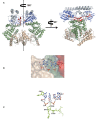
Figure 1.
Figure 1.
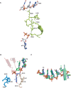
(A) Cartoon of the UvrB dimer with domains 1a, 1b, 2, 3 and the β-hairpin highlighted in green, grey, blue, light brown and red, respectively. (B) View of nucleotides A2 to T4 (sticks) associated with monomer A (depicted as van der Waals spheres) that extend across the domain 1a-domain 3 interface (coloured as above) similar to the analogous nucleotides in monomer B. (C) The domain 3–DNA interactions (domain 3 is shown in green). Arg506 donates a hydrogen bond to O2P of C3, while also donating a hydrogen bond to the carbonyl oxygen of Ser477. A2 is stabilized by a stacking interaction involving Phe527.
Figure 2.
Figure 2.
(A) Arg506 directly connects the DNA and ATP binding sites via a network of hydrogen bonds and salt bridges. This involves Arg540, Asp510 and Arg543; all of which are essential for repair. (B) T7 is extra-helical in monomer A (slate), but intra-helical in monomer B (green). The extra-helical thymine is stabilized in a recess formed by residues Val250, Phe249 and Ala248 in domain 2 and Gly147, Leu148 and Gly149 in domain 1a that contribute largely van der Waals contacts. The exocyclic oxygens are stabilized by the guanadinium group of Arg123 (also in domain 1a) and the main chain NH group of Gly149. All domains are coloured as above. (C) Superposition of nucleotides C3 to T7 in monomer A (extraS, slate) and monomer B (intraS, green). A slight translocation of extraS towards the β-hairpin could potentially disfavour the intra-helical conformation of T7 present in intraS as a result of steric clashes between the furanose rings of T6 and T7 (red arrow).
Figure 3.
Figure 3.
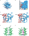
(A) Superposition of domain 2 from monomer B with domain 2 from the trithymine UvrB–DNA complex (2D7D.pdb) reveals considerable re-modeling and a relative rotation of ∼30°. (B) The re-configuration of domain 2 in each monomer gives rise to partial encapsulation of the β-hairpin motifs that (C) mediate a range of electrostatic, van der Waals and hydrophobic interactions. These include hydrogen bonds donated by the guanadinium group of Arg194 (domain 2) to the carbonyl oxygens of Asp106 and Thr105, respectively (β-hairpin), the amide nitrogen of Gln97 (β-hairpin) to the carboxylate group of Glu209 (domain 2) and the carboxylate group of Asp187 to the guanadinium moiety of Arg285. This configuration of the β-hairpin and domain 2 is further stabilized by hydrophobic interactions involving Thr107, Ile109, Val102 (β-hairpin) and Phe203, Cys211, Ala229 and Leu230 (domain 2). (D) The domain 3 dimer interface comprises the highly conserved helix spanning residues Ser481–Lys495 and is largely mixed where Thr481, Leu482, Ile485, Leu493 contribute hydrophobic interactions. (E) Superposition of the domain 3 interface with the B. caldotenax UvrB co-ordinates (1D9X.pdb) reveals that the C-terminal residues (magenta) in the wild-type monomer would prevent full dimerization of UvrB consistent with their proposed auto inhibitory role.
Figure 4.
Figure 4.
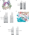
(A) Left panel: the relative positions of the NCS related T481 residues whose OG1 atoms are separated by 14.9 Å, consistent with the cross-linking agent BM(PEG)2. Right panel: the structure of BM(PEG)2. (B) Gel shift assay of the T481C mutant using the G10 duplex. The T481C mutant has comparable binding activity to wild-type UvrB. (C) SDS–PAGE gel of WT UvrB and the T481C mutant after treatment with the cross-linking agent BM(PEG)2.
Figure 5.
Figure 5.
(A) Cw-EPR spectra of WT UvrB (blue) and the T481C mutant (red) recorded at room temperature. (B) Distance distribution obtained by Tikhonov regularization of the 4 pulse DEER spectrum (Supplementary Figure S3D). (C) Docking of the UvrA UvrB interacting domain (magenta) onto the UvrB dimer (via superposition of the domain 2 residues in the UvrA(interacting domain)–UvrB(interacting domain) complex (3FPN.pdb) results in no major steric clashes.
Figure 6.
Figure 6.
(A) A native PAGE gel demonstrating that both UvrB and UvrB2X form dimeric complexes with UvrA that co-elute. (B) UvrA–UvrB (UvrB2X) gel shift assay using the T50 substrate. The UvrA–UvrB2X complex remains trapped on the DNA unlike wild-type UvrB where a UvrB–DNA complex is formed. (C) Gel shift assay illustrating that UvrB2X is not defective in its ability to associate with DNA. (D) ATP-ase assay of UvrB2X in the presence and absence of DNA. Although UvrB2X has residual ATP-ase activity, it is no longer stimulated by ssDNA which is the most likely cause of the handover defect. (E) Putative model of a UvrA2Bdimer complex. The UvrB dimer can be successfully docked against UvrA with the UvrB interacting domains rotated by 30° relative to their positions in the B. stearothermophilus and T. maritima structures. In this configuration, the UvrA and UvrB dimer 2-fold axes are aligned and the β-hairpin positioned close to the UvrA dimer interface, the location of the DNA binding site.
Figure 7.
Figure 7.
(A) A putative UvrB2–DNA complex derived by applying the operator relating the two monomers in our dimer to the UvrB–stem loop DNA monomer co-ordinates (2FDC.pdb). (B) Gel shift assays performed with the R489P mutant in the presence of UvrA and the T50 substrate (left) and in the presence of the G10 self-loading substrate (right). In both instances, R489P is entirely defective in its ability to form the B–DNA complex. (C) Left: gel shift assays of the R194E/D187R and R194E/I485E interfacial mutants in the presence of UvrA and the T50 substrate (the β-hairpin mutant F101A/F108A is provided as a control since it is entirely defective in its capacity to form a UvrB–DNA complex either in the presence or absence of UvrA). The R194E/D187R mutant exhibits greater affinity for DNA in the UvrB–DNA complex compared to native UvrB while the R194E/I485E has almost wild type affinity. Right: gel shift assays of the same mutants (excluding F101A/F108A) performed using the G10 substrate. The R194E/D187R mutant similarly displays a higher affinity for DNA that contrasts with R194E/I485E that is highly defective. (D) The putative UvrB2–DNA model viewed from the interfacial region between the two monomers. Asp187 and Arg190 are both appropriately positioned to interact with the highly distorted duplex. In the model, Tyr95 at the base of the β-hairpin is also predicted to intercalate between the disrupted bases. (E) Gel shift assays performed using the R190E mutant in the presence of UvrA and the T50 substrate (left) and in the presence of the G10 substrate (right). In both instances, R190E is defective in its capacity to form the UvrB–DNA complex although there is partial rescue in the presence of UvrA.

Similar articles

Cited by

References

    1. Van Houten B, Croteau DL, DellaVecchia MJ, Wang H, Kisker C. ‘Close-fitting sleeves’: DNA damage recognition by the UvrABC nuclease system. Mutat. Res. 2005;577:92–117. - PubMed
    1. Truglio JJ, Croteau DL, Van Houten B, Kisker C. Prokaryotic nucleotide excision repair: the UvrABC system. Chem. Rev. 2006;106:233–252. - PubMed
    1. Friedberg EC, Walker GC, Siede W. DNA Repair and Mutagenesis. Washington DC: ASM Press; 1995.
    1. Seeley TW, Grossman L. Mutations in the Escherichia coli UvrB ATPase motif compromise excision repair capacity. Proc. Natl Acad. Sci. USA. 1989;86:6577–6581. - PMC - PubMed
    1. Verhoeven EE, Wyman C, Moolenaar GF, Hoeijmakers JH, Goosen N. Architecture of nucleotide excision repair complexes: DNA is wrapped by UvrB before and after damage recognition. EMBO J. 2001;20:601–611. - PMC - PubMed

Publication types