. 2012 Aug;60(2):476-86.
doi: 10.1161/HYPERTENSIONAHA.111.189571. Epub 2012 Jul 2.

Role of copper transport protein antioxidant 1 in angiotensin II-induced hypertension: a key regulator of extracellular superoxide dismutase

Affiliations

Role of copper transport protein antioxidant 1 in angiotensin II-induced hypertension: a key regulator of extracellular superoxide dismutase

Kiyoshi Ozumi et al. Hypertension. 2012 Aug.

Abstract

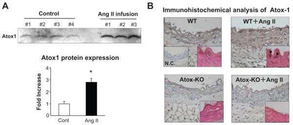
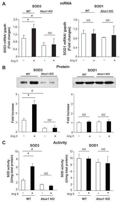
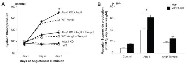
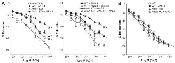
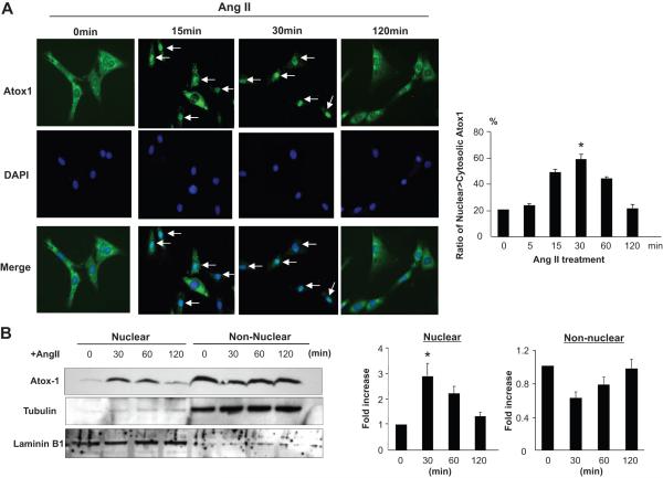
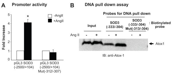
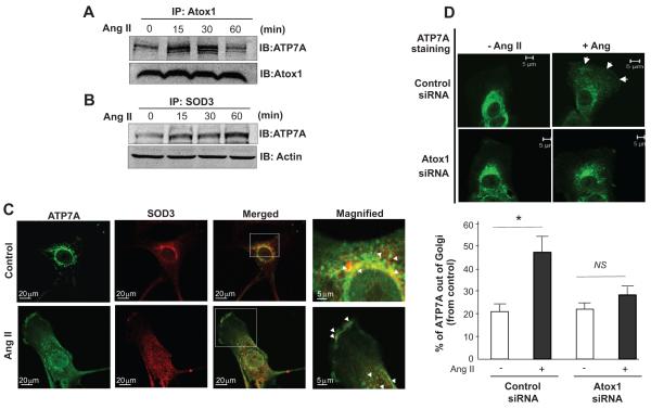
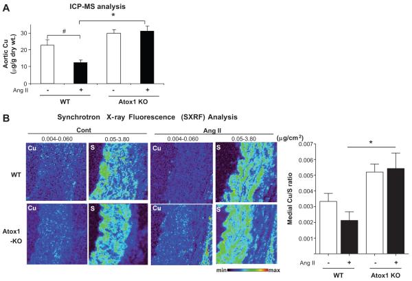
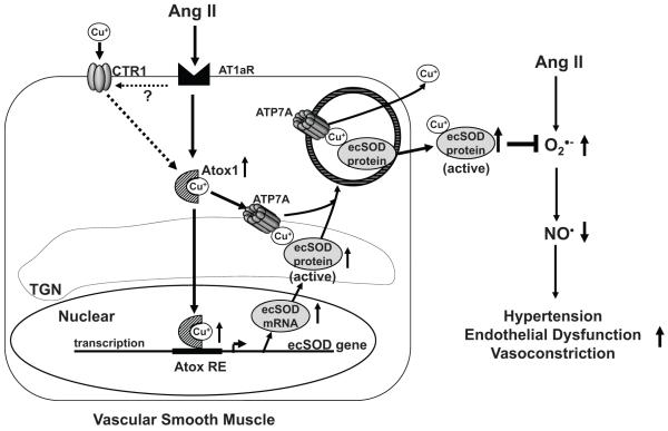
Extracellular superoxide dismutase (SOD3) is a secretory copper enzyme involved in protecting angiotensin II (Ang II)-induced hypertension. We found previously that Ang II upregulates SOD3 expression and activity as a counterregulatory mechanism; however, underlying mechanisms are unclear. Antioxidant 1 (Atox1) is shown to act as a copper-dependent transcription factor, as well as a copper chaperone, for SOD3 in vitro, but its role in Ang II-induced hypertension in vivo is unknown. Here we show that Ang II infusion increases Atox1 expression, as well as SOD3 expression and activity, in aortas of wild-type mice, which are inhibited in mice lacking Atox1. Accordingly, Ang II increases vascular superoxide production, reduces endothelium-dependent vasodilation, and increases vasoconstriction in mesenteric arteries to a greater extent in Atox1(-/-) than in wild-type mice. This contributes to augmented hypertensive response to Ang II in Atox1(-/-) mice. In cultured vascular smooth muscle cells, Ang II promotes translocation of Atox1 to the nucleus, thereby increasing SOD3 transcription by binding to Atox1-responsive element in the SOD3 promoter. Furthermore, Ang II increases Atox1 binding to the copper exporter ATP7A, which obtains copper from Atox1, as well as translocation of ATP7A to plasma membranes, where it colocalizes with SOD3. As its consequence, Ang II decreases vascular copper levels, which is inhibited in Atox1(-/-) mice. In summary, Atox1 functions to prevent Ang II-induced endothelial dysfunction and hypercontraction in resistant vessels, as well as hypertension, in vivo by reducing extracellular superoxide levels via increasing vascular SOD3 expression and activity.


Figures

Figure 1
Figure 1. Effect of angiotensin II (Ang II) infusion on Atox1 expression in aortas of WT and Atox1−/− mice in vivo by immunoblotting (A) and immunohistochemistry (B)
Aortas of C57Bl/6 mice were harvested after a 7-day infusion of Ang II (0.7 mg/ kg/ day) or vehicle subcutaneously with osmotic minipumps, and subjected to Western analysis with antibodies specific to Atox1. Representative blots were from 3 individual experiments. *P<0.001 vs. WT mice. B, Immunohistochemical analysis of Atox1 expression in aortas from Ang II-treated C57Bl/6 (WT) and Atox1−/− (Atox1-KO) mice. Arrows in inset shows nuclear staining of Atox1. All images were taken at 5 different fields and the cell images are representative of >3 different experiments.
Figure 2
Figure 2. Effect of Ang II infusion on mRNA expression (A), protein level (B), and activity (C) of SOD3 and SOD1 in aortas from Atox1−/− mice
Mice were infused with Ang II as described in Figure 1. A and B, mRNA expression and protein levels of SOD1 and SOD3 in aortas of C57Bl/6 and Atox1−/− mice were measured by real-time PCR and by western analysis with antibodies specific to either SOD1 or SOD3. C, Activity of SOD3 and SOD1 in aortas were analyzed by the inhibition of cytochrome c reduction by xanthine/xanthine oxidase. SOD3 activity was determined after separation with Con A-Sepharose. The results are presented as mean ± SE from four separate experiments. Representative blots are from 3 individual experiments. *P<0.001; #P<0.01 vs. either vehicle-infused WT or Atox1−/− mice; NS, not significant
Figure 3
Figure 3. Effect of Ang II on blood pressure and vascular O2•− production in Atox1−/− mice
A, Either Ang II (0.7 mg/kg/day) or Ang II and the SOD mimetic Tempol (50 mg/kg/day) or vehicles were infused, and blood pressure was measured before, 4 and 7 days after minipumps implantation (n=5 per group). #P<0.01 vs. WT or Atox1−/− mice with Tempol treatment. B, O2•− production in aorta from C57Bl/6 (WT) and Atox1−/− mice as determined with lucigenin-enhanced chemiluminescence (5 μmol/L). #P < 0.01 vs. WT mice (n=6).
Figure 4
Figure 4. Effect of Ang II infusion on endothelium-dependent vascular relaxation in mesenteric arteries from Atox1−/− and WT mice
A and B, First or second-order mesenteric resistance arteries was studied using a wire myograph and precontracted with phenylephrine. Vasodilation was evoked by acetylcholine (Ach) in absence or presence of the SOD mimetic tempol (A) and sodium nitroprusside (SNP) (B). Data are expressed as mean ± SE (n=6-8 per group). * P<0.001 vs. remaining three groups, #P<0.01 vs. either untreated WT mice or Ang II-treated WT mice with Tempol treatment. NS, not significant.
Figure 5
Figure 5. Effect of Ang II on nuclear translocation of Atox1 in vascular smooth muscle cells (VSMCs)
A, VSMCs were stimulated with Ang II (100 nM) for indicated time and cells were stained with anti-Atox 1 antibody and the nuclear marker, DAPI. In each image, ratio of Nuclear >Cytosolic Atox1 (immunofluorescence of nuclear Atox1 is higher than that of cytosolic Atox1) was calculated from 5 randomized view and the cell images are representative of >3 different experiments. *P<0.001vs. no treated cells. White arrows indicate nuclear Atox1 predominant cells. B, Nuclear and cytoplasmic fractions were immunoblotted with anti-Atox 1 antibody, cytoplasmic marker, tubulin and nuclear marker, laminin B1. Right panel shows averaged data for nuclear and non-nuclear expression of Atox1 protein, expressed as fold increased against control VSMCs (Mean ± SE, n=3). *P<0.001vs. untreated cells.
Figure 6
Figure 6. Role of Atox1 in Ang II-induced SOD3 promoter activity in VSMCs
A, Role of Atox1 on Ang II-induced transactivation of the SOD3 gene promoter in VSMCs. Cells were transiently transfected with SOD3 promoter luciferase reporter constructs (pGL3 SOD3 (−2500/+104) or mutated SOD3 promoter luciferase reporter constructs (pGL3 SOD3, Mut(−312/−307) along with Ang II (Mean ± SE, n=3). *P<0.001 vs untreated cells. B, DNA pull-down assay, showing Ang II-induced binding of Atox1 to the GAAAGA sequence (Atox1-reponsive element (Atox1-RE)) in the SOD3 promoter. Nuclear extract from VSMCs treated with or without Ang II was incubated with biotinylated oligonuclotide probes and streptavidin-Sepharose. The Protein-DNA complexes were subjected to SDS-PAGE, followed by immunoblotting with anti-Atox1 antibody. The experiments was performed using two different oligonucleotide, SOD3 (−333/−304) and SOD3 (−333/−304) Mut (−313/−304). Representative figure was from three independent experiments.
Figure 7
Figure 7. Effect of Ang II on interaction among Atox1, ATP7A, and ecSOD in VSMCs
A and B, VSMCs were stimulated with Ang II (100 nM), and lysates were immunoprecipitated (IP) with either anti-Atox1 or anti-ecSOD antibody, followed by immunoblotting (IB) with anti-ATP7A antibody. C, Effect of Ang II on subcellular localization of ATP7A (green) and SOD3 (red) in human aortic smooth muscle cells (HASMs). D, HASMs transfected with Atox1 or control siRNAs were stained with anti-ATP7A antibody. Images were representative of 3 different experiments taken at 5 different fields/well.
Figure 8
Figure 8. Effect of Ang II-induced hypertension on abundance and spatial distribution of copper in the vascular tissue from Atox1−/− and WT mice
A and B,, Aortic copper content in Atox1−/− and WT mice infused with Ang II or vehicle for 7 days, as described in Figure 1 was measured by inductively coupled plasma mass spectrometry (ICP-MS) or Synchrotron-based X-ray Fluorescence (SXRF). SXRF scans (1-2 sec per pixel) were performed in paraffin-embedded tissue (left). The maximum and minimum threshold values in microgram per square centimeter are given above each frame. Map of copper shows areas of the lowest to the highest content scaled to a rainbow color (bottom). Total sulfur is used as a surrogate for total cellular protein and to visualize the morphology of tissue sections. Data are quantified using three samples for each group, two to three scans/sample (right).
Figure 9
Figure 9. Proposed model for protective role of Atox1 in Ang II-induced hypertension through regulating SOD3 expression and activity
Atox1 plays an important role in Ang II-induced transcription and activity of SOD3 by increasing Atox1 binding to the Atox1 response element in SOD3 promoter as well as to copper transporter ATP7A protein required for copper delivery to SOD3 for enhancing its specific activity. As its consequence, Atox1 contributes to the decrease in extracellular O2•− levels in the vessel wall, thereby increasing available NO to preserve endothelial function, and reducing blood pressure.

Comment in

Similar articles

Cited by

References

    1. Garrido AM, Griendling KK. NADPH oxidases and angiotensin II receptor signaling. Mol Cell Endocrinol. 2009;302:148–158. - PMC - PubMed
    1. Fukai T, Ushio-Fukai M. Superoxide Dismutases: Role in Redox Signaling, Vascular Function and Diseases. Antioxid Redox Signal. 2011;15(6):1583–1606. - PMC - PubMed
    1. Chu Y, Alwahdani A, Iida S, Lund DD, Faraci FM, Heistad DD. Vascular effects of the human extracellular superoxide dismutase R213G variant. Circulation. 2005;112:1047–1053. - PubMed
    1. Jung O, Marklund SL, Geiger H, Pedrazzini T, Busse R, Brandes RP. Extracellular superoxide dismutase is a major determinant of nitric oxide bioavailability: in vivo and ex vivo evidence from ecSOD-deficient mice. Circ Res. 2003;93:622–629. - PubMed
    1. Gongora MC, Qin Z, Laude K, Kim HW, McCann L, Folz JR, Dikalov S, Fukai T, Harrison DG. Role of extracellular superoxide dismutase in hypertension. Hypertension. 2006;48:473–481. - PubMed

Publication types

MeSH terms