. 2012 Sep;14(9):1136-45.
doi: 10.1093/neuonc/nos139. Epub 2012 Jun 29.

Signal transducer and activator of transcription 3 promotes angiogenesis and drives malignant progression in glioma

Affiliations

Signal transducer and activator of transcription 3 promotes angiogenesis and drives malignant progression in glioma

Tiffany A Doucette et al. Neuro Oncol. 2012 Sep.

Abstract

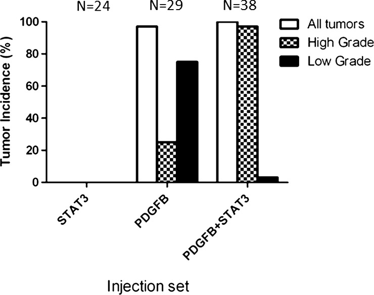
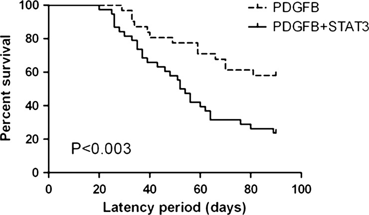
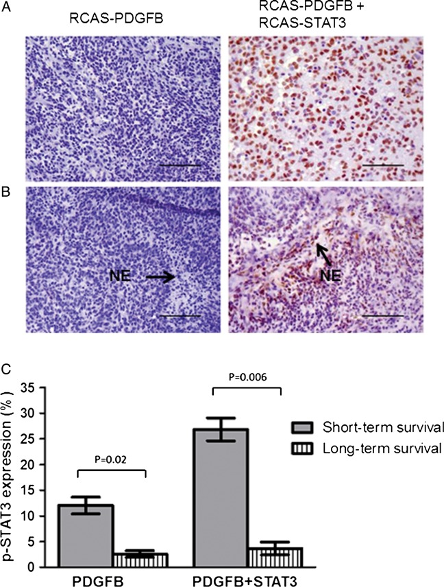
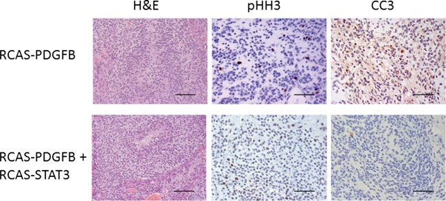
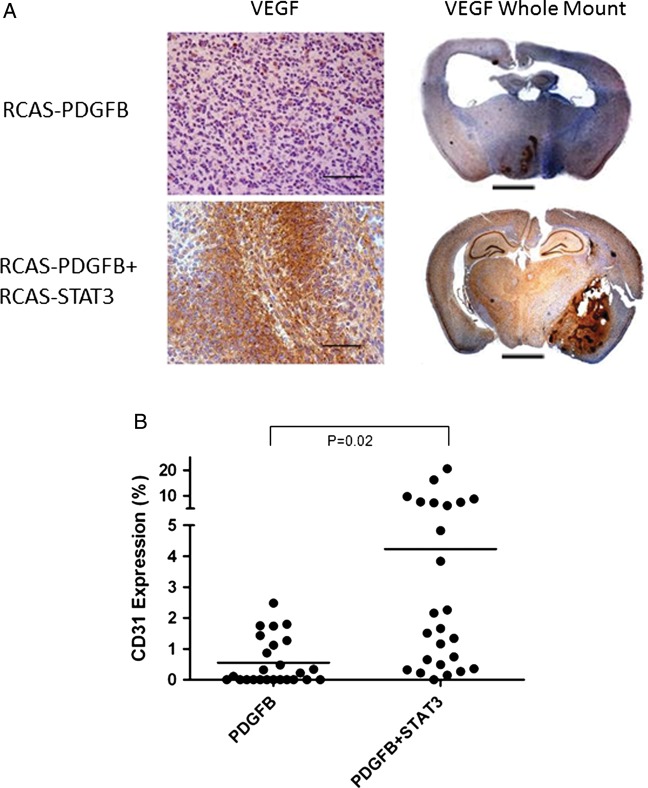
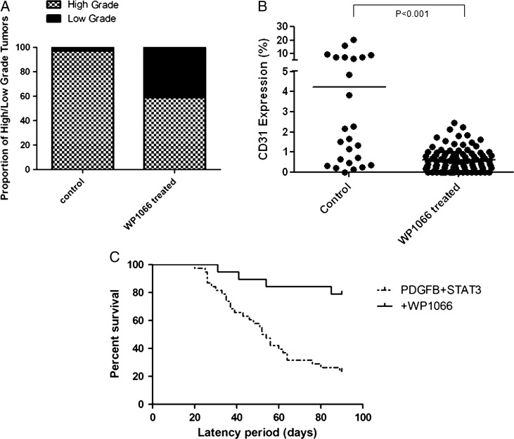
Signal transducer and activator of transcription (STAT) 3 has been described as a "master regulator" of signaling pathways involved in the transition from low-grade glioma (LGG) to high-grade glioma (HGG). Although STAT3 is overexpressed in HGGs, it remains unclear whether its overexpression is sufficient to induce or promote the malignant progression of glioma. To characterize the effect of STAT3 expression on tumor progression in vivo, we expressed the STAT3 gene in glioneuronal progenitor cells in mice. STAT3 was expressed alone or concurrently with platelet-derived growth factor B (PDGFB), a well-described initiator of LGG. STAT3 alone was insufficient to induce tumor formation; however, coexpression of STAT3 with PDGFB in mice resulted in a significantly higher incidence of HGGs than PDGFB alone. The median symptomatic tumor latency in mice coexpressing STAT3 and PDGFB was significantly shorter, and mice that developed symptomatic tumors demonstrated significantly higher expression of phosphorylated STAT3 intratumorally. In HGGs, expression of STAT3 was associated with suppression of apoptosis and an increase in tumor cell proliferation. HGGs induced by STAT3 and PDGFB also displayed frequent foci of necrosis and microvascular proliferation. The expression of CD31 (a marker of endothelial proliferation) was significantly higher in tumors induced by coexpression of STAT3 and PDGFB. When mice injected with PDGFB and STAT3 were treated with a STAT3 inhibitor, median survival increased and the incidence of HGG and CD31 expression decreased significantly. These results demonstrate that STAT3 promotes the malignant progression of glioma. Inhibiting STAT3 expression mitigates tumor progression and improves survival, validating it as a therapeutic target.


Figures

Fig. 1.
Fig. 1.
The incidence of high-grade gliomas was significantly higher in mice injected with RCAS-PDGFB + RCAS-STAT3 than in those injected with RCAS-PDGFB alone. RCAS-STAT3 was insufficient to induce tumor formation.
Fig. 2.
Fig. 2.
The median tumor latency was significantly longer in mice injected with RCAS-PDGFB alone (90 days, range 29–90 days) than in those injected with RCAS-PDGFB + RCAS-STAT3 (53 days, range 20–90 days).
Fig. 3.
Fig. 3.
Expression of pSTAT3 is increased in tumors induced by RCAS-PDGFB + RCAS-STAT3 and pSTAT3-expressing cells appear to localize to areas of necrosis. (A) Photomicrograph (400× magnification) showing relatively minimal pSTAT3 expression in the nuclei within an HGG induced by RCAS-PDGFB compared with a tumor induced by RCAS-PDGFB + RCAS-STAT3 (scale bar = 50 µm). (B) Photomicrograph (400×) showing pSTAT3-expressing cells preferentially localizing to areas of necrosis (denoted as NE) in HGGs induced by RCAS-PDGFB + RCAS-STAT3 but not in the only HGG induced by RCAS-PDGFB alone that displayed necrosis (scale bar = 50 µm). (C) Percentage of cells expressing pSTAT3 in mice killed due to symptomatic tumor formation before the end of the 90-day observation period (labeled “short-term survival”) compared with those who survived to 90 days (labeled “long-term survival”) in RCAS-PDGFB (left) and RCAS-PDGFB + RCAS-STAT3 (right) injection sets. Error bars indicate SEM.
Fig. 4.
Fig. 4.
STAT3 promotes tumor cell proliferation and suppression of apoptosis. Representative hematoxylin and eosin (H&E) photomicrographs (400× magnification) of HGGs induced by RCAS-PDGFB or by RCAS-PDGFB + RCAS-STAT3. These tumors were studied for expression of pHH3 as a marker of tumor cell proliferation and cleaved caspase 3 (CC3) as a marker of apoptosis. HGGs induced by RCAS-PDGFB + RCAS-STAT3 showed higher expression of pHH3 and lower expression of CC3 compared with tumors induced by RCAS-PDGFB alone (scale bar = 50 µm).
Fig. 5.
Fig. 5.
Tumors induced by RCAS-PDGFB + RCAS-STAT3 display increased microvascular proliferation. (A) Photomicrograph (400×) of VEGF staining in high-grade tumors induced by RCAS-PDGFB or by RCAS-PDGFB + RCAS-STAT3. Tumors induced by RCAS-PDGFB + RCAS-STAT3 demonstrate large foci of VEGF staining throughout the tumor mass, whereas there is minimal staining in high-grade tumors induced by RCAS-PDGFB alone. Whole-mount photomicrograph (50×) of VEGF staining in a high-grade tumor, showing the localization of VEGF to the high-grade areas (areas surrounding necrosis or microvascular proliferation) (scale bar = 2.5 mm). (B) Expression of CD31 in tumors induced by RCAS-PDGFB or by RCAS-PDGFB + RCAS-STAT3. Horizontal lines indicate the mean.
Fig. 6.
Fig. 6.
Tumors induced by RCAS-PDGFB and RCAS-STAT3 after treatment with WP1066. (A) WP1066 treatment decreases the percentage of high-grade gliomas compared with untreated RCAS-PDGFB+ RCAS-STAT3 injected mice (control). (B) WP1066 decreases microvascular proliferation as determined by staining for CD31 in tumors (horizontal line indicates the mean). (C) The median tumor latency was significantly longer in mice injected with RCAS-PDGFB + RCAS-STAT3 treated with WP1066 compared with the original group of untreated mice.

References

    1. Cairncross G, Berkey B, Shaw E, et al. Phase III trial of chemotherapy plus radiotherapy compared with radiotherapy alone for pure and mixed anaplastic oligodendroglioma: Intergroup Radiation Therapy Oncology Group Trial 9402. J Clin Oncol. 2006;24:2707–2714. - PubMed
    1. Jaeckle KA, Ballman KV, Rao RD, Jenkins RB, Buckner JC. Current strategies in treatment of oligodendroglioma: evolution of molecular signatures of response. J Clin Oncol. 2006;24:1246–1252. - PubMed
    1. Stupp R, Mason WP, van den Bent MJ, et al. Radiotherapy plus concomitant and adjuvant temozolomide for glioblastoma. N Engl J Med. 2005;352:987–996. - PubMed
    1. van den Bent MJ, Carpentier AF, Brandes AA, et al. Adjuvant procarbazine, lomustine, and vincristine improves progression-free survival but not overall survival in newly diagnosed anaplastic oligodendrogliomas and oligoastrocytomas: a randomized European Organisation for Research and Treatment of Cancer phase III trial. J Clin Oncol. 2006;24:2715–2722. - PubMed
    1. Louis DN, Ohgaki H, Wiestler OD, et al. The 2007 WHO classification of tumours of the central nervous system. Acta Neuropathol. 2007;114:97–109. - PMC - PubMed

Publication types

MeSH terms

Substances