. 2012 Sep;80(9):3206-14.
doi: 10.1128/IAI.00645-12. Epub 2012 Jul 2.

Biofilm-infected intracerebroventricular shunts elicit inflammation within the central nervous system

Affiliations

Biofilm-infected intracerebroventricular shunts elicit inflammation within the central nervous system

Jessica N Snowden et al. Infect Immun. 2012 Sep.

Abstract

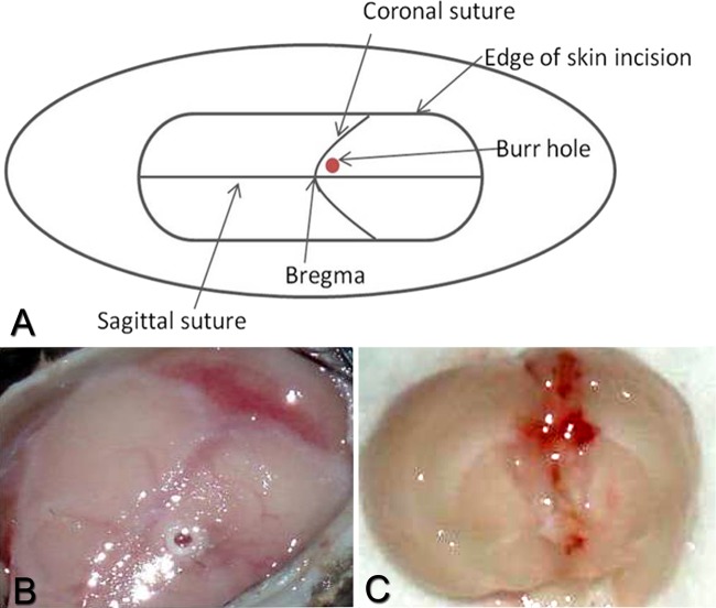
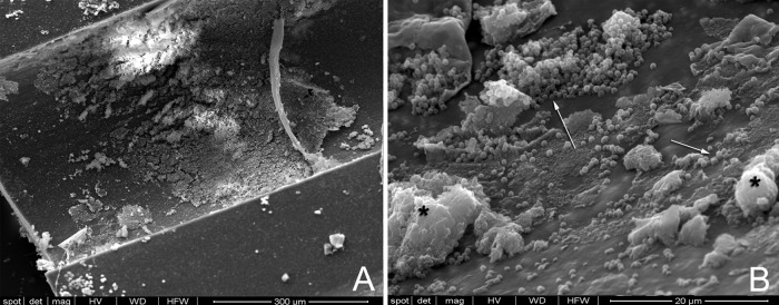
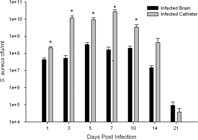
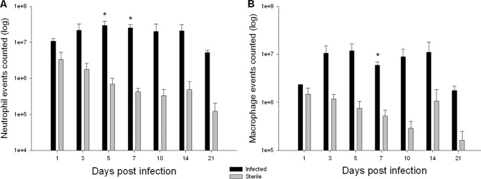
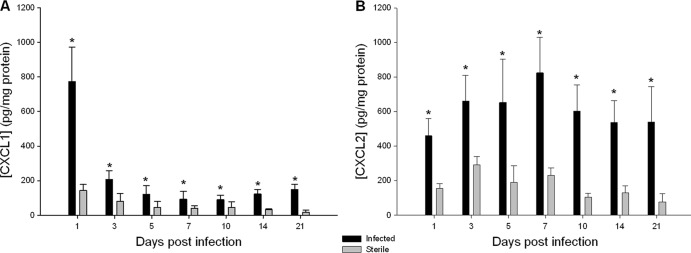
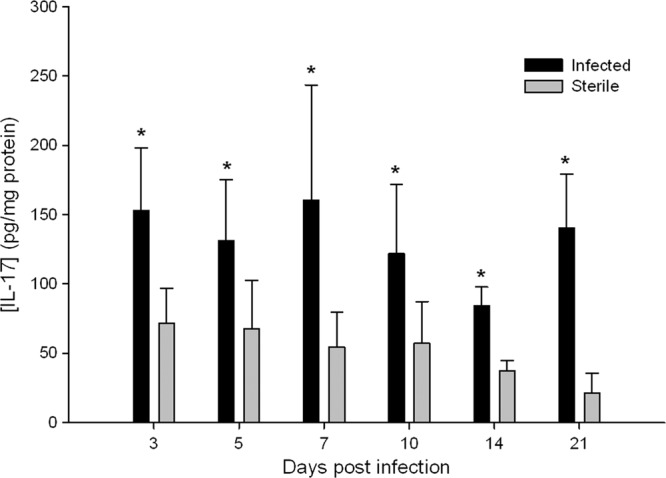
Central nervous system catheter infections are a serious complication in the treatment of hydrocephalus. These infections are commonly caused by Staphylococcus epidermidis and Staphylococcus aureus, both known to form biofilms on the catheter surface. Our objective was to generate a novel murine model of central nervous system catheter-associated biofilm infection using a clinical S. aureus isolate and characterize the nature of the inflammatory response during biofilm growth. Silicone catheters were precoated with S. aureus to facilitate bacterial attachment, whereupon infected or sterile catheters were stereotactically inserted into the lateral ventricle of the brain in C57BL/6 mice and evaluated at regular intervals through day 21 postinsertion. Animals tolerated the procedure well, with no clinical signs of illness or bacterial growth seen in the control group. Bacterial titers associated with central nervous system catheters were significantly elevated compared to those from the surrounding parenchyma, consistent with biofilm formation and minimal planktonic spread of infection. Catheter-associated bacterial burdens progressively increased, with maximal colonization achieved at day 7 postinfection. Analysis of inflammatory infiltrates by fluorescence-activated cell sorting (FACS) revealed significant macrophage and neutrophil influx, which peaked at days 3 and 5 to 7, respectively. In contrast, there were no detectable immune infiltrates associated with tissues surrounding sterile catheters. Biofilm infection led to significant increases in chemokine (CXCL1 and CCL2) and proinflammatory cytokine (interleukin 17 [IL-17]) expression in tissues surrounding infected central nervous system catheters. Based on these results, we propose this approach is a valid animal model for further investigations of catheter-associated central nervous system shunt infections.


Figures

Fig 1
Fig 1
S. aureus strain ACH1719 is capable of forming biofilm in vitro. (A) In microtiter plate cultures (performed in triplicate), ACH1719 forms biofilm grossly equivalent to that observed with USA300 LAC (not shown) and UAMS-1. Static glass chamber biofilm culture stained with Syto9 (green) and scanned at 1-μm intervals demonstrates a thick lawn of biofilm growth with ACH1719. (B) Orthogonal view; (C) three-dimensional Z-stack reconstruction.
Fig 2
Fig 2
Overview of implantation procedure. Silicone catheters are precoated with mouse serum and incubated with 2 × 10⁴ CFU/ml S. aureus for 4 h to facilitate bacterial attachment. Catheter fragments coated with S. aureus or serum alone were stereotactically inserted into the lateral ventricle of C57BL/6 mice. This location (A and B; from bregma: +0.02 mm rostral, +1 mm lateral, −2 mm deep) was chosen because the lateral ventricles are fairly large in diameter and more easily accessible at this location (C).
Fig 3
Fig 3
Visualization of S. aureus biofilm growth on explanted catheters. Catheters were removed 10 days postinfection and visualized with scanning election microscopy. While some biofilm disruption occurred following catheter removal and processing, a large portion of the catheter surface is covered with bacteria (A; 200× magnification). Higher magnification of this area shows bacteria adhering to the catheter surface (white arrows) as well as white blood cells (*) (B; 3,000× magnification).
Fig 4
Fig 4
Catheter-associated bacterial growth is significantly elevated in comparison to infection in surrounding brain parenchyma. Catheters were removed from the brain tissue, rinsed in PBS, and then sonicated in 500 μl of PBS. The titer of this solution was then determined, and the solution was cultured on blood agar plates. Supernatants from homogenates of catheter-associated tissue were also cultured on blood agar plates. Results represent three replicates of independent experiments with 4 to 5 mice per group (sterile, infected) in each replicate (n = 12 to 15 total animals per group). The cultures from the catheters and parenchyma of the mice implanted with sterile catheters were negative and are not represented. *, P < 0.05.
Fig 5
Fig 5
Central nervous system catheter infection induces robust neutrophil and macrophage recruitment. Catheter-associated cells were recovered from infected and sterile mice at multiple time points following catheter implantation using a Percoll gradient method and analyzed by FACS. Results are presented as the absolute number of positive cells for each population, averaged from independent experiments. Results represent three replicates of independent experiments with 4 to 5 mice per group (sterile, infected) in each replicate (n = 12 to 15 total animals per group). *, P < 0.05.
Fig 6
Fig 6
Elevated levels of proinflammatory chemokines CXCL1 (A) and CXCL2 (B). Supernatants from homogenates of catheter-associated tissue were analyzed for CXCL1 and CXCL2 by ELISA. Results were normalized to the amount of total protein recovered to account for differences in tissue sampling size. Results represent three replicates of independent experiments with 4 to 5 mice per group (sterile, infected) in each replicate (n = 12 to 15 total animals per group). *, P < 0.05.
Fig 7
Fig 7
Antimicrobial cytokine IL-17 levels are elevated in infected central nervous system catheters. Supernatants from homogenates of catheter-associated tissue were analyzed for IL-17 by ELISA. Results were normalized to the amount of total protein recovered to account for differences in tissue sampling size. Results represent three replicates of independent experiments with 4 to 5 mice per group (sterile, infected) in each replicate (n = 12 to 15 total animals per group). *, P < 0.05.

Similar articles

Cited by

References

    1. Almansa R, et al. 2011. Imbalanced pro- and anti-Th17 responses (IL-17/granulocyte colony-stimulating factor) predict fatal outcome in 2009 pandemic influenza. Crit. Care 15:448. - PMC - PubMed
    1. Baldwin AC, Kielian T. 2004. Persistent immune activation associated with a mouse model of Staphylococcus aureus-induced experimental brain abscess. J. Neuroimmunol. 151:24–32 - PubMed
    1. Beenken KE, Blevins JS, Smeltzer MS. 2003. Mutation of sarA in Staphylococcus aureus limits biofilm formation. Infect. Immun. 71:4206–4211 - PMC - PubMed
    1. Beenken KE, et al. 2004. Global gene expression in Staphylococcus aureus biofilms. J. Bacteriol. 186:4665–4684 - PMC - PubMed
    1. Bell MD, et al. 1996. Recombinant human adenovirus with rat MIP-2 gene insertion causes prolonged PMN recruitment to the murine brain. Eur. J. Neurosci. 8:1803–1811 - PubMed

Publication types

MeSH terms