. 2012 Aug 10;287(33):27480-9.
doi: 10.1074/jbc.M112.358929. Epub 2012 Jun 29.

Deficiency of Capn4 gene inhibits nuclear factor-κB (NF-κB) protein signaling/inflammation and reduces remodeling after myocardial infarction

Affiliations

Deficiency of Capn4 gene inhibits nuclear factor-κB (NF-κB) protein signaling/inflammation and reduces remodeling after myocardial infarction

Jian Ma et al. J Biol Chem. .

Abstract

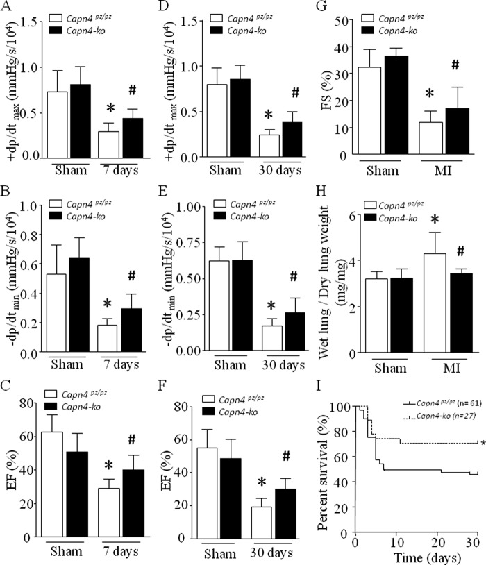
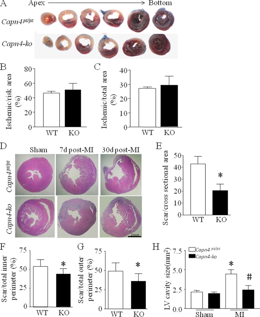
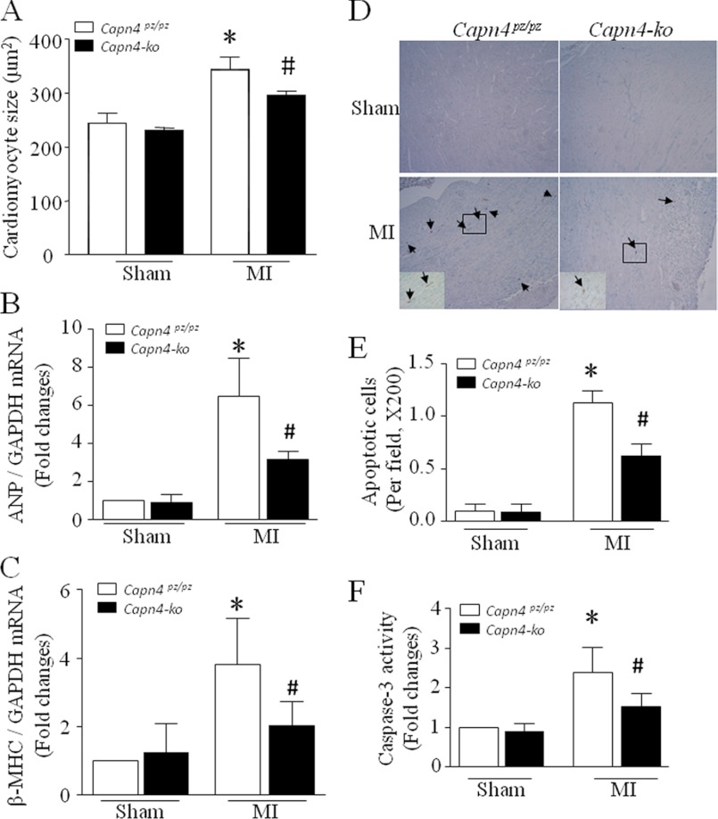
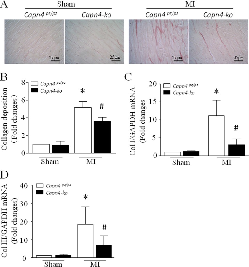
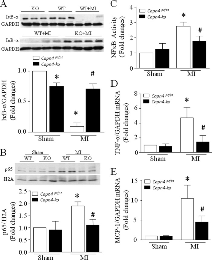
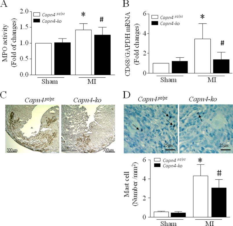
Calpain has been implicated in acute myocardial injury after myocardial infarction (MI). However, the causal relationship between calpain and post-MI myocardial remodeling has not been fully understood. This study examined whether deletion of Capn4, essential for calpain-1 and calpain-2 activities, reduces myocardial remodeling and dysfunction following MI, and if yes, whether these effects of Capn4 deletion are associated with NF-κB signaling and inflammatory responses in the MI heart. A novel mouse model with cardiomyocyte-specific deletion of Capn4 (Capn4-ko) was employed. MI was induced by left coronary artery ligation. Deficiency of Capn4 dramatically reduced the protein levels and activities of calpain-1 and calpain-2 in the Capn4-ko heart. In vivo cardiac function was relatively improved in Capn4-ko mice at 7 and 30 days after MI when compared with their wild-type littermates. Deletion of Capn4 reduced apoptosis, limited infarct expansion, prevented left ventricle dilation, and reduced mortality in Capn4-ko mice. Furthermore, cardiomyocyte cross-sectional areas and myocardial collagen deposition were significantly attenuated in Capn4-ko mice, which were accompanied by down-regulation of hypertrophic genes and profibrotic genes. These effects of Capn4 knock-out correlated with restoration of IκB protein and inhibition of NF-κB activation, leading to suppression of proinflammatory cytokine expression and inflammatory cell infiltration in the Capn4-ko heart after MI. In conclusion, deficiency of Capn4 reduces adverse myocardial remodeling and myocardial dysfunction after MI. These effects of Capn4 deletion may be mediated through prevention of IκB degradation and NF-κB activation, resulting in inhibition of inflammatory responses.


Figures

FIGURE 1.
FIGURE 1.
Assessment of myocardial function. A–F, hemodynamic measurement. Maximal positive and minimal negative first derivative of LV pressure (+dP/dtmax and −dP/dtmin) and ejection fraction (EF) were measured in Capn4-ko and Capn4pz/pz mice 7 days (A–C) and 30 days after MI (D–F). G, echocardiographic analysis. The fractional shortening (FS) value was determined by echocardiography 30 days after MI. H, index of lung congestion. The ratio of dry lung to wet lung weight was measured 30 days after MI. Data are mean ± S.D., n = 6–10. *, p < 0.05 versus sham group; #, p < 0.05 versus MI in Capn4pz/pz mice. I, adult Capn4PZ/PZ and Capn4-ko mice received left coronary artery ligation surgery. Twenty-four hours after surgery, surviving mice including 61 Capn4PZ/PZ and 27 Capn4-ko mice were monitored, and mortality was recorded for 30 days. *, p < 0.05 versus Capn4pz/pz mice.
FIGURE 2.
FIGURE 2.
Infarct size and LV cavity size after MI. A, representative TTC staining showing no changes in infarct size between Capn4pz/pz and Capn4-ko mice 24 h after MI. After perfusion with Evans blue dye, the hearts were excised, and the left ventricle was sectioned into six slices for TTC staining of infarct areas. B and C, quantifications of the ratios of ischemic/risk area and ischemia/total area. Data are mean ± S.D., n = 5. D, representative photomicrographs of hematoxylin and eosin-stained sections at the papillary muscle level in Capn4pz/pz (WT) and Capn4-ko (KO) mice 7 and 30 days after MI (magnification ×2). E–G, quantification of scar size and ratios of inner (outer) infarct to total inner (outer) perimeter at the papillary muscle level 7 days after MI. H, LV cavity size was determined at the papillary muscle level 30 days after MI. Data are mean ± S.D., n = 6–7. *, p < 0.05 versus WT or sham group; #, p < 0.05 versus MI in WT mice.
FIGURE 3.
FIGURE 3.
Cardiac hypertrophy and apoptosis in Capn4-ko MI mice. A, quantification of myocyte cross-sectional areas in Capn4pz/pz and Capn4-ko mice 30 days after MI. B and C, the mRNA levels of atrial natriuretic peptide (ANP) (B) and β-MHC (C) were measured in noninfarcted areas. D, representatives of TUNEL staining (yellow-brown signal, arrows showing nuclear localization) in Capn4pz/pz and Capn4-ko hearts 7 days after MI (magnification ×10; inset, magnification ×40). E, quantification of TUNEL-positive cells in Capn4pz/pz and Capn4-ko hearts. F, caspase-3 activity was measured in noninfarcted areas 7 days after MI. Data are mean ± S.D., n = 6–8. *, p < 0.05 versus sham group; #, p < 0.05 versus MI in Capn4pz/pz mice.
FIGURE 4.
FIGURE 4.
Myocardial fibrosis in noninfarcted areas of MI hearts. A, representative photomicrographs of picrosirius red-stained sections at the papillary muscle level in Capn4pz/pz and Capn4-ko mice 30 days after MI (red signal, magnification ×40). B, quantification of collagen deposition in noninfarcted areas of Capn4pz/pz and Capn4-ko mice after MI. C and D, the mRNA levels of collagen I (ColI) (C) and collagen III (ColIII) (D) were measured in noninfarcted areas 30 days after MI. Data are mean ± S.D., n = 6–7. *, p < 0.05 versus sham group; #, p < 0.05 versus MI in Capn4pz/pz mice.
FIGURE 5.
FIGURE 5.
Effects of Capn4 knock-out on IκB degradation and NF-κB signaling after MI. A, the upper two panels are representative Western blots for IκB protein from 3–4 out of 6–8 different hearts in each group. The lower panel is the quantification of IκB protein relative to GAPDH. B, the upper panel is a representative Western blot for p65 protein in the nucleus from 2 out of 6 different hearts in each group, and the lower panel is the quantification of p65 protein relative to histone 2A (H2A). C, NF-κB activity was measured in Sham and MI Capn4pz/pz and Capn4-ko hearts. D and E, the mRNA levels of TNF-α (D) and Mcp-1 (E) were measured in noninfarcted areas 7 days after MI. Data are mean ± S.D., n = 6–8. *, p < 0.05 versus sham group; #, p < 0.05 versus MI in Capn4pz/pz mice.
FIGURE 6.
FIGURE 6.
Effects of Capn4 knock-out on inflammation after MI. A, myeloperoxidase activity was measured in the infarct areas of Capn4pz/pz and Capn4-ko hearts 7 days after MI. B, the mRNA levels of CD68 were measured in noninfarcted areas 7 days after MI. C, the representative immunohistological staining for macrophages in cardiac frozen tissues from six different MI hearts in each group (red-purple signal, arrows showing mast cell, magnification ×4). D, the upper panel is the representative staining for mast cells from six different MI hearts in each group (yellow-brown signal, magnification ×40), and the lower panel is the quantification of mast cell numbers per section. Data are mean ± S.D., n = 6–8. *, p < 0.05 versus sham group; #, p < 0.05 versus MI in Capn4pz/pz mice.

Similar articles

Cited by

References

    1. Sutton M. G., Sharpe N. (2000) Left ventricular remodeling after myocardial infarction: pathophysiology and therapy. Circulation 101, 2981–2988 - PubMed
    1. Frangogiannis N. G., Smith C. W., Entman M. L. (2002) The inflammatory response in myocardial infarction. Cardiovasc. Res. 53, 31–47 - PubMed
    1. Nian M., Lee P., Khaper N., Liu P. (2004) Inflammatory cytokines and postmyocardial infarction remodeling. Circ. Res. 94, 1543–1553 - PubMed
    1. Sorimachi H., Suzuki K. (2001) The structure of calpain. J. Biochem. 129, 653–664 - PubMed
    1. Suzuki K., Hata S., Kawabata Y., Sorimachi H. (2004) Structure, activation, and biology of calpain. Diabetes 53, Suppl. 1, S12–S18 - PubMed

Publication types

MeSH terms