. 2012 Sep 14;287(38):31894-904.
doi: 10.1074/jbc.M112.341230. Epub 2012 Jun 29.

Myosin and tropomyosin stabilize the conformation of formin-nucleated actin filaments

Affiliations

Myosin and tropomyosin stabilize the conformation of formin-nucleated actin filaments

Zoltán Ujfalusi et al. J Biol Chem. .

Abstract

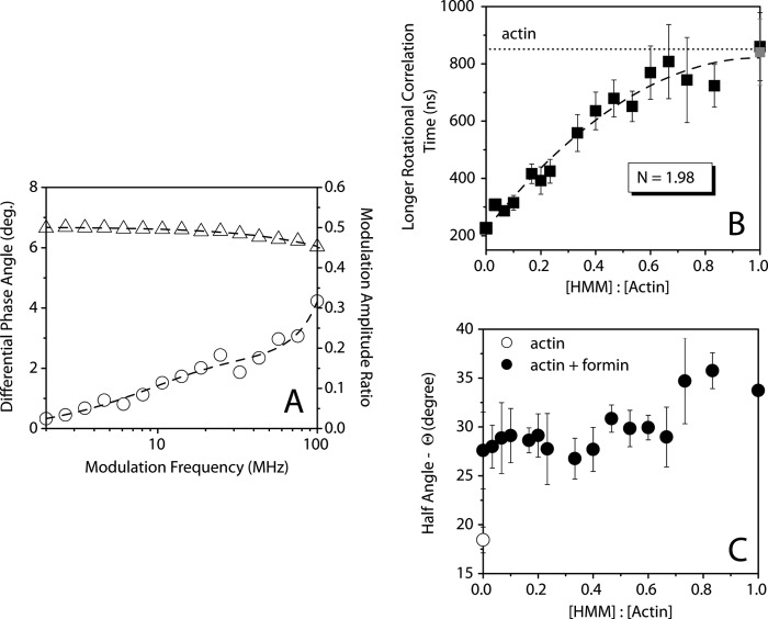
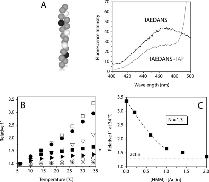
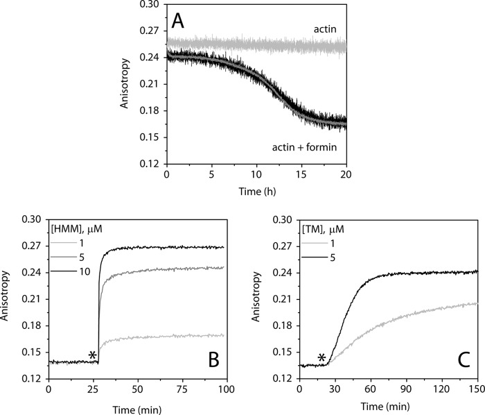
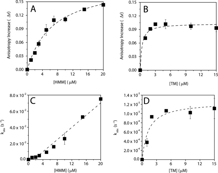
The conformational elasticity of the actin cytoskeleton is essential for its versatile biological functions. Increasing evidence supports that the interplay between the structural and functional properties of actin filaments is finely regulated by actin-binding proteins; however, the underlying mechanisms and biological consequences are not completely understood. Previous studies showed that the binding of formins to the barbed end induces conformational transitions in actin filaments by making them more flexible through long range allosteric interactions. These conformational changes are accompanied by altered functional properties of the filaments. To get insight into the conformational regulation of formin-nucleated actin structures, in the present work we investigated in detail how binding partners of formin-generated actin structures, myosin and tropomyosin, affect the conformation of the formin-nucleated actin filaments using fluorescence spectroscopic approaches. Time-dependent fluorescence anisotropy and temperature-dependent Förster-type resonance energy transfer measurements revealed that heavy meromyosin, similarly to tropomyosin, restores the formin-induced effects and stabilizes the conformation of actin filaments. The stabilizing effect of heavy meromyosin is cooperative. The kinetic analysis revealed that despite the qualitatively similar effects of heavy meromyosin and tropomyosin on the conformational dynamics of actin filaments the mechanisms of the conformational transition are different for the two proteins. Heavy meromyosin stabilizes the formin-nucleated actin filaments in an apparently single step reaction upon binding, whereas the stabilization by tropomyosin occurs after complex formation. These observations support the idea that actin-binding proteins are key elements of the molecular mechanisms that regulate the conformational and functional diversity of actin filaments in living cells.


Figures

FIGURE 1.
FIGURE 1.
Schematic representation of the interactions of actin, myosin, and tropomyosin. Left panel, the structure of an actin monomer with the accurate position of Cys374 (represented by a black circle), the site to which the fluorescent labels (IAEDANS or IAF) were attached. The TM (65) and S1 (66) binding sites in actin are highlighted in black and gray, respectively. Right panel, the structural view of an actin filament (F-actin) with bound S1. The figure is based on Protein Data Bank codes 2ZWH (for actin monomer) and 1MVW (for S1-decorated actin filament). The figure was made using WebLab ViewerPro 3.7 software.
FIGURE 2.
FIGURE 2.
HMM affects the anisotropy decay of formin-nucleated IAEDANS-actin filaments. A, representative frequency-dependent phase (empty circles) and amplitude (empty triangles) data on a logarithmic plot and a corresponding fit (dashed line) with a double exponential function (Equation 5) from anisotropy decay measurements. B, the longer rotational correlation time measured as a function of the HMM:actin concentration ratio. The dotted line indicates the rotational correlation time characteristic for actin filaments in the absence of actin-binding proteins. The rotational correlation time of actin filaments measured in the absence of mDia1-FH2 and in presence of HMM is indicated by a gray square. Fitting the linear lattice model (Equation 7; shown as dashed line) gives N = 1.98 ± 0.2 actin protomers for the length of the cooperative unit. C, the HMM:actin concentration ratio dependence of the half-angle of the cone (ϴ) within which IAEDANS at Cys374 performs wobbling motion in formin-nucleated actin filaments (black circles). The half-angle calculated for actin filaments in the absence of actin-binding proteins is represented by an empty circle. The actin and mDia1-FH2 concentrations were 30 and 1.25 μm, respectively. The error bars presented are standard errors from at least three independent experiments. deg., degree.
FIGURE 3.
FIGURE 3.
HMM decreases the conformational flexibility of formin-nucleated actin filaments. A, left panel, schematic representation of the position of the donors (dark gray circles) and acceptors (light gray circles) within the actin filament. Empty circles represent unlabeled actin protomers. This arrangement allows the characterization of the interprotomer conformational dynamics of the actin filaments. Right panel, representative fluorescence emission spectra of the donor measured in the absence (IAEDANS; black line) or presence (IAEDANS-IAF; gray line) of the acceptor in the absence of actin-binding proteins. B, temperature dependence of the relative f′. The experiments were carried out with 5 μm actin in the absence of actin-binding proteins (empty circles), in the presence of 500 nm mDia1-FH2 (empty squares), and in the presence of 500 nm mDia1-FH2 and 1 (filled circles), 3 (empty triangles), 5 (filled squares), or 10 μm (filled triangles) HMM. The arrow at the right indicates the increase of the HMM concentration and the decrease of actin filament flexibility. As a control, the temperature dependence of the relative f′ characteristic for the actin-myosin complex is represented by asterisks (our previous unpublished data) showing that myosin binding does not significantly affect the temperature dependence of the relative f′. C, the relative f′ at 34 °C as a function of the HMM:actin concentration ratio. The dotted line indicates the relative f′ characteristic for actin filaments in the absence of actin-binding proteins. HMM concentrations are indicated as HMM head concentrations. Fitting the linear lattice model (Equation 7; shown as dashed line) gives N = 1.30 ± 0.1 actin protomers for the length of the cooperative unit. The experiments were carried out with 5 μm actin, 500 nm mDia1-FH2, and HMM as indicated.
FIGURE 4.
FIGURE 4.
The effect of HMM and TM on the conformational dynamics of formin-nucleated actin filaments revealed by steady-state anisotropy. A, the kinetics of the change in the steady-state anisotropy of IAEDANS-actin filaments (5 μm) measured in the absence (light gray curve) or in the presence of 500 nm mDia1-FH2 (black curve). Black and dark gray curves show the results obtained when mDia1-FH2 was added to actin prior or after polymerization, respectively. Note that the curves are superimposable. B, the kinetics of the change in the steady-state anisotropy of mDia1-FH2-nucleated (500 nm) IAEDANS-actin filaments (5 μm) measured at different HMM concentrations as indicated Representative data are shown. Note that the steady-state anisotropy started to increase right after the addition of HMM (indicated by an asterisk) and plateaued within ∼1–5 min. C, the kinetics of the change in the steady-state anisotropy of mDia1-FH2-nucleated (500 nm) IAEDANS-actin filaments (5 μm) measured in the presence of different TM concentrations as indicated. Representative data are shown. Note that the steady-state anisotropy started to increase right after the addition of TM (indicated by an asterisk) and plateaued within ∼30 min.
FIGURE 5.
FIGURE 5.
The analysis of the effects of HMM and TM on the formin-induced conformational changes in actin filaments. A and B, Δr as a function of HMM (A) or TM (B) concentration. The anisotropy increase was derived from the exponential fit (Equation 3) of the data presented in Fig. 4, B and C. Note that a higher increase in the steady-state anisotropy from the low value of ∼0.14 (characteristic for mDia1-FH2-nucleated actin filaments) indicates a greater stabilization of the conformational dynamics of mDia1-FH2-nucleated actin filaments. The data were analyzed by hyperbolic fits (Equation 4) (dashed lines). The maximum values (Max) were 0.198 ± 0.01 and 0.103 ± 0.00 for HMM and TM, respectively. The half-saturation concentrations (K½) were determined to be 7.07 ± 1.3 and 0.33 ± 0.1 μm for HMM and TM, respectively. C and D, the observed rate constants (kobs) as a function of HMM (C) or TM (D) concentration. The values of kobs were derived from the exponential fit (Equation 3) of the data presented in Fig. 4, B and C. The stabilizing effect of HMM on mDia1-FH2-nucleated actin filaments was rapid, and the observed rate constant increased linearly with increasing amount of HMM. The apparent on-rate constant of anisotropy change determined from the slope of the linear fit was 0.004 μm−1 s−1. The stabilizing effect of TM on mDia1-FH2-nucleated actin filaments was much slower than that of HMM and followed a hyperbolic tendency with increasing TM concentration. The error bars presented on this figure are standard errors from at least two independent experiments.

Similar articles

Cited by

References

    1. Pollard T. D., Cooper J. A. (2009) Actin, a central player in cell shape and movement. Science 326, 1208–1212 - PMC - PubMed
    1. Le Clainche C., Carlier M. F. (2008) Regulation of actin assembly associated with protrusion and adhesion in cell migration. Physiol. Rev. 88, 489–513 - PubMed
    1. Chhabra E. S., Higgs H. N. (2007) The many faces of actin: matching assembly factors with cellular structures. Nat. Cell Biol. 9, 1110–1121 - PubMed
    1. Miki M., Wahl P., Auchet J. C. (1982) Fluorescence anisotropy of labeled F-actin: influence of divalent cations on the interaction between F-actin and myosin heads. Biochemistry 21, 3661–3665 - PubMed
    1. Hild G., Nyitrai M., Somogyi B. (2002) Intermonomer flexibility of Ca- and Mg-actin filaments at different pH values. Eur. J. Biochem. 269, 842–849 - PubMed

Publication types

MeSH terms

LinkOut - more resources