. 2012 Aug 17;287(34):28966-74.
doi: 10.1074/jbc.M112.345629. Epub 2012 Jun 29.

Amino-terminal cysteine residues differentially influence RGS4 protein plasma membrane targeting, intracellular trafficking, and function

Affiliations

Amino-terminal cysteine residues differentially influence RGS4 protein plasma membrane targeting, intracellular trafficking, and function

Guillaume Bastin et al. J Biol Chem. .

Abstract

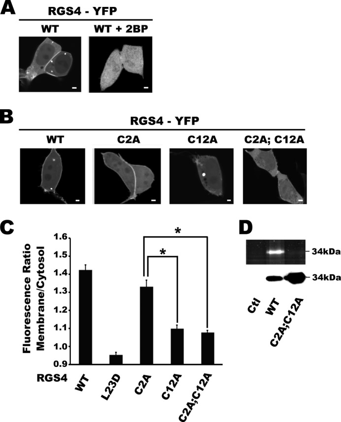
Regulator of G-protein signaling (RGS) proteins are potent inhibitors of heterotrimeric G-protein signaling. RGS4 attenuates G-protein activity in several tissues. Previous work demonstrated that cysteine palmitoylation on residues in the amino-terminal (Cys-2 and Cys-12) and core domains (Cys-95) of RGS4 is important for protein stability, plasma membrane targeting, and GTPase activating function. To date Cys-2 has been the priority target for RGS4 regulation by palmitoylation based on its putative role in stabilizing the RGS4 protein. Here, we investigate differences in the contribution of Cys-2 and Cys-12 to the intracellular localization and function of RGS4. Inhibition of RGS4 palmitoylation with 2-bromopalmitate dramatically reduced its localization to the plasma membrane. Similarly, mutation of the RGS4 amphipathic helix (L23D) prevented membrane localization and its G(q) inhibitory function. Together, these data suggest that both RGS4 palmitoylation and the amphipathic helix domain are required for optimal plasma membrane targeting and function of RGS4. Mutation of Cys-12 decreased RGS4 membrane targeting to a similar extent as 2-bromopalmitate, resulting in complete loss of its G(q) inhibitory function. Mutation of Cys-2 did not impair plasma membrane targeting but did partially impair its function as a G(q) inhibitor. Comparison of the endosomal distribution pattern of wild type and mutant RGS4 proteins with TGN38 indicated that palmitoylation of these two cysteines contributes differentially to the intracellular trafficking of RGS4. These data show for the first time that Cys-2 and Cys-12 play markedly different roles in the regulation of RGS4 membrane localization, intracellular trafficking, and G(q) inhibitory function via mechanisms that are unrelated to RGS4 protein stabilization.


Figures

FIGURE 1.
FIGURE 1.
Disruption of RGS4 plasma membrane targeting domain abrogates its Gq inhibitory function in HEK293 cells. A, localization of YFP fusion constructs was examined by transient transfection and confocal microscopy in live HEK293 cells. Shown are YFP fluorescence images of the basal side (relative to the nuclear equator) of the cells with low to intermediated fluorescence. Images are representative of at least 80 cells examined in each of 3 independent experiments. Scale bars represent 1 μm. B, inositol phosphate production was measured using [3H]myoinositol labeling of transfected cultures. HEK293 cells were co-transfected with vector control or constitutively active Gq(R183C) and the indicated HisStrep-tagged RGS4 construct. Relative expression levels of RGS4-HisStrep proteins and Gq(R183C) were determined by immunoblotting. After overnight labeling, inositol phosphate production was measured as described under “Experimental Procedures.” Values indicate absolute inositol phosphate/total soluble inositol ratios and are the means of five independent experiments each performed in triplicate. Raw cpm data are presented in supplemental Table IA. S.E. are indicated by error bars. One-way ANOVA with a Tukey's post hoc test was used to determine differences between groups (*, p < 0.001).
FIGURE 2.
FIGURE 2.
Defining the effect of palmitoylation and amino-terminal cysteine residues mutation on the plasma membrane targeting of RGS4. A, localization of the wild type RGS4-YFP construct in the presence and absence of the palmitoyl-CoA transferase inhibitor 2-BP was examined by confocal microscopy as described above. B, localization of different YFP-tagged cysteine mutants of RGS4 fusion constructs in HEK293 cells is shown. Scale bars represent 1 μm. C, the ratio of the RGS4-YFP signal between the cytosol and plasma membrane was analyzed by densitometry using ImageJ software. Shown are the means ratio of n > 80 cells. S.E. are indicated by error bars. D, ODYA-17 (palmitic acid analog) labeling of wild type and cysteine mutants of RGS4 is shown. The upper panel shows the extent of palmitoylation labeling in the indicated RGS4 constructs as measured by epitope-tag pulldown, click chemistry adduction of Alexa-488 and in-gel fluorescence. The control sample is from HEK cells not transfected with RGS4. The lower panel shows Western blot analysis of the pulldown eluates analyzed in the panel above. Where necessary, one-way ANOVA with a Tukey's post hoc test was used to determine differences between groups (*, p < 0.01).
FIGURE 3.
FIGURE 3.
Localization of RGS4 wild type and mutant constructs is relatively unaffected by Gq(R183C). A, localization of different YFP-tagged cysteine mutants of RGS4 fusion constructs in HEK293 cells in the presence of co-expressed Gq(R183C) was examined by confocal microscopy as described above. Scale bars represent 1 μm. B, the ratio of the RGS4-YFP signal between the cytosol and plasma membrane was analyzed as above (n > 30). One-way ANOVA with a Tukey's post hoc test was used to determine differences between groups. S.E. are indicated by arrow bars (*, p < 0.05).
FIGURE 4.
FIGURE 4.
Mutation of Cys-2 and Cys-12 exert differential effects on RGS4 Gq inhibitory activity. Inositol phosphate production was measured using [3H]myoinositol labeling from triplicate wells for each transfection condition. Briefly, HEK293 cells were co-transfected with constitutively active Gq(R183C) and the indicated HisStrep-tagged RGS4 construct. Relative expression levels of RGS4-HisStrep proteins and Gq(R183C) were determined by immunoblotting. After overnight labeling, inositol phosphate production was measured as described under “Experimental Procedures.” Values indicate the mean inositol phosphate/total soluble inositol ratio relative to that for the internal RGS4-inactive control (L23D) and are the mean of five independent experiments performed on separate days. Raw cpm data are presented in supplemental Table IB. S.E. are indicated by error bars. NS, not significant. One-way ANOVA with a Tukey's post hoc test was used to determine differences between groups (*, p < 0.05).
FIGURE 5.
FIGURE 5.
Helical net modeling of the hydrophobic surface on the RGS4 amphipathic helix in non-palmitoylated (left) and mono-palmitoylated states. Shown is a two-schematic representation of the α helical RGS4 membrane association domain. Arrows denote putative palmitoylation sites (Cys-2 and Cys-12) in the RGS4 amino terminus. Shading indicates the length of the hydrophobic surface on the amphipathic α-helix of RGS4. Aliphatic and nonpolar aromatic residues are shown as black, polar residues are in white, and palmitoylated cysteine residues are indicated by a filled black box surrounding the residue. Note how Cys-12 palmitoylation may be predicted to increase the length of the hydrophobic surface by at least one turn of the helix.
FIGURE 6.
FIGURE 6.
RGS4 requires the amino-terminal amphipathic α-helix and amino-terminal cysteine residues for endosome localization. The indicated RGS4-YFP fusion constructs were examined for the presence of RGS4-containing endosomes. For each construct, cells with low to intermediate fluorescence intensity were scored for the presence or absence of endosome structures. Shown are the mean of the percentage of cells with RGS4-containing endosomes determined during four independent experiments (n > 40 cells/experiment, n > 180 total cells/construct). One-way ANOVA with a Tukey's post hoc test was used to determine differences between groups (*, p < 0.01). S.E. are indicated by error bars.
FIGURE 7.
FIGURE 7.
Colocalization of RGS4-containing endosomes with TGN38 is differentially affected by Cys-2 and Cys-12. Localization of RGS4-containing endosomal structures with the trans-Golgi-endosome marker TGN38 (TGN38-CFP) was examined by co-transfection in HEK293 cells followed by fluorescent microscopy of live cells. A, WT, upper and lower panels highlight the existence of both strongly (cell 1) and poorly (cell 2) colocalized endosomes in different cells). B, C2A typically showed poorly colocalized endosomes. C, C12A typically showed well colocalized endosomes. Using the excitation and emission discrimination capabilities of the Olympus FV1000 confocal microscope, RGS4 (red pseudocolor) and TGN38 (green pseudocolor) images were collected from the same confocal plane. Merged images indicate areas of potential colocalization (shown in yellow). Scale bars represent 1 μm. D, PCCs for RGS4-containing endosomes with TGN38 expression were determined using Olympus Fluo-View software. Shown are the mean PCC values (WT, n = 84; C12A, n = 70; C2A, n = 12; C2AC12A, n = 12) pooled from four independent experiments. Fisher's r to z′ transformation was employed to determine differences between correlation coefficients of RGS4WT and C12A. *, p < 0.0001. S.E. are indicated by error bars.
FIGURE 8.
FIGURE 8.
Distribution of RGS4-containing endosomes by endosome size and TGN38 colocalization coefficient. Scatter plot for RGS4-containing endosomes comparing diameter and extent of colocalization with TGN38 colocalization (PCC). Plotted data were only available for WT and C12A, as C2A and C2A/C12A constructs localized very poorly to the endosome pool. The plot represents pooled data from four independent experiments. Each data point represents a single endosome. All RGS4-containing endosomes identified by microscopy were examined for their colocalization with TGN38. RGS4-containing endosomes were arbitrarily sorted into strongly (PCC > 0.4) and weakly (PCC < 0.2) TGN38-colocalized pools. Pool distribution profiles of RGS4-containing endosomes varied greatly between the WT and C12A constructs.

Similar articles

Cited by

References

    1. Ross E. M., Wilkie T. M. (2000) GTPase-activating proteins for heterotrimeric G proteins. Regulators of G protein signaling (RGS) and RGS-like proteins. Annu. Rev. Biochem. 69, 795–827 - PubMed
    1. Berman D. M., Wilkie T. M., Gilman A. G. (1996) GAIP and RGS4 are GTPase-activating proteins for the Gi subfamily of G protein α subunits. Cell 86, 445–452 - PubMed
    1. Watson N., Linder M. E., Druey K. M., Kehrl J. H., Blumer K. J. (1996) RGS family members. GTPase-activating proteins for heterotrimeric G-protein α-subunits. Nature 383, 172–175 - PubMed
    1. Pacey L. K., Heximer S. P., Hampson D. R. (2009) Increased GABA(B) receptor-mediated signaling reduces the susceptibility of fragile X knockout mice to audiogenic seizures. Mol. Pharmacol. 76, 18–24 - PubMed
    1. Cifelli C., Rose R. A., Zhang H., Voigtlaender-Bolz J., Bolz S. S., Backx P. H., Heximer S. P. (2008) RGS4 regulates parasympathetic signaling and heart rate control in the sinoatrial node. Circ. Res. 103, 527–535 - PubMed

Publication types

MeSH terms

LinkOut - more resources