. 2012 Jul 17;109(29):11848-53.
doi: 10.1073/pnas.1205553109. Epub 2012 Jul 2.

Mirror neurons encode the subjective value of an observed action

Affiliations

Mirror neurons encode the subjective value of an observed action

Vittorio Caggiano et al. Proc Natl Acad Sci U S A. .

Abstract

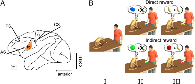
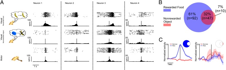
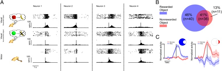
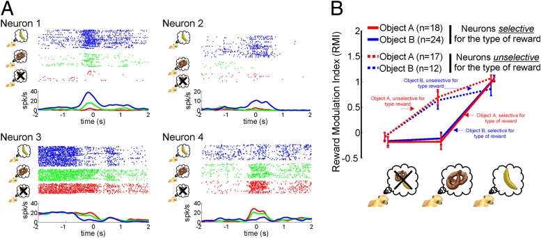
Objects grasped by an agent have a value not only for the acting agent, but also for an individual observing the grasping act. The value that the observer attributes to the object that is grasped can be pivotal for selecting a possible behavioral response. Mirror neurons in area F5 of the monkey premotor cortex have been suggested to play a crucial role in the understanding of action goals. However, it has not been addressed if these neurons are also involved in representing the value of the grasped object. Here we report that observation-related neuronal responses of F5 mirror neurons are indeed modulated by the value that the monkey associates with the grasped object. These findings suggest that during action observation F5 mirror neurons have access to key information needed to shape the behavioral responses of the observer.


Conflict of interest statement

The authors declare no conflict of interest.

Figures

Fig. 1.
Fig. 1.
Recording region and paradigm. (A) Lateral view of the left hemisphere of the monkey brain. The highlighted sector denotes the part of the ventral premotor cortex (area F5) from which neurons were recorded (AS, arcuate sulcus; CS, central sulcus; PS, principal sulcus). (B) Schematic view of the experimental paradigm. In each session we first tested the motor responses of neurons during active movements of the monkey (I). The visual responses of these neurons were further tested with the experimenter executing goal-directed motor acts in front of the monkey. Direct Reward Condition: The experimenter grasped either nonfood (II) or food objects (III). After a food object was grasped, the monkey was rewarded by delivering a raisin in a randomly chosen subset of trials. No reward was given following the grasping of a nonfood object (a metal object). Indirect Reward Condition: The experimenter grasped two objects. One object was randomly followed by a reward (typically a raisin), whereas the other object was not associated with any reward (for further details, see Experimental Procedures). Note that the color of the nonrewarded object used in experiments was actually gray. In the figure it is rendered green to make it easier to distinguish it from the hand. Please also note that the shape and size of the objects shown do not correspond to their actual properties. For details about shape and size, please refer to the Experimental Procedures.
Fig. 2.
Fig. 2.
Responses of exemplary mirror neurons and population activity during the observation of actions predicting later rewards or their absence. (A) Examples of neurons tested in Exp. 1. All four neurons responded to active movements of the monkey. Their visual responses exhibited either a preference for the grasping of a food object (neurons 1 and 2), no preference for the type of object (food or nonfood objects) on which actions were executed (neuron 3), or a preference for the grasping of the nonfood object (neuron 4). (B) Venn diagram illustrating the number of mirror neurons significantly activated in the rewarded and the nonrewarded conditions. The intersection of the two circles represents neurons whose discharges exhibited no statistically significant difference between the two experimental conditions. Neurons not contained in the intersection responded with a significantly stronger discharge during one of the two experimental conditions. (C) Normalized population responses of mirror neurons exhibiting selectivity for either food or nonfood conditions. The shaded regions represent SEM. Vertical lines in A and C represent the time of contact between the experimenter’s hand and the object.
Fig. 3.
Fig. 3.
Responses of exemplary mirror neurons and population activity during the observation of actions predicting later rewards or their absence. (A) Examples of the four types of neurons recorded in Exp. 2. (B) Venn diagram. (C) Normalized population activity. Same conventions as Fig. 2.
Fig. 4.
Fig. 4.
Impact of different subjective values on the visual responses of mirror neurons. (A) Exemplary mirror neurons recorded in the Exp. 3. Neuron 1 showed a monotonic excitatory modulation, with the strongest response to the most relished reward (symbolized by a banana and blue coloring). Neuron 2 exhibited a response only for the most relished reward. Neuron 3 showed a monotonic inhibitory modulation (less-relished reward, indicated by the pretzel and green coloring). Finally, neuron 4 demonstrated again a monotonic excitatory modulation but, unlike neuron 1, its strongest response was in the nonrewarded condition (red coloring). (B) The plot shows average ± SE data, based on an index (RMI) that captures the normalized absolute size of the reward associated discharge. Neurons showing a significant difference between the favorite and the less relished reward were classified as “selective for the type of reward.” The remaining neurons were classified as “unselective for the type of reward” (see Experimental Procedures).

References

    1. di Pellegrino G, Fadiga L, Fogassi L, Gallese V, Rizzolatti G. Understanding motor events: A neurophysiological study. Exp Brain Res. 1992;91:176–180. - PubMed
    1. Rizzolatti G, Fadiga L, Gallese V, Fogassi L. Premotor cortex and the recognition of motor actions. Brain Res Cogn Brain Res. 1996;3:131–141. - PubMed
    1. Gallese V, Fadiga L, Fogassi L, Rizzolatti G. Action recognition in the premotor cortex. Brain. 1996;119:593–609. - PubMed
    1. Fogassi L, et al. Parietal lobe: From action organization to intention understanding. Science. 2005;308:662–667. - PubMed
    1. Rozzi S, Ferrari PF, Bonini L, Rizzolatti G, Fogassi L. Functional organization of inferior parietal lobule convexity in the macaque monkey: Electrophysiological characterization of motor, sensory and mirror responses and their correlation with cytoarchitectonic areas. Eur J Neurosci. 2008;28:1569–1588. - PubMed

Publication types