. 2012 Jul 31;109(31):E2101-9.
doi: 10.1073/pnas.1209397109. Epub 2012 Jul 2.

Melanoregulin regulates a shedding mechanism that drives melanosome transfer from melanocytes to keratinocytes

Affiliations

Melanoregulin regulates a shedding mechanism that drives melanosome transfer from melanocytes to keratinocytes

Xufeng S Wu et al. Proc Natl Acad Sci U S A. .

Abstract

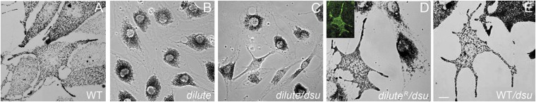
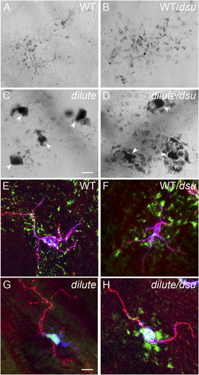
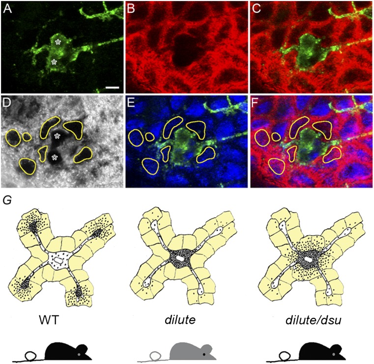
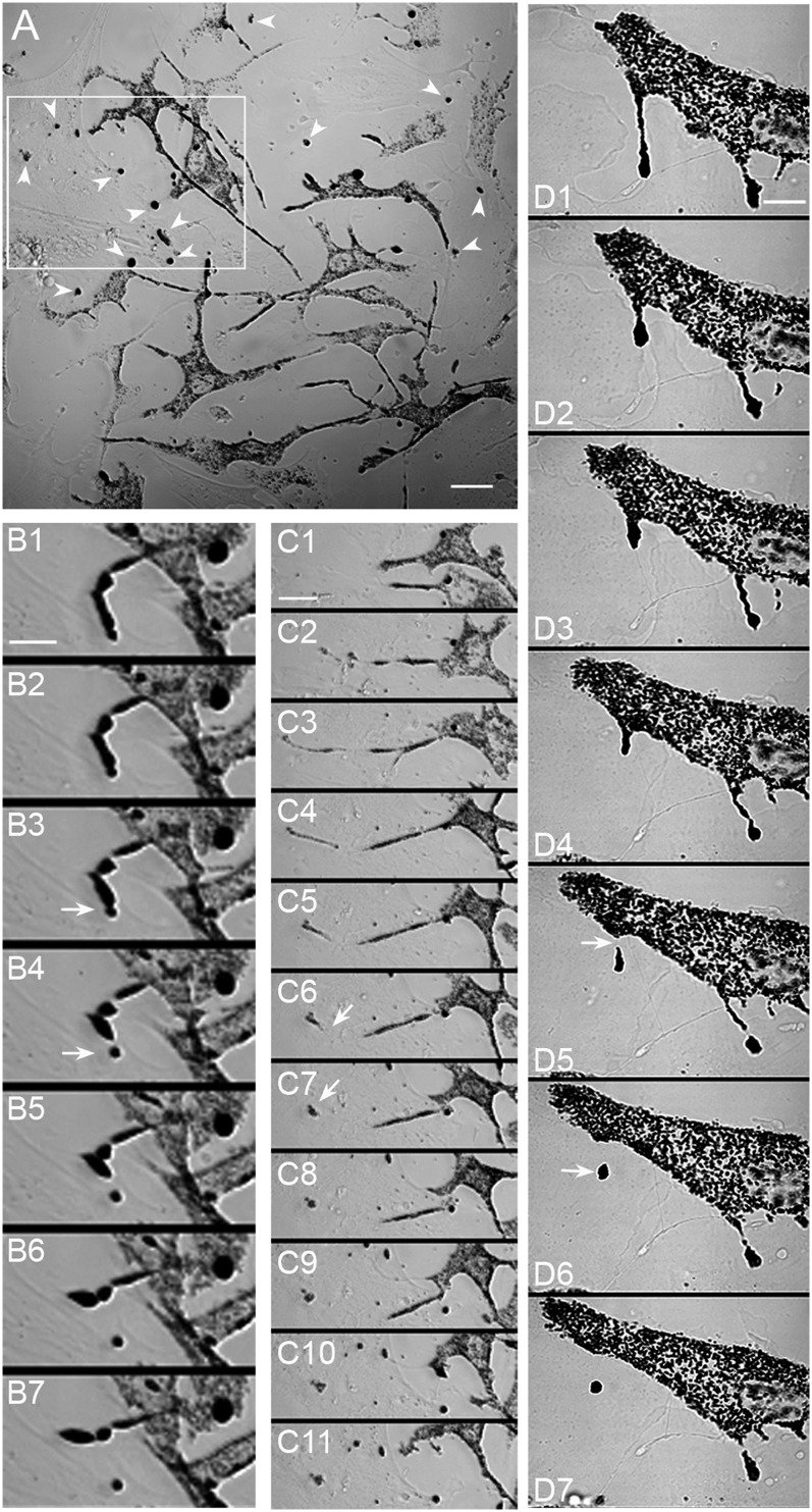
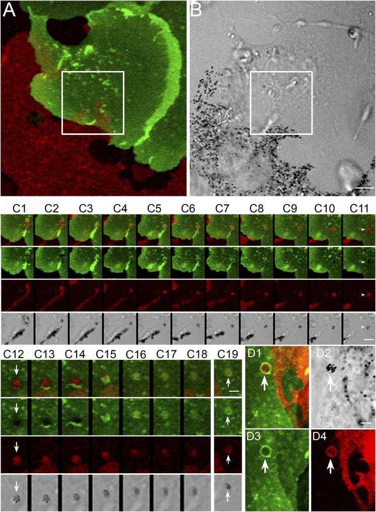
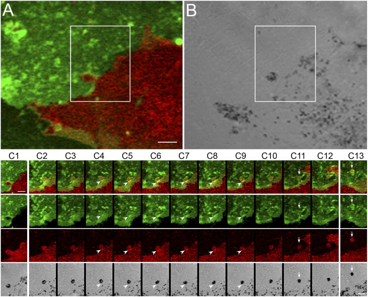
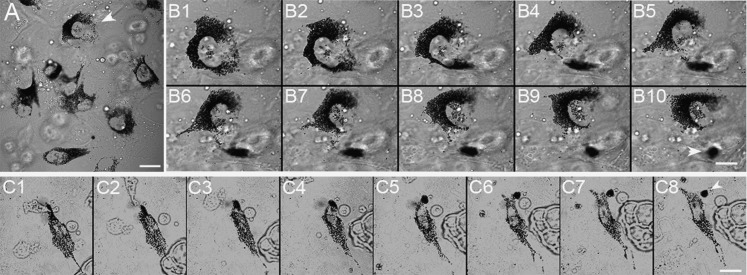
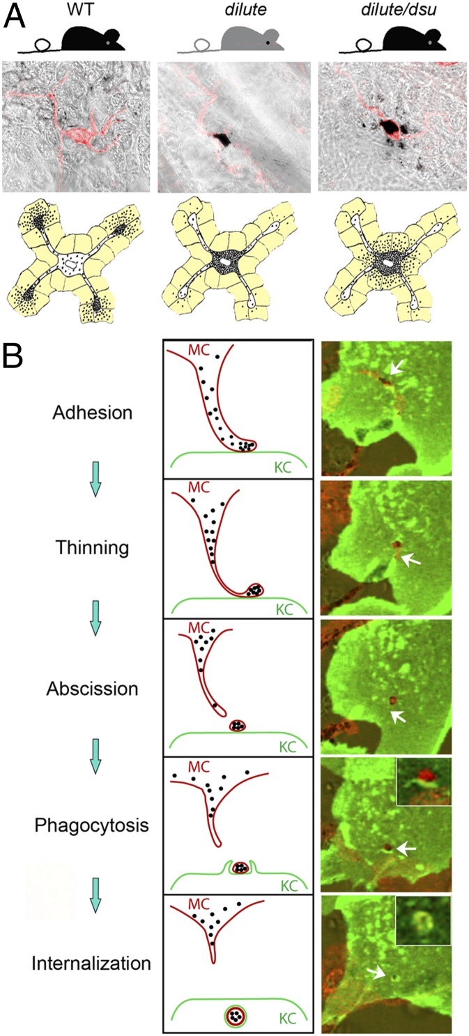
Mammalian pigmentation is driven by the intercellular transfer of pigment-containing melanosomes from the tips of melanocyte dendrites to surrounding keratinocytes. Tip accumulation of melanosomes requires myosin Va, because melanosomes concentrate in the center of melanocytes from myosin Va-null (dilute) mice. This distribution defect results in inefficient melanosome transfer and a dilution of coat color. Dilute mice that simultaneously lack melanoregulin, the product of the dilute suppressor locus, exhibit a nearly complete restoration of coat color, but, surprisingly, melanosomes remain concentrated in the center of their melanocytes. Here we show that dilute/dsu melanocytes, but not dilute melanocytes, readily transfer the melanosomes concentrated in their center to surrounding keratinocytes in situ. Using time-lapse imaging of WT melanocyte/keratinocyte cocultures in which the plasma membranes of the two cells are marked with different colors, we define an intercellular melanosome transfer pathway that involves the shedding by the melanocyte of melanosome-rich packages, which subsequently are phagocytosed by the keratinocyte. Shedding, which occurs primarily at dendritic tips but also from more central regions, involves adhesion to the keratinocyte, thinning behind the forming package, and apparent self-abscission. Finally, we show that shedding from the cell center is sixfold more frequent in cultured dilute/dsu melanocytes than in dilute melanocytes, consistent with the in situ data. Together, these results explain how dsu restores the coat color of dilute mice without restoring intracellular melanosome distribution, indicate that melanoregulin is a negative regulator of melanosome transfer, and provide insight into the mechanism of intercellular melanosome transfer.


Conflict of interest statement

The authors declare no conflict of interest.

Figures

Fig. 1.
Fig. 1.
dsu does not rescue the defect in melanosome distribution exhibited by dilute melanocytes. Shown are bright-field images of WT (A), dilute (B), dilute/dsu (C), dilute/dsu transfected with GFP-tagged myosin Va (melanocyte-spliced isoform) i.e., diluteR/dsu (D) and WT/dsu (E) melanocytes in primary culture (see text for specifics on the genotypes). Inset in D shows the distribution of GFP-tagged myosin Va in the transfected, rescued diluteR/dsu melanocyte. (Scale bar: 10 μm.)
Fig. 2.
Fig. 2.
Melanosomes escape readily from the center of dilute/dsu ear skin melanocytes but not from the center of dilute ear skin melanocytes. Shown are bright-field images of ear skin from WT (A), WT/dsu (B), dilute (C), and dilute/dsu (D) mice. Also shown are images of ear skin from WT (E), WT/dsu (F), dilute (G), and dilute/dsu (H) mice that had been stained with an antibody to the plasma membrane receptor for Kit to define the shape of melanocytes and with an antibody to the melanosomal membrane protein TRP1. The anti-Kit signal is in red, black pigment is pseudo colored green, and the anti-TRP1 signal is in blue (yielding a blue-white color inside melanocytes because of the overlay with the red and green signals). (Scale bars: 10 μm in AD; 7 μm in EH.)
Fig. 3.
Fig. 3.
Pigment that escapes the center of dilute/dsu melanocytes is inside adjacent keratinocytes. Shown are images of ear skin from a dilute/dsu mouse that had been stained for Kit to reveal the shape of melanocytes (green signal in A, C, E, and F), keratin 14 to reveal the presence of keratin filaments within keratinocytes (red signal in B, C, and E), and DAPI to reveal the positions of nuclei (blue signal in E and F) and the distribution of pigment (black in the transmitted-light image in D). (AC) Two adjacent melanocyte cell bodies in green (asterisks in A) surrounded by keratinocytes, which appear as red ovals because of the keratin filaments present in their peripheral cytoplasm (D) In the transmitted-light image, the dramatic accumulations of pigment that surround the two melanocyte cell bodies (marked with asterisks) are circled in yellow. (E) Overlay of this transmitted-light image (including the yellow circles) with the DAPI and Kit signals. (F) Overlay of this transmitted-light image (including the yellow circles) with the DAPI, cKit, and keratin 14 signals. (Scale bar: 7 μm.) (G) Cartoon depicting the basic concept supported by the images in Figs. 2 and 3, i.e., that dsu rescues the coat color of dilute mice without rescuing the defect in intracellular melanosome distribution exhibited by dilute melanocytes by allowing the melanosomes accumulated in their central cytoplasm to be transferred readily to the keratinocytes that immediately surround the melanocyte’s cell body (the keratinocytes in the cartoon are colored light yellow).
Fig. 4.
Fig. 4.
Melanocytes present in primary skin cultures appear to shed melanosome-rich packages from the tips of their dendrites. (A) Low-magnification bright-field image of a primary skin culture. In addition to melanocytes (evident because of their pigmentation) and keratinocytes (barely visible but evident in cultures stained for keratin; see Fig. S1), these cultures contain numerous round black “balls” (arrowheads) that appear to be outside melanocytes. The creation of these packages via some sort of shedding mechanism can be seen at low magnification in Movie S1. The still images in B and in C 1–8 show at higher magnification two apparent shedding events that occurred from the tips of a melanocyte’s dendrite within the boxed region in A (see also Movies S2 and S3, from which these stills were taken). The still images in D (and Movie S4, from which these stills were taken) show another tip shedding event that occurred in a different culture. The white arrows in these sequential still images mark the position of the forming package in the frame most likely immediately preceding (B 3, C 6, and D 5) and the frame most likely immediately following (B 4, C 7, and D 6) the release of the package. The still images in B and D are 2 and 4 min apart, respectively. The still images in C 4–11 are 4 min apart; the still images in C 1 3 are 30 min apart. (Scale bars: 18 μm in A; 8 μm in B; 15 μm in C; 9.5 μm in D.)
Fig. 5.
Fig. 5.
Package shedding drives the intercellular transfer of melanosomes via a process involving adhesion, dendrite thinning, and abscission to create the package, followed by phagocytosis of the package by the keratinocyte. (A and B) A melanocyte/keratinocyte pair from a Holly Skin mouse in which a shedding/transfer event occurs within the boxed region from the tip of the melanocyte’s dendrite (see also Movie S6, from which these two images were taken). The still images in C 1–11, which were taken from the “Thinning/Abscission” portion of Movie S7, demonstrate the thinning of the melanocyte’s dendrite behind the tip to be shed and the subsequent abscission step, which together result in the shedding of a melanosome package that initially sits on the surface of the keratinocyte (arrowheads in C 11 and arrows in C 12). The still images in C 12–18, which were taken from the “Phagocytosis” portion of Movie S7, demonstrate the subsequent phagocytosis of the extracellular melanosome package by the keratinocyte. C 19 shows this package shortly after it had been engulfed by the keratinocyte (arrows). The still images in C 1–11 are 4 min apart; the still images in C 12–18 are 2 min apart; and the image in C 19 is 16 min after C 18. (D) An engulfed package from a separate transfer event (arrows). (Scale bars: 5 μm in A and B; 3.8 μm in C 1–11; 2 μm in C12–19; 2.9 μm in D.)
Fig. 6.
Fig. 6.
Melanosome transfer also occurs from central regions of melanocytes. (A and B) A melanocyte/keratinocyte pair in which a shedding/transfer event occurs within the boxed region at the side of the melanocyte rather than at a dendritic tip (see also Movie S10, from which these two images were taken). The still images in C, which also were taken from Movie S10, demonstrate this side-shedding/transfer event in more detail. Arrowheads in C 4–9 point to transient increases at the base of the forming package of the plasma membrane signals from both the melanocyte and the keratinocyte, which occurred just before the abscission step (arrows in C 11). C 13 shows the package (arrows) 30 min after its engulfment by the keratinocyte. This internalized package can be seen to move toward the center of the keratinocyte in subsequent video frames (see Movie S10). The still images in C 2–12 are 2 min apart. The image in C 1 precedes the image in C 2 by 20 min. (Scale bars: 5 μm in A and B; 3 μm in C).
Fig. 7.
Fig. 7.
Cultured dilute/dsu melanocytes shed packages from their cell center. A shows a field of cultured dilute/dsu melanocytes. Still images in B show at higher magnification a shedding event that occurred from the center of the cell marked with an arrowhead in A (see also Movie S12, Example 1, from which these stills were taken). The still images in C show a second example of a shedding event from the center of a dilute/dsu melanocyte (see also Movie S12, Example 2, from which these stills were taken). The white arrowheads in B 10 and C 8 indicate the shed packages. The still images in B are 8 min apart. The still images in C are 10 min apart. (Scale bars: 12 μm in A; 8 μm in B; 15 μm in C.)
Fig. P1.
Fig. P1.
(A) The cartoon depicts how dsu restores the coat color of dilute mice without restoring the defect in intracellular melanosome distribution with dilute melanocytes (keratinocytes are shaded yellow). Representative examples of pigment distribution and melanocyte shape (red signal) in ear skin are shown also. (B) (Left) Schematic of the proposed mechanism of melanosome transfer as gleaned from time-lapse imaging of melanocyte (MC)/keratinocyte (KC) cocultures in which the plasma membranes of these cells are red and green, respectively. Transfer involves adhesion between the side or tip of the MC’s dendrite and the surface of the KC, thinning of the MC’s dendrite behind the site of contact, an apparent self-abscission event in which the MC sheds a plasma membrane-enclosed package containing multiple melanosomes, and the eventual uptake of the package by the KC via phagocytosis. The internalized package appears as a cluster of black melanosomes surrounded by a red membrane (the remnants of the MC’s plasma membrane) that in turn is surrounded by a green membrane (the phagosomal membrane derived from the KC’s plasma membrane). (Right) Representative video frames for each step. Because melanoregulin negatively regulates this shedding mechanism, the dsu mutation increases melanosome transfer on both WT and dilute backgrounds.

Similar articles

Cited by

References

    1. Kondo T, Hearing VJ. Update on the regulation of mammalian melanocyte function and skin pigmentation. Expert Rev Dermatol. 2011;6(1):97–108. - PMC - PubMed
    1. Wu X, Bowers B, Rao K, Wei Q, Hammer JA., 3rd Visualization of melanosome dynamics within wild-type and dilute melanocytes suggests a paradigm for myosin V function In vivo. J Cell Biol. 1998;143:1899–1918. - PMC - PubMed
    1. O’Sullivan TN, et al. dsu functions in a MYO5A-independent pathway to suppress the coat color of dilute mice. Proc Natl Acad Sci USA. 2004;101:16831–16836. - PMC - PubMed
    1. Moore KJ, et al. Dilute suppressor dsu acts semidominantly to suppress the coat color phenotype of a deletion mutation, dl20J, of the murine dilute locus. Proc Natl Acad Sci USA. 1988;85:8131–8135. - PMC - PubMed
    1. Moore KJ, Swing DA, Copeland NG, Jenkins NA. The murine dilute suppressor gene encodes a cell autonomous suppressor. Genetics. 1994;138:491–497. - PMC - PubMed

MeSH terms

Substances