. 2012 Jul 31;109(31):E2110-6.
doi: 10.1073/pnas.1209414109. Epub 2012 Jul 2.

MicroRNAs bind to Toll-like receptors to induce prometastatic inflammatory response

Affiliations

MicroRNAs bind to Toll-like receptors to induce prometastatic inflammatory response

Muller Fabbri et al. Proc Natl Acad Sci U S A. .

Abstract

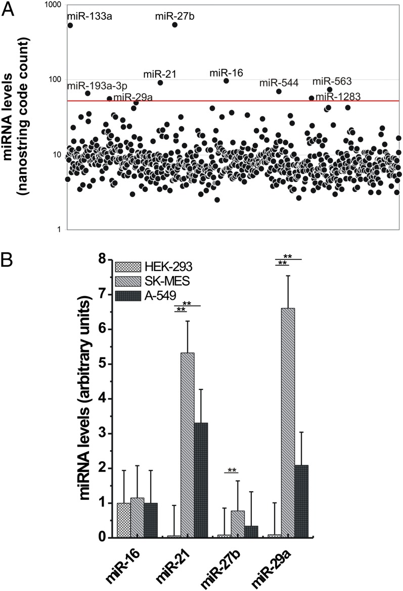
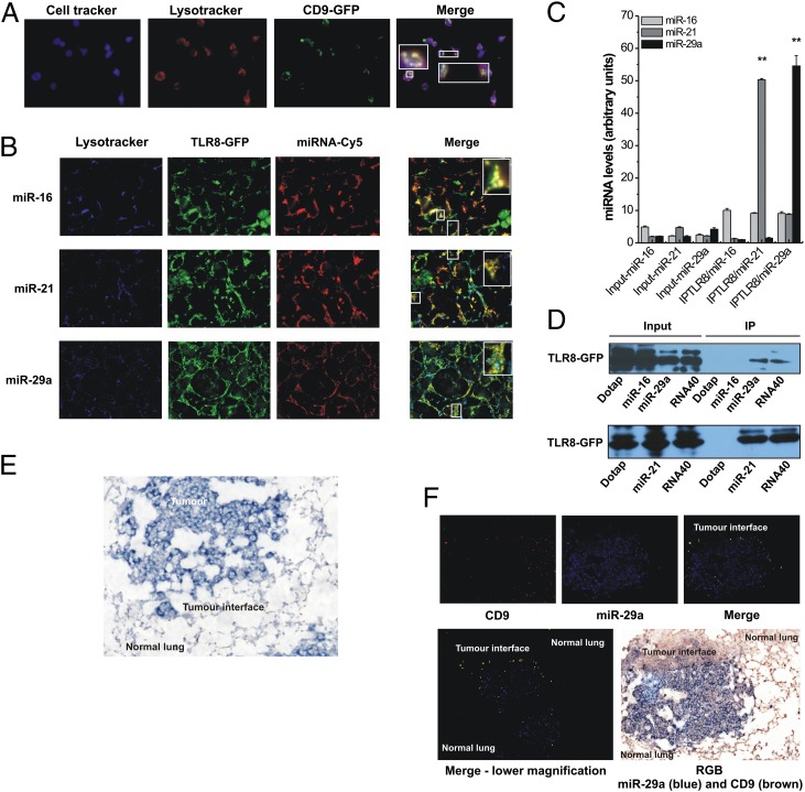
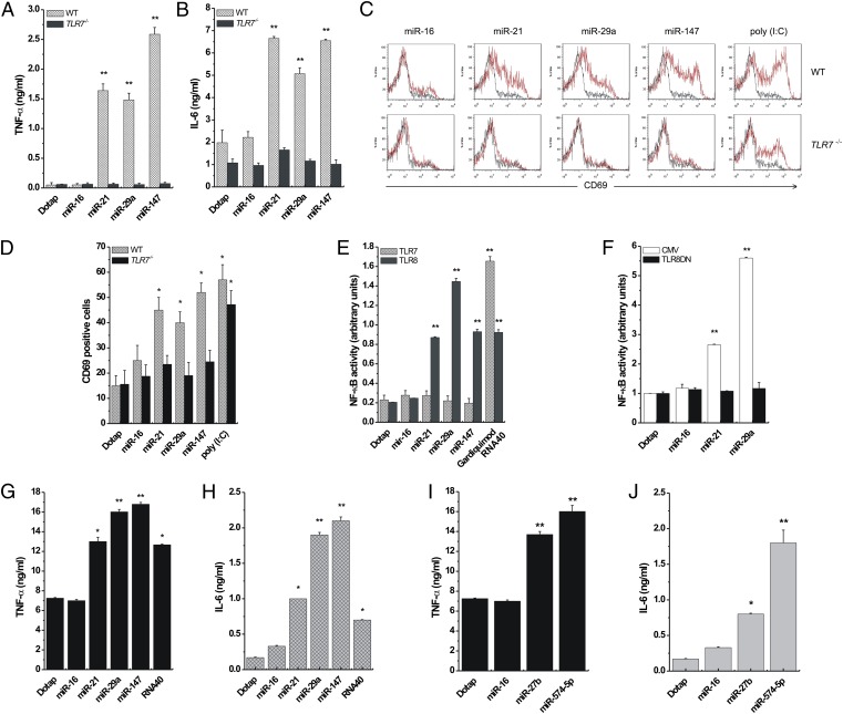
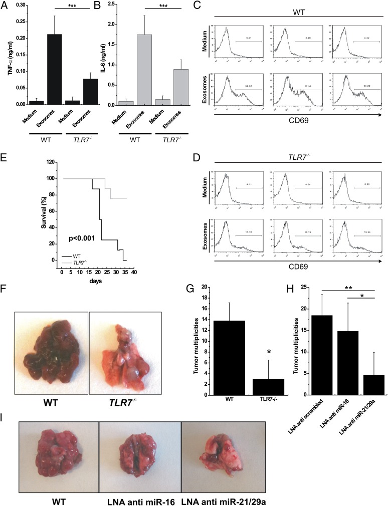
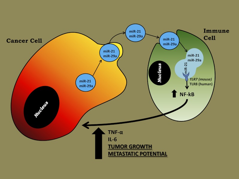
MicroRNAs (miRNAs) are small noncoding RNAs, 19-24 nucleotides in length, that regulate gene expression and are expressed aberrantly in most types of cancer. MiRNAs also have been detected in the blood of cancer patients and can serve as circulating biomarkers. It has been shown that secreted miRNAs within exosomes can be transferred from cell to cell and can regulate gene expression in the receiving cells by canonical binding to their target messenger RNAs. Here we show that tumor-secreted miR-21 and miR-29a also can function by another mechanism, by binding as ligands to receptors of the Toll-like receptor (TLR) family, murine TLR7 and human TLR8, in immune cells, triggering a TLR-mediated prometastatic inflammatory response that ultimately may lead to tumor growth and metastasis. Thus, by acting as paracrine agonists of TLRs, secreted miRNAs are key regulators of the tumor microenvironment. This mechanism of action of miRNAs is implicated in tumor-immune system communication and is important in tumor growth and spread, thus representing a possible target for cancer treatment.


Conflict of interest statement

The authors declare no conflict of interest.

Figures

Fig. 1.
Fig. 1.
Levels of miRNAs in exosomes derived from lung cancer cell lines and HEK-293 cells. (A) Scatter plot representing the NanoString miRNA profile obtained from A-549 purified exosomes. The red line indicates the threshold of 50 code counts. (B) Validation of the NanoString results in A-549 and SK-MES purified exosomes compared with HEK-293 purified vesicles by quantitative real-time PCR. The experiments were conducted in hextuplicate; results are presented as average ± SD. **P < 0.0001.
Fig. 2.
Fig. 2.
miR-21 and miR-29a interact with murine TLR7 and human TLR8 in the endosomes. (A) Confocal images of RAW 264.7 cells stained with cell tracker (blue) and with LysoTracker endosome marker (red) and cocultured with HEK-293–secreted CD9-GFP exosomes (green). Colocalization is indicated in yellow (merged image). (B) Confocal images of HEK-293 cells cotransfected with endosome LysoTracker (blue), GFP-tagged TLR8 (TLR8-GFP) (green), and Cy5-conjugated mature miRNAs (miRNA-Cy5) (red). Colocalization is indicated in yellow (merged image). (C) Levels of miR-16, miR-21, and miR-29a in the coimmunoprecipitates for TLR8 (IPTLR8/miR-16, IPTLR8/miR-21, and IPTLR8/miR-29a, respectively) in TLR8-HEK-293 cells detected by quantitative real-time PCR. Results are shown as means ± SD. **P < 0.001. (D) Immunoblotting with anti-GFP antibody for TLR8-GFP complex performed on immunoprecipitates derived from TLR8-GFP-HEK-293 cells. (E) LNA-ISH for miR-29a (blue) performed on mice tumors. (F) (Upper) ISH of CD9 (red) and miR-29a (blue) in mouse tumors. Coexpression is indicated in yellow (merged image). (Lower Left) Merged image with lower magnification indicates coexpression of CD9 and miR-29a at the tumor interface. (Lower Right) Corresponding red/green/blue image (miR-29a is stained in blue and CD9 in brown).
Fig. 3.
Fig. 3.
miR-21, -29a, and -147 induce TLR activation. (A and B) ELISA for TNF-α (A) and IL-6 (B) performed on peritoneal macrophages isolated from WT (n = 4) and TLR7−/− (n = 4) mice and treated with Dotap formulations of the indicated miRNAs. (C) Flow-cytometric analysis of CD69 in spleen cells of WT and TLR7−/− mice treated with the indicated miRNAs. (D) Graphic representation of the results presented in C. Poly (I:C) was used as a positive control for TLR3-mediated CD69 activation. (E) NF-κB activity in TLR7- and TLR8-HEK-293 cells treated with Dotap alone or with Dotap formulations of the indicated miRNAs. Gardiquimod and ssRNA40 were used as positive controls for TLR7- and TLR8-mediated NF-κB activation, respectively. (F) NF-κB activity in TLR8-HEK-293 cells transfected with a plasmid encoding a dominant negative form of TLR8 (TLR8DN), or its empty vector counterpart (CMV) and treated with Dotap alone or with Dotap formulations of the indicated mature miRNAs. (G and H), ELISA for TNF-α (G) and IL-6 (H) performed on human PBMC isolated from the blood of two healthy donors and treated with Dotap alone or with Dotap formulations of the indicated mature miRNAs. ssRNA40 sequence was used as positive control for TLR8-mediated cytokine secretion. Results in AH are shown as means ± SD. *P < 0.05; **P < 0.01. (I and J) ELISA for TNF-α and IL-6 performed on human PBMCs treated with Dotap formulations of mature miR-27b and –574–5p for 24 h. Incubation with Dotap alone and with miR-16 was used as negative control. The experiments were conducted in triplicate. Results are presented as average ± SD. *P < 0.01; **P < 0.0001.
Fig. 4.
Fig. 4.
miRNA-induced TLR7 activation increases formation of lung multiplicities in mice. (A and B) ELISA for TNFα (A) and IL-6 (B) performed on conditioned medium of peritoneal macrophages isolated from WT (n = 3) and TLR7−/− (n = 3) mice, incubated with RPMI (Medium; negative control) or exosomes purified from LLC cells for 48 h. (C and D) Flow-cytometric analysis of CD69 in spleen cells isolated from WT and TLR7−/− mice treated as in A and B. (E) Kaplan–Meier curves for WT (n = 7) and TLR7−/− (n = 7) mice after tail injection of LLC cells. (F) Representative images of different tumor multiplicities detected in lungs in the WT and the TLR7−/− mouse groups. (G) Tumor multiplicities in the WT and TLR7−/− mouse groups, after tail injection of LLC cells. (H) Tumor multiplicities in B6 mice injected with LLC cells transfected with LNA anti-scrambled (control; n = 6), LNA anti–miR-16 (n = 6), or LNA anti–miR-21/29a (n = 6). (I) Representative images of lungs in mice injected with LLC cells transfected as indicated. Results in AE, G, and H are shown as means ± SD. *P < 0.05; **P < 0.01; ***P ≤ 0.005.
Fig. P1.
Fig. P1.
MiRNAs are secreted by cancer cells in exosomes and can reach and bind TLR7 (in mice) or TLR8 (in humans) in the endosomes of surrounding immune cells. As a result, TLRs are activated, and the immune cells release cytokines, such as TNF-α and IL-6, which promote cancer growth and dissemination.

Comment in

  • Cancer. Malicious exosomes.
    Anastasiadou E, Slack FJ. Anastasiadou E, et al. Science. 2014 Dec 19;346(6216):1459-60. doi: 10.1126/science.aaa4024. Science. 2014. PMID: 25525233 No abstract available.

References

    1. Ambros V. microRNAs: Tiny regulators with great potential. Cell. 2001;107:823–826. - PubMed
    1. Bartel DP. MicroRNAs: Target recognition and regulatory functions. Cell. 2009;136:215–233. - PMC - PubMed
    1. Croce CM. Causes and consequences of microRNA dysregulation in cancer. Nat Rev Genet. 2009;10:704–714. - PMC - PubMed
    1. Fabbri M, Croce CM. Role of microRNAs in lymphoid biology and disease. Curr Opin Hematol. 2011;18:266–272. - PMC - PubMed
    1. Lawrie CH, et al. Detection of elevated levels of tumour-associated microRNAs in serum of patients with diffuse large B-cell lymphoma. Br J Haematol. 2008;141:672–675. - PubMed

Publication types

MeSH terms