

JNK and PTEN cooperatively control the development of invasive adenocarcinoma of the prostate
- PMID: 22753496
- PMCID: PMC3409732
- DOI: 10.1073/pnas.1209660109
JNK and PTEN cooperatively control the development of invasive adenocarcinoma of the prostate
Abstract
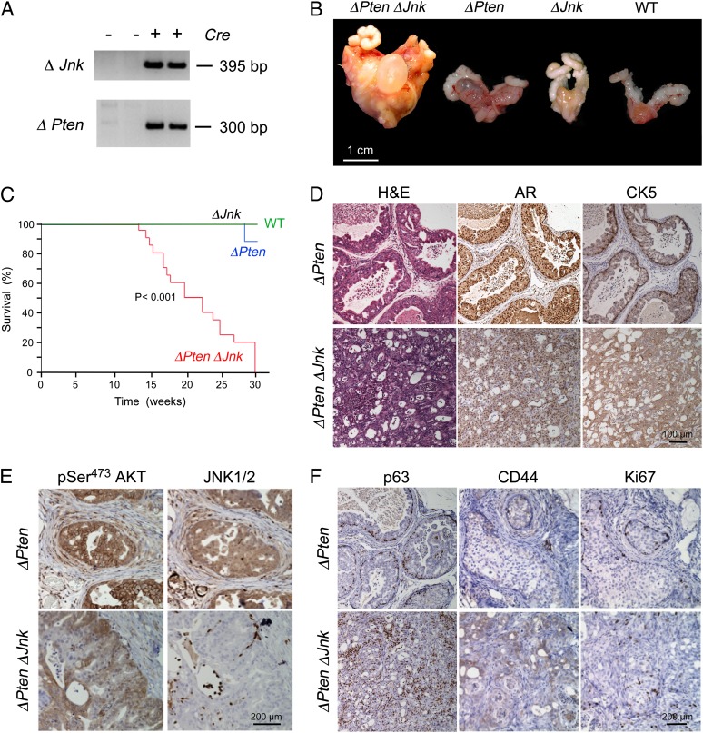
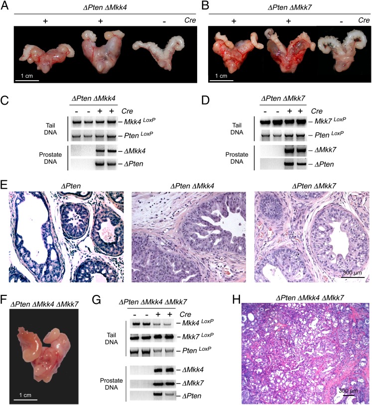
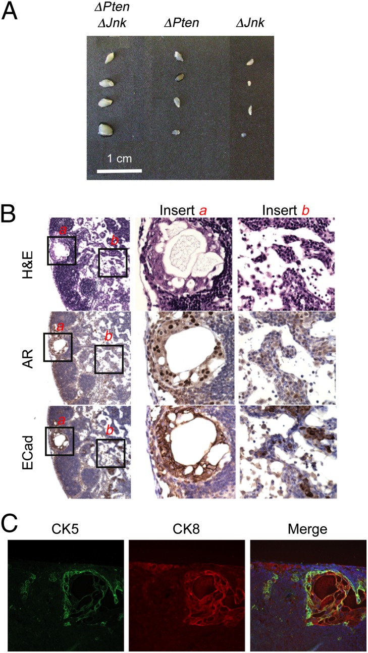
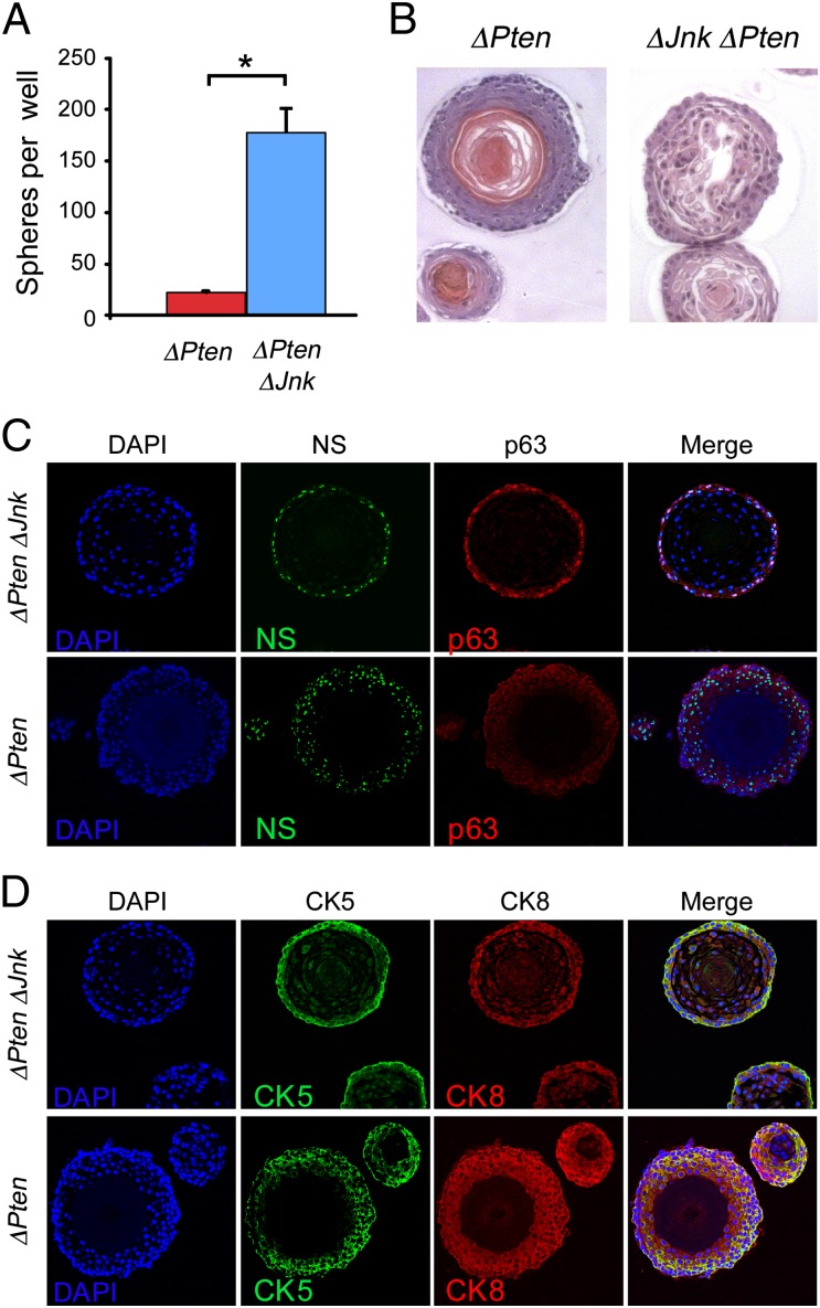
The c-Jun NH(2)-terminal kinase (JNK) signal transduction pathway is implicated in cancer, but the role of JNK in tumorigenesis is poorly understood. Here, we demonstrate that the JNK signaling pathway reduces the development of invasive adenocarcinoma in the phosphatase and tensin homolog (Pten) conditional deletion model of prostate cancer. Mice with JNK deficiency in the prostate epithelium (ΔJnk ΔPten mice) develop androgen-independent metastatic prostate cancer more rapidly than control (ΔPten) mice. Similarly, prevention of JNK activation in the prostate epithelium (ΔMkk4 ΔMkk7 ΔPten mice) causes rapid development of invasive adenocarcinoma. We found that JNK signaling defects cause an androgen-independent expansion of the immature progenitor cell population in the primary tumor. The JNK-deficient progenitor cells display increased proliferation and tumorigenic potential compared with progenitor cells from control prostate tumors. These data demonstrate that the JNK and PTEN signaling pathways can cooperate to regulate the progression of prostate neoplasia to invasive adenocarcinoma.
Conflict of interest statement
The authors declare no conflict of interest.
Figures
Comment in
-
Re: JNK and PTEN cooperatively control the development of invasive adenocarcinoma of the prostate.J Urol. 2013 May;189(5):1989. doi: 10.1016/j.juro.2013.01.065. Epub 2013 Jan 23. J Urol. 2013. PMID: 23594670 No abstract available.
Similar articles
-
Identification of the JNK signaling pathway as a functional target of the tumor suppressor PTEN.Cancer Cell. 2007 Jun;11(6):555-69. doi: 10.1016/j.ccr.2007.04.021. Cancer Cell. 2007. PMID: 17560336
-
c-Jun NH(2)-terminal kinase mediates leptin-stimulated androgen-independent prostate cancer cell proliferation via signal transducer and activator of transcription 3 and Akt.Biochim Biophys Acta. 2008 Oct;1782(10):593-604. doi: 10.1016/j.bbadis.2008.07.005. Epub 2008 Aug 5. Biochim Biophys Acta. 2008. PMID: 18718531
-
Sprouty genes function in suppression of prostate tumorigenesis.Proc Natl Acad Sci U S A. 2012 Dec 4;109(49):20023-8. doi: 10.1073/pnas.1217204109. Epub 2012 Nov 13. Proc Natl Acad Sci U S A. 2012. PMID: 23150596 Free PMC article.
-
Pten inactivation and the emergence of androgen-independent prostate cancer.Cancer Res. 2007 Jul 15;67(14):6535-8. doi: 10.1158/0008-5472.CAN-07-1271. Cancer Res. 2007. PMID: 17638861 Review.
-
PTEN, more than the AKT pathway.Carcinogenesis. 2007 Jul;28(7):1379-86. doi: 10.1093/carcin/bgm052. Epub 2007 Mar 6. Carcinogenesis. 2007. PMID: 17341655 Review.
Cited by
-
JunB and PTEN in prostate cancer: 'loss is nothing else than change'.Cell Death Differ. 2015 Apr;22(4):522-3. doi: 10.1038/cdd.2014.232. Cell Death Differ. 2015. PMID: 25747853 Free PMC article. No abstract available.
-
Prostate cancer and the unfolded protein response.Oncotarget. 2016 Aug 16;7(33):54051-54066. doi: 10.18632/oncotarget.9912. Oncotarget. 2016. PMID: 27303918 Free PMC article. Review.
-
Mutations in the transcription factor FOXO1 mimic positive selection signals to promote germinal center B cell expansion and lymphomagenesis.Immunity. 2021 Aug 10;54(8):1807-1824.e14. doi: 10.1016/j.immuni.2021.07.009. Immunity. 2021. PMID: 34380064 Free PMC article.
-
miR-30e* is overexpressed in prostate cancer and promotes NF-κB-mediated proliferation and tumor growth.Oncotarget. 2017 Jun 28;8(40):67626-67638. doi: 10.18632/oncotarget.18795. eCollection 2017 Sep 15. Oncotarget. 2017. PMID: 28978058 Free PMC article.
-
Rifampicin reduces advanced glycation end products and activates DAF-16 to increase lifespan in Caenorhabditis elegans.Aging Cell. 2015 Jun;14(3):463-73. doi: 10.1111/acel.12327. Epub 2015 Feb 26. Aging Cell. 2015. PMID: 25720500 Free PMC article.
References
-
- Davis RJ. Signal transduction by the JNK group of MAP kinases. Cell. 2000;103:239–252. - PubMed
-
- Tournier C, et al. Requirement of JNK for stress-induced activation of the cytochrome c-mediated death pathway. Science. 2000;288:870–874. - PubMed
-
- Lamb JA, Ventura JJ, Hess P, Flavell RA, Davis RJ. JunD mediates survival signaling by the JNK signal transduction pathway. Mol Cell. 2003;11:1479–1489. - PubMed
Publication types
MeSH terms
Substances
Grants and funding
- HHMI/Howard Hughes Medical Institute/United States
- P30 DK032520/DK/NIDDK NIH HHS/United States
- R01 CA121110/CA/NCI NIH HHS/United States
- R01 CA107166/CA/NCI NIH HHS/United States
- CA065861/CA/NCI NIH HHS/United States
- P01 AI046629/AI/NIAID NIH HHS/United States
- CA107166/CA/NCI NIH HHS/United States
- F32 CA112988/CA/NCI NIH HHS/United States
- DK032520/DK/NIDDK NIH HHS/United States
- R01 CA065861/CA/NCI NIH HHS/United States
- CA121110/CA/NCI NIH HHS/United States
- CA112988/CA/NCI NIH HHS/United States
- AI046629/AI/NIAID NIH HHS/United States
LinkOut - more resources
Full Text Sources
Medical
Molecular Biology Databases
Research Materials
Miscellaneous
