. 2012 Jul 17;109(29):11705-10.
doi: 10.1073/pnas.1121583109. Epub 2012 Jul 2.

Confinement induces actin flow in a meiotic cytoplasm

Affiliations

Confinement induces actin flow in a meiotic cytoplasm

Mathieu Pinot et al. Proc Natl Acad Sci U S A. .

Abstract

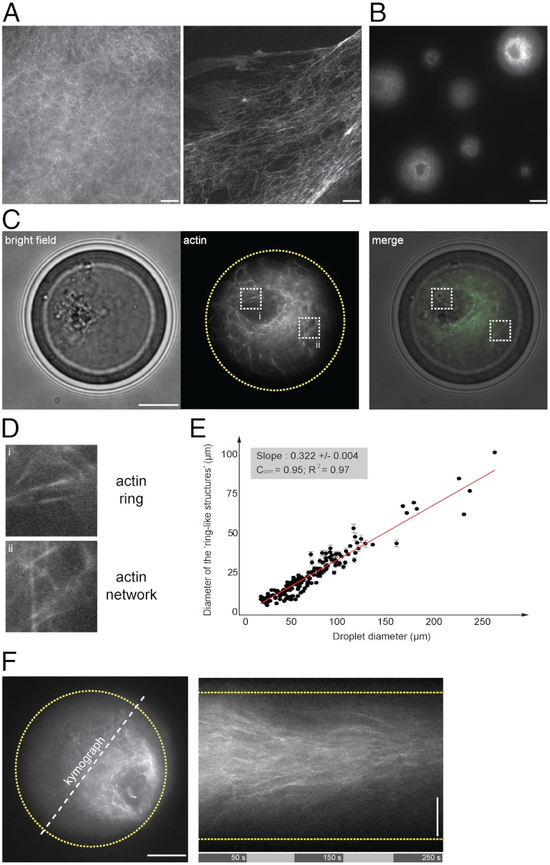
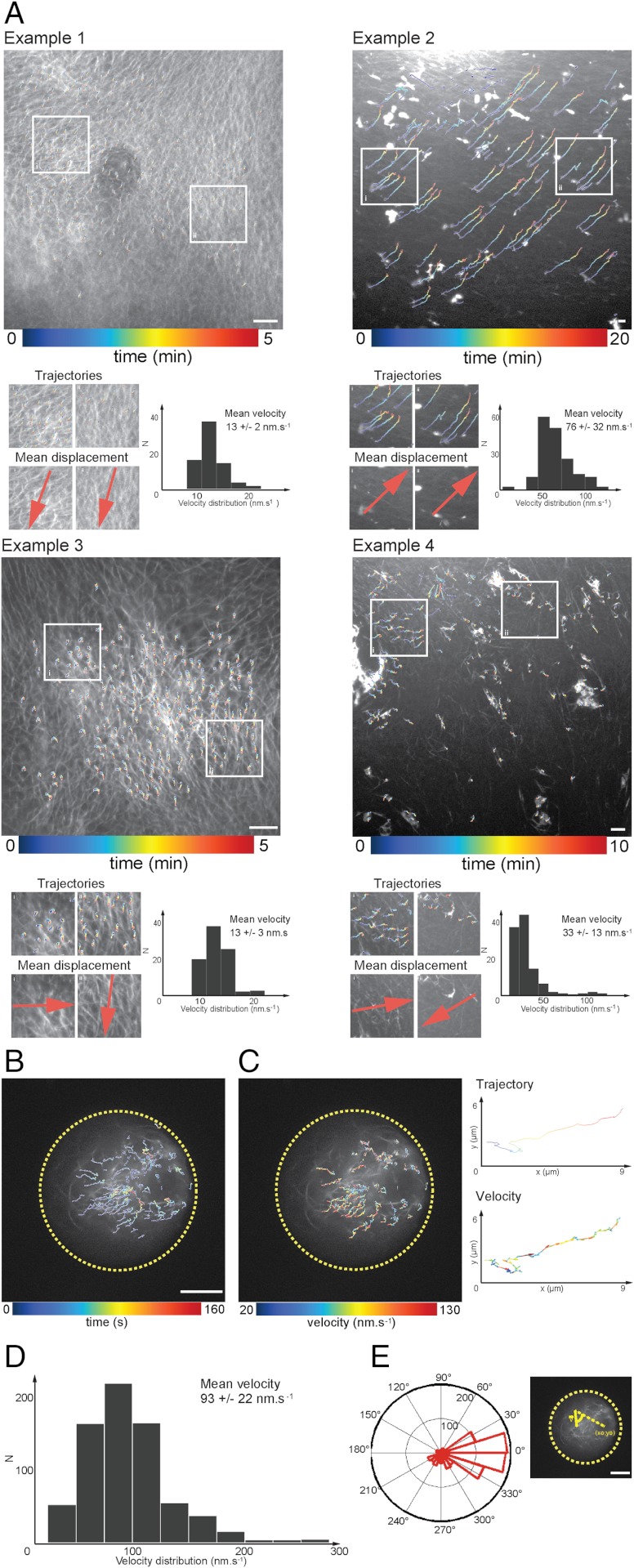
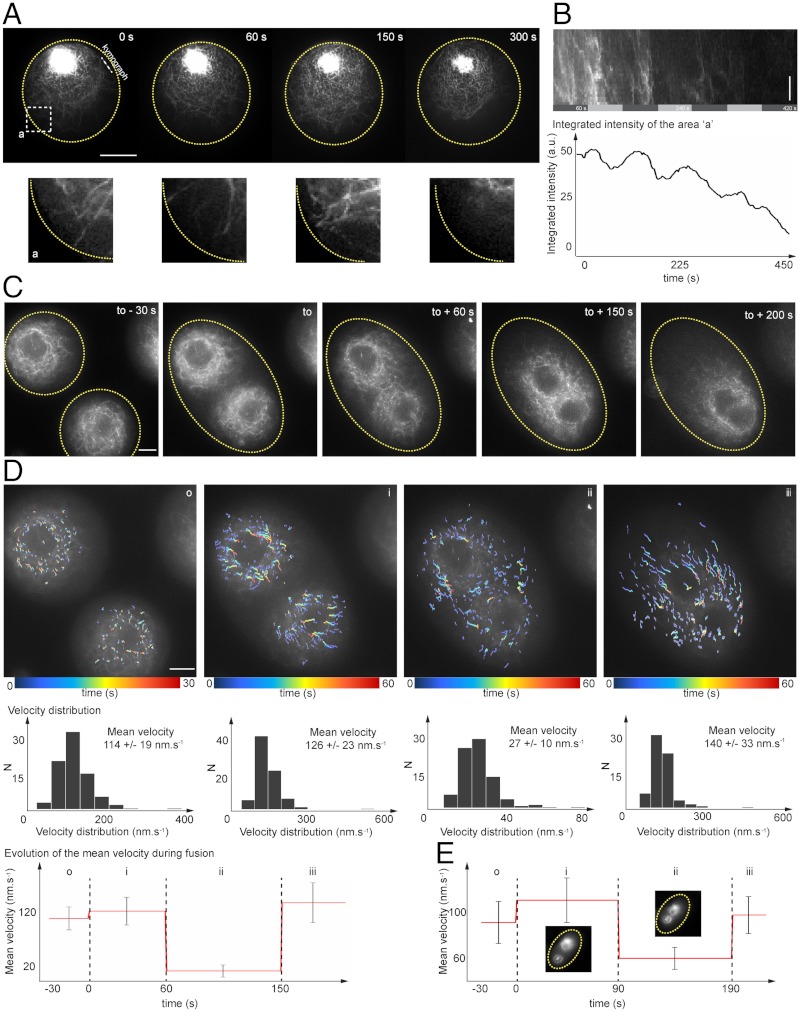
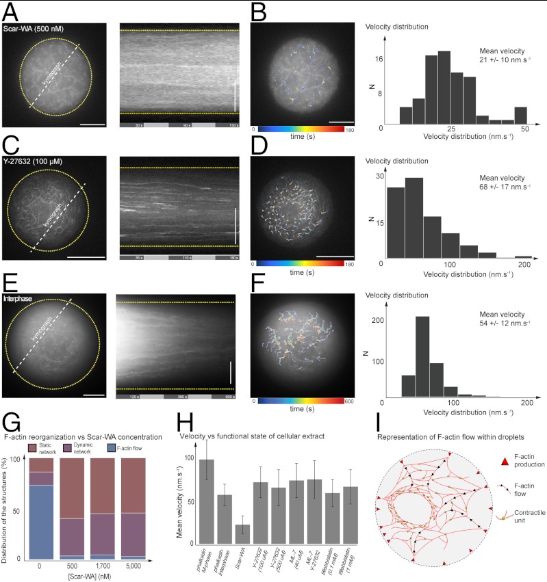
In vivo, F-actin flows are observed at different cell life stages and participate in various developmental processes during asymmetric divisions in vertebrate oocytes, cell migration, or wound healing. Here, we show that confinement has a dramatic effect on F-actin spatiotemporal organization. We reconstitute in vitro the spontaneous generation of F-actin flow using Xenopus meiotic extracts artificially confined within a geometry mimicking the cell boundary. Perturbations of actin polymerization kinetics or F-actin nucleation sites strongly modify the network flow dynamics. A combination of quantitative image analysis and biochemical perturbations shows that both spatial localization of F-actin nucleators and actin turnover play a decisive role in generating flow. Interestingly, our in vitro assay recapitulates several symmetry-breaking processes observed in oocytes and early embryonic cells.


Conflict of interest statement

The authors declare no conflict of interest.

Figures

Fig. 1.
Fig. 1.
Symmetry breaking of confined F-actin network. (A) Fluorescent observation of F-actin network (in presence of Alexa488-conjugated phalloidin) generated in bulk extracts (5 min incubation). (B) Fluorescent observation of F-actin network within extract-in-oil droplets. (C, D) Bright field and fluorescent observations of F-actin network confined within a 34 μm-diameter droplet. (D) Actin filaments organized in a ring-like structure asymmetrically positioned within the droplet and surrounded by an actin cloud (actin network). (E) Plot of the ring-like structure diameter as a function of the droplet diameter shows a linear correlation between the ring and the droplet diameter. Quantifications of the ring diameters were performed by extracting a plot profile of the fluorescent structure. The droplet diameter was measured using bright field illumination. We estimated the precision of such measurements to 1 μm. (F) Representative kymograph illustrating the directional flow dynamics of F-actin network within a 40 μm-diameter droplet. Scale bars, 10 μm.
Fig. 2.
Fig. 2.
F-actin spatiotemporal dynamics in bulk and within droplets: quantitative characterization of the flow velocity. (A) Displacement field of trajectories in bulk extracts shows variability in velocity distribution and orientation of F-actin network. (B) Displacement fields reveal a convergent flow toward the ring-like structure. (C) Velocity field amplitude distribution computed for trajectories tracked within a single droplet. (D) Histogram of mean velocities (mean = 93 ± 22 nm s-1, N = 15 experiments, 700 trajectories, and 20 droplets). (E) Angular orientation of tracked trajectories (N = 20 droplets, approximately 600 trajectories analyzed). Scale bars, 10 μm.
Fig. 3.
Fig. 3.
Dynamical properties of the F-actin network: periodicity in F-actin production at the boundary and actin-based structure fusion. (A) Time series of F-actin production at the droplet boundary. (B) Kymograph representation and time evolution of the integrated intensity of a region of interest localized at the vicinity of the droplet boundary. F-actin production shows periodicity with a period of approximately 100 s. (C) Time series acquisitions of two droplets undergoing fusion. (D) Characterization of velocity distributions for three steps: (i) prior to droplet fusion, (ii) during F-actin network fusion, and (iii) during ring-like structure fusion. (E) Evolution of the mean velocity in a second fusion droplet event. Scale bars, 10 μm.
Fig. 4.
Fig. 4.
Testing the role of the spatial localization of F-actin nucleation sites, the requirement of myosin II, and the regulation of the cell cycle in the generation of symmetry breaking and actin flow. (A) Kymograph of F-actin dynamics within droplet in presence of Scar-WA (500 nM). (B) Velocity field in presence of Scar-WA inhibitor (500 nM) and velocity distribution (N = 3 droplets, approximately 70 trajectories, mean = 21 ± 10 nm). (C) Kymograph of F-actin network in presence of Rho-Kinase inhibitor Y27632 (100 μM). (D) Velocity field of F-actin network dynamics in presence of Rho-Kinase inhibitor Y27632 (100 μM) and velocity distribution (N = 5 droplets, approximately 100 trajectories, mean = 68 ± 17 nm). (E) Kymograph of F-actin dynamics within droplet in interphase state. (F) Velocity field in interphase state and velocity distribution (N = 10 droplets, approximately 500 trajectories, mean = 54 ± 12 nm). (G) Histogram of events characterizing the network dynamic state for increasing concentration of Scar-WA (0, 0.5, 1.7, and 5 μM). (H) Histogram of the mean velocity as a function of the functional state of confined cellular extract. (I) Schematic of F-actin-based structure and flow within droplet. Scale bars, 10 μm.

References

    1. Pollard TD, Cooper JA. Actin, a central player in cell shape and movement. Science. 2009;326:1208–1212. - PMC - PubMed
    1. Li H, Guo F, Rubinstein B, Li R. Actin-driven chromosomal motility leads to symmetry breaking in mammalian meiotic oocytes. Nat Cell Biol. 2008;10:1301–1308. - PubMed
    1. Azoury J, et al. Spindle positioning in mouse oocytes relies on a dynamic meshwork of actin filaments. Curr Biol. 2008;18:1514–1519. - PubMed
    1. Schuh M, Ellenberg J. A new model for asymmetric spindle positioning in mouse oocytes. Curr Biol. 2008;18:1986–1992. - PubMed
    1. Pollard TD, Borisy GG. Cellular motility driven by assembly and disassembly of actin filaments. Cell. 2003;112:453–465. - PubMed

Publication types

LinkOut - more resources