. 2012 Sep 1;590(17):4223-37.
doi: 10.1113/jphysiol.2012.236570. Epub 2012 Jul 2.

Hypoxic regulation of cardiac Ca2+ channel: possible role of haem oxygenase

Affiliations

Hypoxic regulation of cardiac Ca2+ channel: possible role of haem oxygenase

Angelo O Rosa et al. J Physiol. .

Abstract

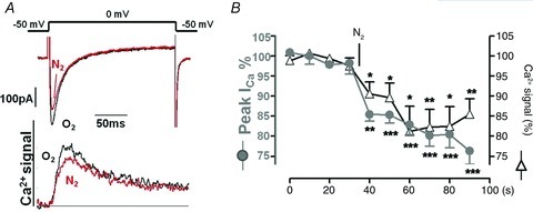
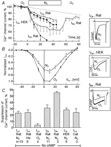
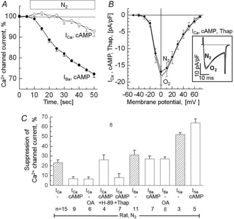
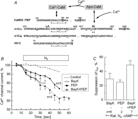
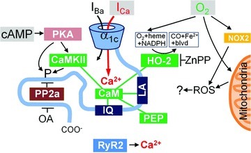
Acute and chronic hypoxias are common cardiac diseases that lead often to arrhythmia and impaired contractility. At the cellular level it is unclear whether the suppression of cardiac Ca(2+) channels (Ca(V)1.2) results directly from oxygen deprivation on the channel protein or is mediated by intermediary proteins affecting the channel. To address this question we measured the early effects of hypoxia (5-60 s, P(O(2)) < 5 mmHg) on Ca(2+) current (I(Ca)) and tested the involvement of protein kinase A (PKA) phosphorylation, Ca(2+)/calmodulin-mediated signalling and the haem oxygenase (HO) pathway in the hypoxic regulation of Ca(V)1.2 in rat and cat ventricular myocytes and HEK-293 cells. Hypoxic suppression of ICa) and Ca(2+) transients was significant within 5 s and intensified in the following 50 s, and was reversible. Phosphorylation by cAMP or the phosphatase inhibitor okadaic acid desensitized I(Ca) to hypoxia, while PKA inhibition by H-89 restored the sensitivity of I(Ca) to hypoxia. This phosphorylation effect was specific to Ca(2+), but not Ba(2+) or Na(+), permeating through the channel. CaMKII inhibitory peptide and Bay K8644 reversed the phosphorylation-induced desensitization to hypoxia. Mutation of CAM/CaMKII-binding motifs of the α(1c) subunit of Ca(V)1.2 fully desensitized the Ca(2+) channel to hypoxia. Rapid application of HO inhibitors (zinc protoporphyrin (ZnPP) and tin protoporphyrin (SnPP)) suppressed the channel in a manner similar to acute hypoxia such that: (1) I(Ca) and I(Ba) were suppressed within 5 s of ZnPP application; (2) PKA activation and CaMKII inhibitors desensitized I(Ca), but not I(Ba), to ZnPP; and (3) hypoxia failed to further suppress I(Ca) and I(Ba) in ZnPP-treated myocytes. We propose that the binding of HO to the CaM/CaMKII-specific motifs on Ca(2+) channel may mediate the rapid response of the channel to hypoxia.


Figures

Figure 2
Figure 2. The suppression of ICa by hypoxia is accompanied by a similar suppression of the ICa-triggered intracellular Ca2+ transients
A, simultaneous measurements of ICa and Ca2+ transients measured with 0.4 mm K4Fura-2 in the patch pipette in non-cAMP dialysed rat cardiomyocytes before (O2) and after 40 s of hypoxia (N2). B, time course of suppression ICa and Ca2+ transients during exposure to hypoxic solution.
Figure 1
Figure 1. Suppression of current through the L-type Ca2+ channel during brief periods of hypoxia
A, time course of hypoxia-induced changes in the peak inward current in rat cardiomyocytes where the permeating ion was Ca2+ (ICa), Ba2+ (IBa) or Na+ (INa) and in HEK cells where the recombinant channel conducted Ca2+ (ICa). B, current–voltage relations for ICa in rat ventricular cardiomyocytes measured under normoxic control conditions (O2) and after 1–2 min exposure to hypoxia (N2). C, average suppression of peak inward current measured with different permeating ions (Ca2+ vs. Ba2+ vs. Na+) and preparations (rat vs. cat cardiomyocytes vs. HEK-cell expression system) after 45–60 s superfusion with solutions equilibrated with N2, He or O2. The inset panels show that currents activated by depolarization from −60 to +10 mV displayed different rates of inactivation, but similar degrees of suppression during hypoxia (ICa vs. IBa in rat cardiomyocytes vs. ICa in HEK cells). All illustrated experiments were conducted in the absence of cAMP or other interventions that might evoke cAMP-dependent phosphorylation. The experiments with cat cardiomyocytes were performed at 30–32°C, the rest at room temperature, 23–25°C.
Figure 3
Figure 3. Phosphorylation of the Ca2+ channel makes it insensitive to hypoxia when the permeating ion is Ca2+ (ICa), but not when it is Ba2+ (IBa) or Na+ (INa)
A, time course of ICa and IBa during 40 s hypoxia (N2) in rat ventricular myocytes dialysed with 0.2 mm cAMP. B, the I–V relation is insensitive to hypoxia even when SR Ca2+ stores are depleted by pre-incubation with 1 μm thapsigargin (Thap). The inset panel shows sample currents recorded with depolarization to +10 mV. C, bar graph showing the reduction in the currents through the Ca2+ channel during 45–60 s of hypoxia in cells dialysed with 0.2 mm cAMP or incubated with 1 μm of the phosphatase inhibitor okadaic acid (OA). Results obtained with the PKA inhibitor H-89 and thapsigargin (Thap) are included. Results obtained under non-phosphorylating conditions are reproduced for comparison from Fig. 1C as hatched bars. All illustrated results were obtained using rat ventricular cells and hypoxic solutions equilibrated with 100% N2.
Figure 4
Figure 4. Inhibition of Ca2+-dependent inactivation of ICa by calmodulin domain peptide290−309 and Bay K is associated with enhanced suppression of ICa by anoxia
A, putative CaM-binding (IQ and hydrophobic 1–5–10 motifs) and phosphorylation sites (grey highlight) in the partial AA sequences of CaMKII, peptide290−309 (PEP), wild-type and mutant α1c subunits of the Ca2+ channel (α1c77 and α1c86) and haem oxygenase (HO-2) (Colbran, 1993; Rhoads & Friedberg, 1997; Soldatov et al. 1997; Boehning et al. 2004). B, time course of suppression of ICa upon removal of extracellular O2 in cAMP-dialysed rat cardiomyocytes that in addition were dialysed with 100 μm PEP, treated with 1 μm Bay K8644 (BayK) or both. For comparison purposes, ICa in cardiac control cells dialysed with 0.2 mm of cAMP is reproduced from Fig. 3A (dashed line). C, comparison of the anoxic suppressions of ICa in cardiomyocytes measured after 60–90 s of hypoxia (during recording of I–V relations). A, amino acid sequences of α1c77 and α1c86 subunits.
Figure 5
Figure 5. The cytoplasmic CaM/CaMKII binding sites of the α1C subunit of the Ca2+ channel is required for its hypoxia-induced suppression
The measured Ca2+ and Ba2+ currents (ICa and IBa) show that Ca2+-dependent inactivation (A) and anoxic suppression (C) are present in HEK-293 cell expressing the α1c77 of the L-type Ca2+ channel, but not in cells expressing the mutant α1c86 subunit lacking CaM/CAMKII binding sites (B and D; cf. Fig. 4B). The currents in panels A and B were normalized to emphasize differences and similarities in kinetics. Extracellular Ba2+ at 20 mm was used to enhance the currents shown in panels C and D. All measurements were done under non-phosphorylating conditions.
Figure 6
Figure 6. Suppression the Ca2+ channel by inhibition of haem oxygenase
A, brief exposures to the haem oxygenase inhibitor ZnPP in concentrations of 10 and 100 nm cause rapid, reversible, dose-dependent suppression of ICa. B, effect of 10 nm ZnCl2 or 100 nm ZnPP on ICa. Insets in panels A and B show traces of ICa measured at the indicated times (a, b and c). C and D, normalized I–V relations of IBa measured in control cells (Control, open circles) and in cells exposed to 100 nm ZnPP (C,filled triangles) that was applied either in the external solution (C) or via the patch pipette by 9 min of dialysis (D). All experiments were performed on rat ventricular cardiomyocytes under normoxic and non-phosphorylating conditions.
Figure 7
Figure 7. Inhibition of the Ca2+ channel in rat cardiomyocytes by haem oxygenase inhibitors with respect to phosphorylation, permeating ion and hypoxia
A and B, effect of 100 nm ZnPP on ICa (A) and IBa (B) measured in the absence of phosphorylating interventions (no cAMP, filled circles) and with dialysis of either 0.2 mm cAMP (cAMP, open circles) or 60 nm okadaic acid (OA, open triangles). C, pretreatment with an inhibitor of CaMKII (10 μm CK59) restored the suppressive effect (double arrow) of 100 nm ZnPP on ICa in cardiomyocytes dialysed with 0.2 mm cAMP. Notice that in this, as in all our experiments, the suppression of ICa was quantified based on a normalization that yielded 100% immediately before the exposure to hypoxia or ZnPP. D, hypoxia (N2) had no additional suppressive effects after IBa had been suppressed by 1 μm ZnPP. E, summary of the quantitative effects of 100 nm ZnPP on ICa and IBa, as measured in panels AC. This bar graph has the same layout as used for hypoxic effects in Fig. 3C, but shows data obtained with a different protein kinase inhibitor (CK59 vs. H-89).
Figure 8
Figure 8. Proposed model for modulation of L-type Ca2+ channel by oxygen
The carboxy-tail of the α1C subunit includes LA and IQ motifs for calmodulin- (CaM) mediated interactions and a binding site for protein phosphatase 2a (PP2a). CaM is shown as also interacting with CaMKII, haem oxygenase-2 (HO-2) and inhibitory peptide290−309 (PEP). The primary inputs to the system are thought to be: cAMP leading to PKA-mediated phosphorylation (left), permeation of Ca2+ through the channel to a restricted space (middle), and molecular oxygen (O2) that binds to haem/HO. PKA may act directly or via CaMKII and may have multiple phoshporylation targets (including HO-2). Similarly okadaic acid (OA) may promote phosphorylation inhibiting different phosphatases. ROS generation by mitochondria or membrane bound NOX proteins, CO-mediated signalling and ICa-triggered release of Ca2+ from the SR are thought to be of little or no importance for the rapid suppression of the Ca2+ channel at the onset of hypoxia.

Similar articles

Cited by

References

    1. Adachi-Akahane S, Cleemann L, Morad M. Cross-signaling between L-type Ca2+ channels and ryanodine receptors in rat ventricular myocytes. J Gen Physiol. 1996;108:435–454. - PMC - PubMed
    1. Adachi-Akahane S, Cleemann L, Morad M. BAY K 8644 modifies Ca2+ cross signaling between DHP and ryanodine receptors in rat ventricular myocytes. Am J Physiol Heart Circ Physiol. 1999;276:H1178–1189. - PubMed
    1. Belmonte S, Morad M. Shear fluid-induced Ca2+ release and the role of mitochondria in rat cardiac myocytes. Ann NY Acad Sci. 2008;1123:58–63. - PubMed
    1. Boehning D, Sedaghat L, Sedlak TW, Snyder SH. Heme oxygenase-2 is activated by calcium-calmodulin. J Biol Chem. 2004;279:30927–30930. - PubMed
    1. Bradshaw JM, Kubota Y, Meyer T, Schulman H. An ultrasensitive Ca2+/calmodulin-dependent protein kinase II-protein phosphatase 1 switch facilitates specificity in postsynaptic calcium signaling. Proc Natl Acad Sci U S A. 2003;100:10512–10517. - PMC - PubMed

Publication types

MeSH terms

Substances

LinkOut - more resources