. 2012 Oct;265(1):186-93.
doi: 10.1148/radiol.12120638. Epub 2012 Jul 2.

A receptor-targeted fluorescent radiopharmaceutical for multireporter sentinel lymph node imaging

Affiliations

A receptor-targeted fluorescent radiopharmaceutical for multireporter sentinel lymph node imaging

Derek K Emerson et al. Radiology. 2012 Oct.

Abstract

Purpose: To determine the imaging and receptor-binding properties of a multireporter probe designed for sentinel lymph node (SLN) mapping via nuclear and fluorescence detection.

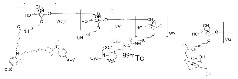
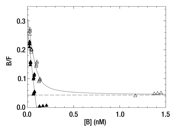
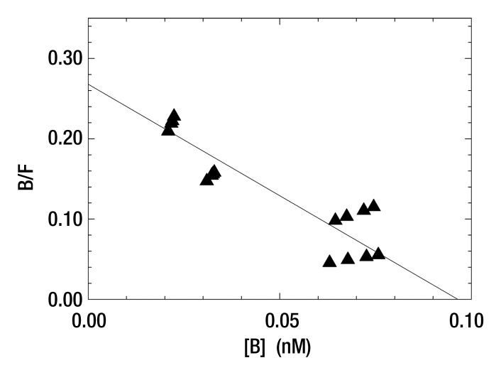
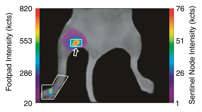
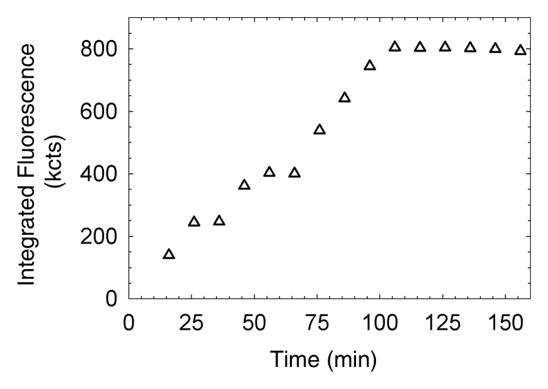
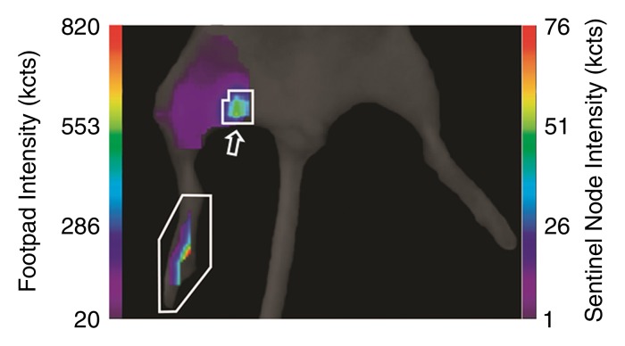
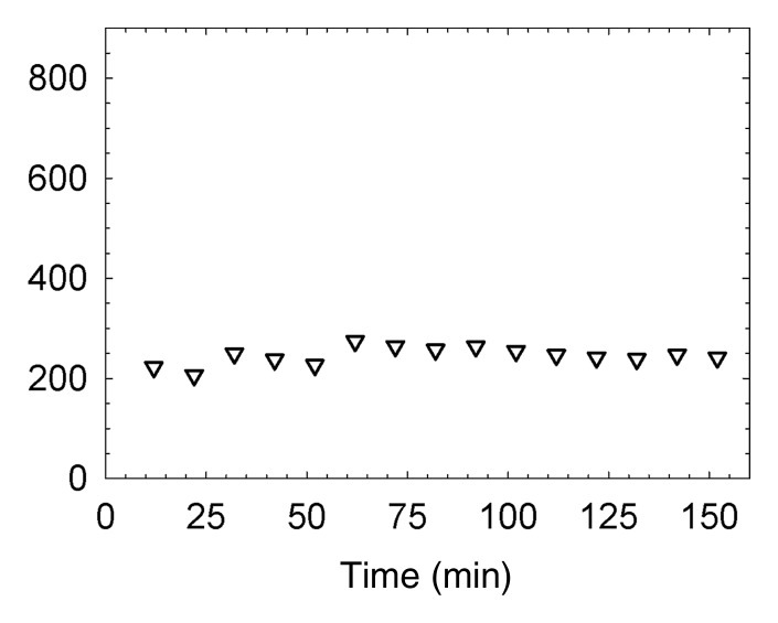
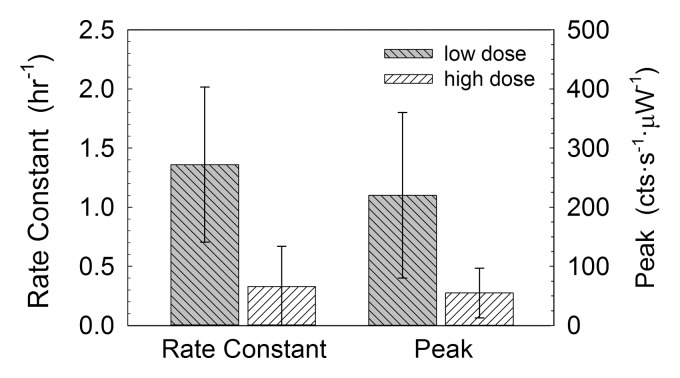
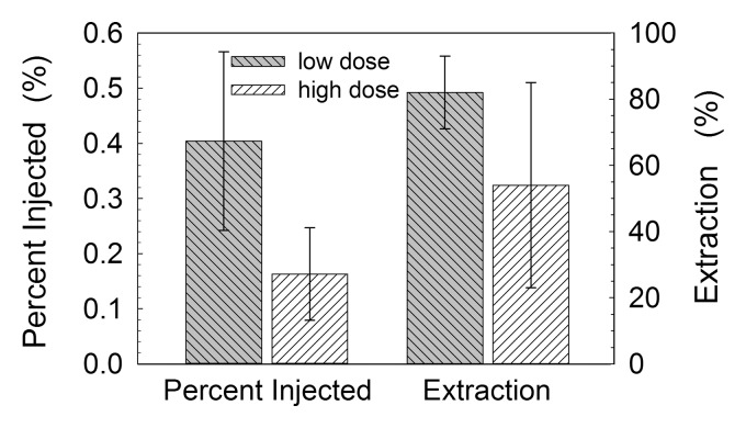
Materials and methods: The animal experiments were approved by the institutional animal care and use committee. A multireporter probe was synthesized by covalently attaching cyanine 7 (Cy7), a near-infrared cyanine dye, to tilmanocept, a radiopharmaceutical that binds to a receptor specific to recticuloendothelial cells. In vitro binding assays of technetium 99m (99mTc)-labeled Cy7 tilmanocept were conducted at 4°C by using receptor-bearing macrophages. Optical SLN imaging after foot pad administration was performed by using two molar doses of Cy7 tilmanocept. Six mice were injected with 0.11 nmol of 99mTc-labeled Cy7 tilmanocept (low-dose group); an additional six mice were injected with 31 nmol of 99mTc-labeled Cy7 tilmanocept (high-dose group) to saturate the receptor sites within the SLN. After 2.5 hours of imaging, the mice were euthanized, and the sentinel and distal lymph nodes were excised and assayed for radioactivity for calculation of SLN percentage of injected dose and extraction. Four mice were used as controls for autofluorescence. Standard optical imaging software was used to plot integrated fluorescence intensity against time for calculation of the SLN uptake rate constant and scaled peak intensity. Significance was calculated by using the Student t test.

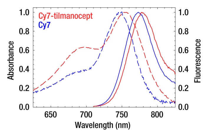
Results: In vitro binding assays showed subnanomolar affinity (mean dissociation constant, 0.25 nmol/L±0.10 [standard deviation]). Fluorescence imaging showed a detection sensitivity of 1.6×10(3) counts·sec(-1)·μW(-1) per picomole of Cy7. All four imaging metrics (percentage of injected dose, SLN extraction, SLN uptake rate constant, and expected peak fluorescence intensity) exhibited higher values (P=.005 to P=.042) in the low-dose group than in the high-dose group; this finding was consistent with receptor-mediated image formation.

Conclusion: The multireporter probe 99mTc-labeled Cy7 tilmanocept exhibits in vitro and in vivo receptor-binding properties for successful receptor-targeted SLN mapping with nuclear and optical imaging.


Figures

Figure 1a:
Figure 1a:
(a) The receptor-targeted fluorescent radiopharmaceutical, Cy7 tilmanocept, is composed of one molecule of 10-kDa dextran, to which are attached multiple units of Cy7, diethylene triamine pentaacetic acid, and mannose. Cy7 is an optical reporter that emits a photon with a near-infrared wavelength; diethylene triamine pentaacetic acid permits radiolabeling with 99mTc; and mannose units impart affinity to a receptor that resides within the SLN. (b) Absorbance spectra of a Cy7 tilmanocept preparation (0.90 Cy7 per dextran) and unconjugated Cy7 (dashed red and blue lines, respectively). Fluorescence spectra of a Cy7 tilmanocept preparation and unconjugated Cy7 (solid red and blue lines, respectively) show an emission peak at 780 nm for each preparation; this is a 7-nm red shift from Cy7.
Figure 1b:
Figure 1b:
(a) The receptor-targeted fluorescent radiopharmaceutical, Cy7 tilmanocept, is composed of one molecule of 10-kDa dextran, to which are attached multiple units of Cy7, diethylene triamine pentaacetic acid, and mannose. Cy7 is an optical reporter that emits a photon with a near-infrared wavelength; diethylene triamine pentaacetic acid permits radiolabeling with 99mTc; and mannose units impart affinity to a receptor that resides within the SLN. (b) Absorbance spectra of a Cy7 tilmanocept preparation (0.90 Cy7 per dextran) and unconjugated Cy7 (dashed red and blue lines, respectively). Fluorescence spectra of a Cy7 tilmanocept preparation and unconjugated Cy7 (solid red and blue lines, respectively) show an emission peak at 780 nm for each preparation; this is a 7-nm red shift from Cy7.
Figure 2a:
Figure 2a:
(a, b) Scatchard plots show results of a single-binding experiment analyzed with LIGAND software (25), which separated the specific- (▲) and nonspecific-bound (△) radioactivity prior to estimation of the dissociation constant. (a) Mean dissociation constant was 0.36 nmol/L ± 0.14 at 4°C (negative slope of straight solid line). (b) Expanded view of the specific-bound component used to calculate the dissociation constant of the individual assay. Mean dissociation constant of five Cy7 tilmanocept assays was 0.25 nmol/L ± 0.10, which was not significantly different from tilmanocept without Cy7 attached (0.42 nmol/L ± 0.31).
Figure 2b:
Figure 2b:
(a, b) Scatchard plots show results of a single-binding experiment analyzed with LIGAND software (25), which separated the specific- (▲) and nonspecific-bound (△) radioactivity prior to estimation of the dissociation constant. (a) Mean dissociation constant was 0.36 nmol/L ± 0.14 at 4°C (negative slope of straight solid line). (b) Expanded view of the specific-bound component used to calculate the dissociation constant of the individual assay. Mean dissociation constant of five Cy7 tilmanocept assays was 0.25 nmol/L ± 0.10, which was not significantly different from tilmanocept without Cy7 attached (0.42 nmol/L ± 0.31).
Figure 3a:
Figure 3a:
(a, c) Examples of fluorescence imaging of mouse SLNs (arrows) within ROIs (white outlines). kcts = Kilocounts. (a) Optical image obtained 150 minutes after administration of a 0.11-nmol dose of Cy7 tilmanocept yielded an SLN with an integrated fluorescence intensity with an ROI of 780 × 103 counts. The pixel with the highest fluorescence intensity held 64 × 103 counts. (b) Time-intensity curve from the SLN shows sustained accumulation from 140 × 103 counts at 15 minutes to 800 × 103 counts at 100 minutes. SLN uptake rate constant, percentage of injected dose, and SLN extraction were 1.65 hours−1, 0.53%, and 89%, respectively. (c) Optical image (150 minutes) of the injection site and popliteal lymph node in a mouse in the high-dose group (31 nmol) shows decreased sentinel node intensity. (d) Time-intensity curve from the SLN showed diminished uptake due to receptor saturation. SLN accumulation rate, percentage of injected dose, and SLN extraction were 0.029 hour−1, 0.28%, and 19%, respectively.
Figure 3b:
Figure 3b:
(a, c) Examples of fluorescence imaging of mouse SLNs (arrows) within ROIs (white outlines). kcts = Kilocounts. (a) Optical image obtained 150 minutes after administration of a 0.11-nmol dose of Cy7 tilmanocept yielded an SLN with an integrated fluorescence intensity with an ROI of 780 × 103 counts. The pixel with the highest fluorescence intensity held 64 × 103 counts. (b) Time-intensity curve from the SLN shows sustained accumulation from 140 × 103 counts at 15 minutes to 800 × 103 counts at 100 minutes. SLN uptake rate constant, percentage of injected dose, and SLN extraction were 1.65 hours−1, 0.53%, and 89%, respectively. (c) Optical image (150 minutes) of the injection site and popliteal lymph node in a mouse in the high-dose group (31 nmol) shows decreased sentinel node intensity. (d) Time-intensity curve from the SLN showed diminished uptake due to receptor saturation. SLN accumulation rate, percentage of injected dose, and SLN extraction were 0.029 hour−1, 0.28%, and 19%, respectively.
Figure 3c:
Figure 3c:
(a, c) Examples of fluorescence imaging of mouse SLNs (arrows) within ROIs (white outlines). kcts = Kilocounts. (a) Optical image obtained 150 minutes after administration of a 0.11-nmol dose of Cy7 tilmanocept yielded an SLN with an integrated fluorescence intensity with an ROI of 780 × 103 counts. The pixel with the highest fluorescence intensity held 64 × 103 counts. (b) Time-intensity curve from the SLN shows sustained accumulation from 140 × 103 counts at 15 minutes to 800 × 103 counts at 100 minutes. SLN uptake rate constant, percentage of injected dose, and SLN extraction were 1.65 hours−1, 0.53%, and 89%, respectively. (c) Optical image (150 minutes) of the injection site and popliteal lymph node in a mouse in the high-dose group (31 nmol) shows decreased sentinel node intensity. (d) Time-intensity curve from the SLN showed diminished uptake due to receptor saturation. SLN accumulation rate, percentage of injected dose, and SLN extraction were 0.029 hour−1, 0.28%, and 19%, respectively.
Figure 3d:
Figure 3d:
(a, c) Examples of fluorescence imaging of mouse SLNs (arrows) within ROIs (white outlines). kcts = Kilocounts. (a) Optical image obtained 150 minutes after administration of a 0.11-nmol dose of Cy7 tilmanocept yielded an SLN with an integrated fluorescence intensity with an ROI of 780 × 103 counts. The pixel with the highest fluorescence intensity held 64 × 103 counts. (b) Time-intensity curve from the SLN shows sustained accumulation from 140 × 103 counts at 15 minutes to 800 × 103 counts at 100 minutes. SLN uptake rate constant, percentage of injected dose, and SLN extraction were 1.65 hours−1, 0.53%, and 89%, respectively. (c) Optical image (150 minutes) of the injection site and popliteal lymph node in a mouse in the high-dose group (31 nmol) shows decreased sentinel node intensity. (d) Time-intensity curve from the SLN showed diminished uptake due to receptor saturation. SLN accumulation rate, percentage of injected dose, and SLN extraction were 0.029 hour−1, 0.28%, and 19%, respectively.
Figure 4:
Figure 4:
Box plots show mouse fluorescence imaging studies of 99mTc-labeled Cy7 tilmanocept demonstrated receptor binding to SLNs. Cy7 tilmanocept SLN uptake rate constant in the low-dose group (1.36 hours−1 ± 0.66) was significantly (P = .005) greater than that in the high-dose group (0.33 hours−1 ± 0.34). Uptake peak as measured by peak scaled fluorescence intensity was significantly (P = .018) higher in the low-dose group (220 counts · sec−1 · μW−1 ± 140) than in the high-dose group (55 counts · sec−1 · μW−1 ± 42).
Figure 5:
Figure 5:
Box plots show radioactivity measurements that enabled us to confirm the findings of mouse fluorescence imaging studies. At 3 hours after injection, the percentage of injected dose accumulated by SLNs in the low-dose group (0.11 nmol) accumulated a significantly (P = .007) greater percentage (0.40% ± 0.16) of the injected dose than did the high-dose group (31 nmol) (0.16% ± 0.08). SLN extraction in the low-dose group (83% ± 11) was greater (P = .042) than that in the high-dose group (54% ± 31).

Similar articles

Cited by

References

    1. Morton DL, Wen D-R, Wong JH, et al. Technical details of intraoperative lymphatic mapping for early stage melanoma. Arch Surg 1992;127(4):392–399 - PubMed
    1. Krag DN, Weaver DL, Alex JC, Fairbank JT. Surgical resection and radiolocalization of the sentinel lymph node in breast cancer using a gamma probe. Surg Oncol 1993;2(6):335–339; discussion 340 - PubMed
    1. Giuliano AE, Kirgan DM, Guenther JM, Morton DL. Lymphatic mapping and sentinel lymphadenectomy for breast cancer. Ann Surg 1994;220(3):391–398; discussion 398–401 - PMC - PubMed
    1. Morton DL, Thompson JF, Essner R, et al. Validation of the accuracy of intraoperative lymphatic mapping and sentinel lymphadenectomy for early-stage melanoma: a multicenter trial—Multicenter Selective Lymphadenectomy Trial Group. Ann Surg 1999;230(4):453–463; discussion 463–465 - PMC - PubMed
    1. Turner RR, Ollila DW, Krasne DL, Giuliano AE. Histopathologic validation of the sentinel lymph node hypothesis for breast carcinoma. Ann Surg 1997;226(3):271–276; discussion 276–278 - PMC - PubMed

LinkOut - more resources