. 2012 Jul;32(7):2479-2485.

siRNA-mediated down-regulation of ceramide synthase 1 leads to apoptotic resistance in human head and neck squamous carcinoma cells after photodynamic therapy

Affiliations

siRNA-mediated down-regulation of ceramide synthase 1 leads to apoptotic resistance in human head and neck squamous carcinoma cells after photodynamic therapy

Duska Separovic et al. Anticancer Res. 2012 Jul.

Abstract

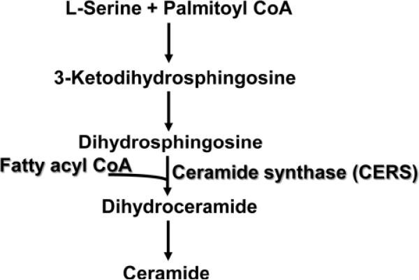
Background: The effectiveness of photodynamic therapy (PDT) for cancer treatment correlates with apoptosis. We previously observed that the knockdown of ceramide synthase 6, an enzyme from the de novo sphingolipid biosynthesis pathway, is associated with marked reduction in C18-dihydroceramide and makes cells resistant to apoptosis post-PDT. Down-regulation of ceramide synthase 1 (CERS1) can also render cells resistant to anticancer drugs.

Aim: To explore the impact of CERS1 knockdown on apoptosis and the sphingolipid profile, post-PDT, with the silicone phthalocyanine Pc 4, in a human head and neck squamous carcinoma cell line.

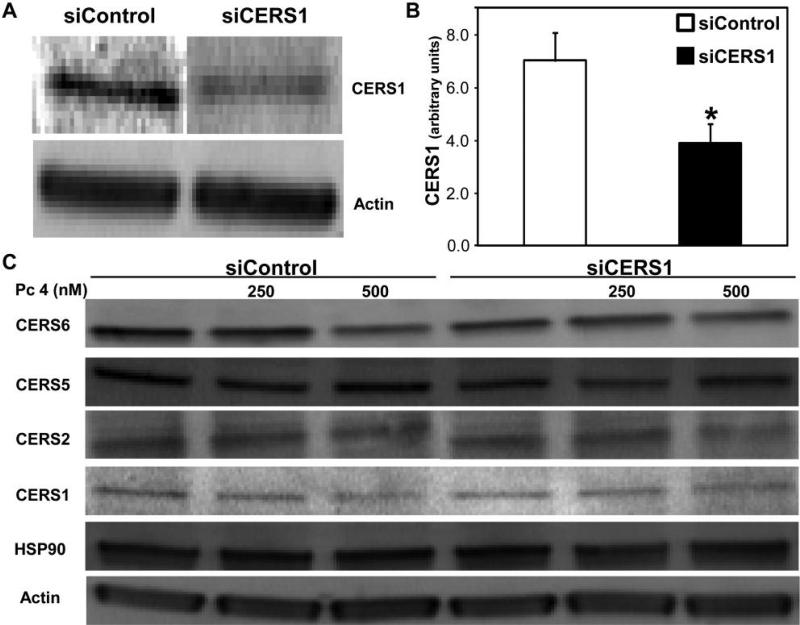
Materials and methods: Besides siRNA transfection and PDT treatment, the following methods were used: immunoblotting for protein expression, mass spectrometry for sphingolipid analysis, spectroflurometry and flow cytometry for apoptosis detection, and trypan blue assay for cell viability evaluation.

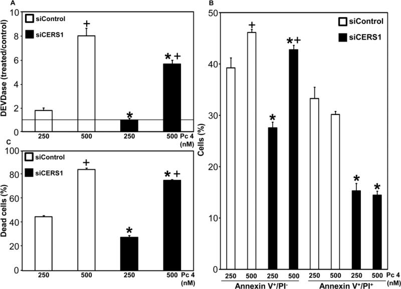
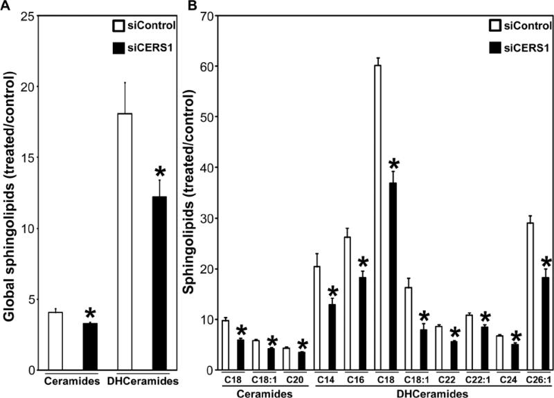
Results: CERS1 knockdown led to inhibition of PDT-induced caspase 3-like (DEVDase) activation, of apoptosis and cell death. CERS1 knockdown was associated with global and selective decreases in ceramides and dihydroceramides, in particular C18-, C18:1- and C20-ceramide post-PDT.

Conclusion: Our novel findings are consistent with the notion that CERS1 regulates apoptotic resistance to PDT, partly via C18- and C20-ceramide, and that CERS1 is a molecular target for controlling resistance to PDT.


Figures

Figure 1
Figure 1
The de novo sphingolipid biosynthesis pathway.
Figure 2
Figure 2
Effect of ceramide synthase 1 (CERS1) knockdown and photodynamic therapy (PDT) on expression of ceramide synthases. UM-SCC-22A cells were transfected with siRNA targeted against non-targeted control (siControl; 25 nM) or CERS1 (siCERS6; 25 nM). Twenty-four hours after transfection, cells were collected and seeded in fresh growth medium. A and B: Cells were incubated at 37°C for an additional 24 hours prior to collection. C: After overnight exposure to the photosensitizer Pc 4 (250 and 500 nM), cells were irradiated with red light (2 mW/cm2). Following PDT, cells were incubated at 37°C for a further 2 h, collected on ice and processed for PAGE/Western immunoblotting. Equal protein loading was verified using anti-actin and anti-HSP90. A: Knockdown of CERS1 was confirmed in two independent experiments. B: CERS1 protein levels were quantified from the blots and expressed in arbitrary units. The data are shown as the mean±SEM, *p≤0.05, n=5-6. C: Western blots of CERS1, -2, -5 and -6 in siControl- and siCERS1-tranfected cells are shown. Representative blots from 2-5 independent determinations are shown.
Figure 3
Figure 3
Ceramide synthase 1 (CERS1) knockdown suppressed caspase-3-like (DEVDase) activation, apoptosis and cell death after photodynamic therapy (PDT). A: Following 2 h-incubation post-PDT, cells were collected, cell lysates were prepared and DEVDase activity was measured using acetyl-Asp-Glu-Val-Asp-7-amino-4-methylcoumarin (Ac-DEVD-AMC), as the fluorogenic substrate. B: Following 24 h-incubation post-PDT, cells were collected and processed for flow cytometry. Annexin V/propidium iodide (PI) staining was used to detect apoptosis. C: Following 24 h-incubation post-PDT, cells were collected, stained with trypan blue and counted. A: The data are expressed as ratios of PDT-treated versus untreated controls. In all panels, data are shown as the mean±SEM, n=3-11. The significance (p≤0.05) is indicated as follows: *CERS1 knockdown suppressed PDT-induced DEVDase activation, apoptosis and cell death; +DEVDase activation, apoptosis and cell death was different at two PDT doses.
Figure 4
Figure 4
Effect of ceramide synthase 1 (CERS1) knockdown on photodynamic therapy (PDT)-induced accumulation of global (A) or individual (B) ceramides and dihydroceramides (DHceramides). Following 2 h-incubation post-PDT (500 nM Pc 4 + 200 mJ/cm2), cells were collected and processed for mass spectrometry. The data are expressed as ratios of PDT-treated versus untreated controls. In both panels, the data are shown as the mean±SEM, n=3-5. *Indicates that CERS1 knockdown suppressed PDT-induced accumulation of corresponding ceramides or dihydroceramides at p≤0.05.

Similar articles

Cited by

References

    1. Levy M, Futerman AH. Mammalian ceramide synthases. IUBMB Life. 2010;62:347–356. - PMC - PubMed
    1. Henry B, Moller C, Dimanche-Boitrel MT, Gulbins E, Becker KA. Targeting the ceramide system in cancer. Cancer Lett. 2011 doi: 10.1016/j.canlet.2011.07.010. - PubMed
    1. Mullen TD, Hannun YA, Obeid LM. Ceramide synthases at the centre of sphingolipid metabolism and biology. Biochem J. 2012;441:789–802. - PMC - PubMed
    1. Riebeling C, Allegood JC, Wang E, Merrill AH, Jr, Futerman AH. Two mammalian longevity assurance gene (LAG1) family members, TRH1 and TRH4, regulate dihydroceramide synthesis using different fatty acyl-CoA donors. J Biol Chem. 2003;278:43452–43459. - PubMed
    1. Mizutani Y, Kihara A, Igarashi Y. Mammalian Lass6 and its related family members regulate synthesis of specific ceramides. Biochem J. 2005;390:263–271. - PMC - PubMed

Publication types

MeSH terms