

MEK genomics in development and disease
- PMID: 22753777
- PMCID: PMC3398258
- DOI: 10.1093/bfgp/els022
MEK genomics in development and disease
Abstract
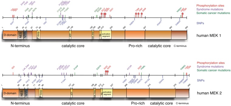
The mitogen-activated protein kinase kinases (the MAPK/ERK kinases; MKKs or MEKs) and their downstream substrates, the extracellular-regulated kinases have been intensively studied for their roles in development and disease. Until recently, it had been assumed any mutation affecting their function would have lethal consequences. However, the identification of MEK1 and MEK2 mutations in developmental syndromes as well as chemotherapy-resistant tumors, and the discovery of genomic variants in MEK1 and MEK2 have led to the realization the extent of genomic variation associated with MEKs is much greater than had been appreciated. In this review, we will discuss these recent advances, relating them to what is currently understood about the structure and function of MEKs, and describe how they change our understanding of the role of MEKs in development and disease.
Figures
References
-
- Depeille PE, Ding Y, Bromberg-White JL, et al. MKK signaling and vascularization. Oncogene. 2007;26:1290–6. - PubMed
-
- Mansfield PJ, Shayman JA, Boxer LA. Regulation of polymorphonuclear leukocyte phagocytosis by myosin light chain kinase after activation of mitogen-activated protein kinase. Blood. 2000;95:2407–12. - PubMed
-
- Widmann C, Gibson S, Jarpe MB, et al. Mitogen-activated protein kinase: conservation of a three-kinase module from yeast to human. Physiol Rev. 1999;79:143–80. - PubMed
-
- Haccard O, Sarcevic B, Lewellyn A, et al. Induction of metaphase arrest in cleaving Xenopus embryos by MAP kinase. Science. 1993;262:1262–5. - PubMed
Publication types
MeSH terms
Substances
Supplementary concepts
Grants and funding
LinkOut - more resources
Full Text Sources
Medical
Miscellaneous
