Comparative Study
. 2012 Aug;18(8):1527-40.
doi: 10.1261/rna.033134.112. Epub 2012 Jun 29.

The box C/D sRNP dimeric architecture is conserved across domain Archaea

Affiliations
Comparative Study

The box C/D sRNP dimeric architecture is conserved across domain Archaea

Kathleen R Bower-Phipps et al. RNA. 2012 Aug.

Abstract

Box C/D small (nucleolar) ribonucleoproteins [s(no)RNPs] catalyze RNA-guided 2'-O-ribose methylation in two of the three domains of life. Recent structural studies have led to a controversy over whether box C/D sRNPs functionally assemble as monomeric or dimeric macromolecules. The archaeal box C/D sRNP from Methanococcus jannaschii (Mj) has been shown by glycerol gradient sedimentation, gel filtration chromatography, native gel analysis, and single-particle electron microscopy (EM) to adopt a di-sRNP architecture, containing four copies of each box C/D core protein and two copies of the Mj sR8 sRNA. Subsequently, investigators used a two-stranded artificial guide sRNA, CD45, to assemble a box C/D sRNP from Sulfolobus solfataricus with a short RNA methylation substrate, yielding a crystal structure of a mono-sRNP. To more closely examine box C/D sRNP architecture, we investigate the role of the omnipresent sRNA loop as a structural determinant of sRNP assembly. We show through sRNA mutagenesis, native gel electrophoresis, and single-particle EM that a di-sRNP is the near exclusive architecture obtained when reconstituting box C/D sRNPs with natural or artificial sRNAs containing an internal loop. Our results span three distantly related archaeal species--Sulfolobus solfataricus, Pyrococcus abyssi, and Archaeoglobus fulgidus--indicating that the di-sRNP architecture is broadly conserved across the entire archaeal domain.


Figures
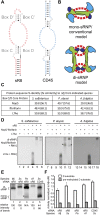
FIGURE 1.
FIGURE 1.
Box C/D sRNPs from four archaeal species migrate as di-sRNPs on native gels. (A) Structural characteristics of a naturally occurring box C/D sRNA, sR8, and a two-stranded box C/D sRNA mimic, CD45. (Gray) Conserved C, C′, and D, D′ boxes. Naturally occurring sRNAs such as sR8 exist as a single, continuous nucleotide strand. The CD45 RNA resembles a box C/D sRNA, but exists as two separate oligonucleotide strands instead of one continuous RNA molecule and therefore lacks an internal loop. (B) Two conflicting models for box C/D sRNP assembly. The core proteins are as follows: fibrillarin (cyan), Nop5 (green), L7Ae (dark blue). The box C/D sRNA is indicated by red dashed lines representing the two guide sequences of each sRNA; sRNA orientation is left intentionally ambiguous. (C) Sequence percent identity (and percent similarity) of box C/D core proteins from three archaeal species as compared to the homologous proteins from the Mj species. (D) Native gel electrophoresis of in vitro–assembled RNP complexes from Ss, Pa, and Af. In the absence of L7Ae, both Ss and Pa proteins Nop5 and fibrillarin bind to sR8, creating several RNP species, one of which (*, lane 11) is present in the assembly of the Pa sRNP (*, lane 12) and has the same mobility as the mono-sRNP from Ss assembled with CD45 sRNA (E, lane 5). (E) All tested archaeal box C/D sRNPs assembled with a naturally occurring box C/D sRNA migrate as di-sRNPs as assayed by native gel electrophoresis. (*) Pa Nop5-fibrillarin heterotetramer associating with the sRNA in the absence of L7Ae (see D). Quantitations of the relative intensity of di- and mono-sRNP sized bands are indicated. (F) Methylation activity assays of assembled box C/D sRNPs show that all tested archaeal sRNPs are catalytically active for site-specific methylation.
FIGURE 2.
FIGURE 2.
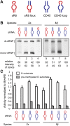
The box C/D sRNA internal loop affects sRNP architecture. (A) The naturally occurring Mj sR8 was mutagenized to contain a stem instead of an internal loop (sR8 faux) but retains all other sequences from the natural sR8 sRNA. The CD45 sRNA (Lin et al. 2011) was mutagenized to contain the internal loop from the Mj sR8 sRNA (CD45 loop). Sequences originating from sR8 (red); sequences originating from CD45 (blue). (B) Native gel electrophoresis showing sRNPs assembled as indicated using core proteins from Ss and Mj with sRNAs from A. Quantitations of the relative intensity of di- and mono-sRNP sized bands are indicated. (C) Activity assays show that all assemblies from B are catalytically active for site-specific 2′-O-ribose methylation.
FIGURE 3.
FIGURE 3.
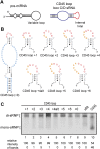
sRNAs with a wide variety of internal loops confer di-sRNP assembly. (A) Hairpin stem–loops are a prevalent secondary structure in at least two classes of small noncoding RNAs: pre-microRNAs (pre-miRNAs) and box C/D s(no)RNAs. The exact number and location of base pairs vary within each class of RNAs—these diagrams are not to scale. (B) Secondary structures of CD45 loop mutants. The internal loop from the CD45 loop sRNA was mutated to include additional nucleotides of the randomized sequence. Box C/D and C′/D′ sequences (gray); sequences originating from CD45 (blue); sequences originating from sR8 (red); randomized sequences inserted in the internal loop (orange). The size of each loop in relation to the 5-nt loop of CD45 is indicated (+1, +2, etc.). (C) Assembly of Ss sRNPs with sRNAs bearing a wide variety of internal loops. Each sRNA mutant was in vitro–transcribed and assembled with Ss box C/D core proteins and assayed for di-sRNP formation by native gel electrophoresis. sRNPs assembled with sR8 and CD45 sRNAs are included as markers for di- and mono-sRNP formation. Quantitations of the relative intensity of di- and mono-sRNP sized bands are indicated.
FIGURE 4.
FIGURE 4.
3D single-particle EM reconstruction of the Ss box C/D di-sRNP. (A) Electron micrograph of negatively stained Ss box C/D sRNPs from glycerol gradient peak fractions. (▸) Examples of particles chosen for analysis. (B) Comparison of experimentally determined class averages (Exp. C.A.) to 2D reprojections of the reconstructed 3D volume (2D Reproj.) and to surface renderings (Surf. Rend.) of the 3D volume. Scale bar, 10 nm. (C) Fourier shell correlation (FSC) curve of the reconstructed Ss box C/D sRNP EM volume. The resolution at 0.5 FSC is indicated. (D) Distribution of the Euler angles assigned to particle images included in the 3D EM reconstruction of the Ss box C/D sRNP. Circle radii are proportional to the number of images assigned to the particular Euler angles. (E) Isodensity of the 3D volume and docking of the crystal structures of the Ss core proteins, L7Ae (dark blue), Nop5 (green), and fibrillarin (cyan), (PDB 3ID5) (Ye et al. 2009) into the isodensity map at the 3σ threshold. Scale bar, 10 nm.
FIGURE 5.
FIGURE 5.
3D single-particle EM reconstruction of the Pa box C/D di-sRNP. (A) Electron micrograph of negatively stained Pa box C/D sRNPs from glycerol gradient peak fractions. (▸) Examples of particles chosen for analysis. (B) Comparison of experimentally determined class averages (Exp. C.A.) to 2D reprojections of the reconstructed 3D volume (2D Reproj.) and to surface renderings (Surf. Rend.) of the 3D volume. Scale bar, 10 nm. (C) FSC curve of the reconstructed Pa box C/D sRNP EM volume. The resolution at 0.5 FSC is indicated. (D) Distribution of the Euler angles assigned to particle images included in the 3D EM reconstruction of the Pa box C/D sRNP. Circle radii are proportional to the number of images assigned to the particular Euler angles. (E) Isodensity of the 3D volume and docking of the crystal structures of the Pa core proteins (PDB 3NMU) (Xue et al. 2010) into the isodensity map at the 3σ threshold. Colors are as in Figure 4. Scale bar, 10 nm.
FIGURE 6.
FIGURE 6.
3D single particle EM reconstruction of the Af box C/D di-sRNP. (A) Electron micrograph of negatively stained Af box C/D sRNPs from glycerol gradient peak fractions. (▸) Examples of particles chosen for analysis. (B) Comparison of experimentally determined class averages (Exp. C.A.) to 2D reprojections of the reconstructed 3D volume (2D Reproj.) and to surface renderings (Surf. Rend.) of the 3D volume. Scale bar, 10 nm. (C) FSC curve of the reconstructed Af box C/D sRNP EM volume. The resolution at 0.5 FSC is indicated. (D) Distribution of the Euler angles assigned to particle images included in the 3D EM reconstruction of the Af box C/D sRNP. Circle radii are proportional to the number of images assigned to the particular Euler angles. (E) Isodensity of the 3D volume and docking of the crystal structures of the Af core proteins (PDB 1NT2) (Aittaleb et al. 2003), (1RLG) (Moore et al. 2004) into the isodensity map at the 3σ threshold. Colors are as in Figure 4. Scale bar, 10 nm.
FIGURE 7.
FIGURE 7.
Distantly related archaeal box C/D sRNPs retain di-sRNP 3D architecture. Front (A) and side (B) views of aligned 3D EM negative stain reconstructions from four species: (Ss) gray; (Pa) yellow; (Af) blue; (Mj) purple. Ss, Pa, and Af are the three reconstructions from this study; Mj is from Bleichert et al. (2009). All reconstructions were set to 3σ thresholds. A c2 pseudosymmetry axis lies directly through the center of the front view (A) of each volume.

Similar articles

Cited by

References

    1. Abramovitz DL, Pyle AM 1997. Remarkable morphological variability of a common RNA folding motif: The GNRA tetraloop–receptor interaction. J Mol Biol 266: 493–506 - PubMed
    1. Aittaleb M, Rashid R, Chen Q, Palmer JR, Daniels CJ, Li H 2003. Structure and function of archaeal box C/D sRNP core proteins. Nat Struct Biol 10: 256–263 - PubMed
    1. Aittaleb M, Visone T, Fenley MO, Li H 2004. Structural and thermodynamic evidence for a stabilizing role of Nop5p in S-adenosyl-L-methionine binding to fibrillarin. J Biol Chem 279: 41822–41829 - PubMed
    1. Balakin AG, Smith L, Fournier MJ 1996. The RNA world of the nucleolus: Two major families of small RNAs defined by different box elements with related functions. Cell 86: 823–834 - PubMed
    1. Bammes BE, Rochat RH, Jakana J, Chen DH, Chiu W 2012. Direct electron detection yields cryo-EM reconstructions at resolutions beyond 3/4 Nyquist frequency. J Struct Biol 177: 589–601 - PMC - PubMed

Publication types

MeSH terms