. 2012 Jul 9;198(1):87-102.
doi: 10.1083/jcb.201202053. Epub 2012 Jul 2.

Z-line formins promote contractile lattice growth and maintenance in striated muscles of C. elegans

Affiliations

Z-line formins promote contractile lattice growth and maintenance in striated muscles of C. elegans

Lei Mi-Mi et al. J Cell Biol. .

Abstract

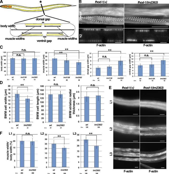
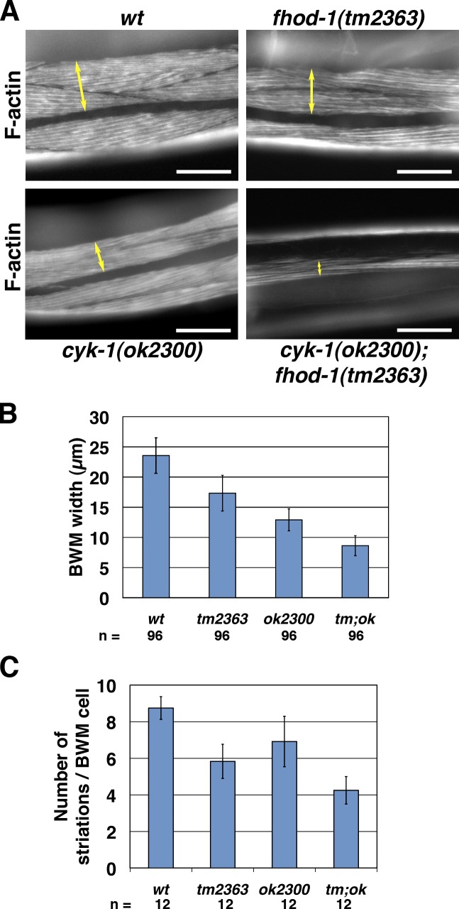
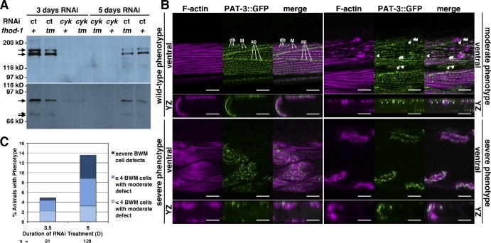
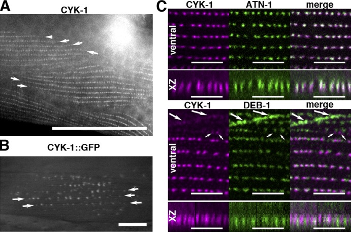
Muscle contraction depends on interactions between actin and myosin filaments organized into sarcomeres, but the mechanism by which actin filaments incorporate into sarcomeres remains unclear. We have found that, during larval development in Caenorhabditis elegans, two members of the actin-assembling formin family, CYK-1 and FHOD-1, are present in striated body wall muscles near or on sarcomere Z lines, where barbed ends of actin filaments are anchored. Depletion of either formin during this period stunted growth of the striated contractile lattice, whereas their simultaneous reduction profoundly diminished lattice size and number of striations per muscle cell. CYK-1 persisted at Z lines in adulthood, and its near complete depletion from adults triggered phenotypes ranging from partial loss of Z line-associated filamentous actin to collapse of the contractile lattice. These results are, to our knowledge, the first genetic evidence implicating sarcomere-associated formins in the in vivo organization of the muscle cytoskeleton.


Figures

Figure 1.
Figure 1.
C. elegans formins. (A) Seven formin genes of C. elegans (left) with protein-coding exons color-coded to match predicted product structural domains (right), including DIA-related formin G domains (dark green), FHOD-specific G domain (G2; olive), DIDs, dimerization domains (DD), helical (H) or coiled-coil (CC) regions, FH1 domains, FH2 domains, core DAD sequences, and zinc finger domain (ZFD). Gray blocks indicate gene sequences deleted by alleles used in this study. (B) Estimated phylogenetic trees based on protein sequence comparisons between mouse (M. Musculus [Mm]; blue), purple sea urchin (S. purpuratus [Sp]; purple), fruit fly (D. melanogaster [Dm]; green), and C. elegans (Ce; red) formin FH2 domains or N termini reveal seven formin subfamilies, five of which (DAAM, DIA, FHOD, FRL, and INF) have C. elegans homologues. N-term, N terminus. Bars, 0.5 substitutions per position.
Figure 2.
Figure 2.
FHOD-1 is expressed in muscle cells. (A) Anti–FHOD-1 Western blot of normalized adult worm extracts from wild type (wt) and indicated formin mutants. 150- and 165-kD signals (arrows) are absent from the fhod-1(tm2363) extract. (B) Superficial and deep views of a wild-type larva (worm anterior is shown on the top, and posterior is shown at the bottom) stained with anti–FHOD-1 reveal puncta near the body surface (small arrows) and within the pharynx in the head (large arrow). Bar, 50 µm. (C) FHOD-1::GFP in a larva coincides with fluorescent phalloidin-stained F-actin–rich pharyngeal muscles (PHA) in the head, vulval muscles (VM) near the middle, and body wall muscles (BWM) extending from nose to tail. Bar, 100 µm.
Figure 3.
Figure 3.
FHOD-1 localizes close to Z lines in BWM cells. (A) Model of BWM sarcomere organization. Side view of one sarcomere shows that Z-line dense bodies (blue) anchor actin filaments (yellow), and M lines (black) anchor myosin filaments (gray). M lines and dense bodies attach to the plasma membrane, placing the contractile lattice between the membrane and the cell body. In top views, sarcomeres can be seen to combine to form oblique striations, in which rows of dense bodies (blue spots) within the F-actin–rich striations form discontinuous Z lines that alternate with M lines. Striations in turn form the spindle-shaped contractile lattice of BWM cells. At boundaries between BWM cells, attachment plaques (larger dark spots) replace dense bodies (smaller dark spots) as Z-line structures. Myosin filaments have been omitted from striation and BWM models for clarity. (B) Ventral view (anterior to the left) of an FHOD-1::GFP–expressing larva stained with fluorescent phalloidin shows FHOD-1–containing puncta along the edges of F-actin–rich BWM cell contractile lattices (large arrows) and in faint striations across the lattices (small arrows). (C) Ventral view of a wild-type larva stained with anti–FHOD-1 reveals endogenous FHOD-1 in similar puncta (large arrows) and striations (small arrows). (D) Animals double-stained for FHOD-1 and either Z-line marker ATN-1 or DEB-1 show that the formin is closely associated with Z lines. In ventral views or in side views of the discontinuous Z line (xz projection), anti–FHOD-1–stained striations are seen in the contractile lattice (cl) of wild-type but not fhod-1(tm2363) animals, and anti–FHOD-1–stained puncta intermingle with DEB-1–stained attachment plaques (arrows) at the cell edge. Staining of the cell body (cb in xz view) or of an elongated structure that parallels BWMs (bottom, arrowheads) is nonspecific. (E) FHOD-1 striations alternate with MYO-3 striations. (F) Anti-GFP–stained striations in FHOD-1::GFP–expressing animals partially overlap with UNC-60B. (G) Proximal to the plasma membrane, LIM-8 occupies two sets of striations, one near Z lines and a fainter one near M lines (arrows), whereas distal to the membrane, LIM-8 detectable near Z lines strongly comingles with FHOD-1::GFP. (H) Worms stained with anti–FHOD-1 and two secondary antibodies with different fluorophores show extensive, but imperfect, comingling of signal along FHOD-1 striations. Bars: (B) 10 µm; (C) 25 µm; (D–H) 5 µm.
Figure 4.
Figure 4.
FHOD-1–containing structures appear in BWMs during mid- and late-larval development. (A) FHOD-1::GFP–expressing embryos stained with fluorescent phalloidin show that FHOD-1 is present diffusely in the elongated bands of F-actin–rich embryonic myoblasts (arrows). (B) In ventral views of FHOD-1::GFP–expressing larvae stained with fluorescent phalloidin, FHOD-1 is not detectable in F-actin–rich BWMs of L1 larvae but appears as puncta along muscle edges and as striations across the BWM contractile lattice in L2 and L3 larvae. Bars, 10 µm.
Figure 5.
Figure 5.
BWM cell growth and positioning are aberrant in fhod-1 mutants. (A) A schematic lateral view of C. elegans (anterior to the left) with a cross section view at the asterisk shows positions of pharyngeal muscles (orange) and BWMs (yellow). The squashed body outline in cross section (dashed line) is consistent with a fixed animal mounted for microscopy, as in B. Dimensions quantified in C are indicated in the cross section schematic. (B) Dorsal and cross section views (yz projections) of fluorescent phalloidin-stained young adults show that fhod-1(tm2363) BWMs are narrower and spaced further apart than wild type. Dotted lines indicate individual BWM cell boundaries. Bars: (dorsal) 50 µm; (yz) 10 µm. (C) Young adult fhod-1 mutants (tm3138 and tm2363) have similar body widths as wild type (wt) but narrower muscles, and fhod-1(tm2363) animals have wider gaps between paired muscles at dorsal and ventral surfaces. Results shown are representative of two independent experiments. (D) Fluorescent phalloidin-stained fhod-1 (tm2363) BWM cells have normal anterior/posterior lengths and radial thicknesses but reduced lateral widths. (E) Ventral views of BWMs in fluorescent phalloidin-stained L1-, L2-, and L3-stage larvae. Bars, 10 µm. (F) Discrepancy between wild-type and fhod-1 mutant (tm2363) BWM widths, expressed as a percentage of total body width, is apparent in L2 and L3 but not L1 stages. Results shown are representative of two independent experiments. For all quantitative results, mean values of n measurements are presented. Error bars indicate one standard deviation. n.s. indicates not significant, P > 0.05. **, P < 0.001.
Figure 6.
Figure 6.
cyk-1 mutation exacerbates BWM defects of fhod-1 mutants. (A) Dorsal views of fluorescent phalloidin-stained animals show the lateral widths of BWMs (double arrows) in fhod-1(tm2363) and cyk-1(ok2300) mutant animals are smaller than wild type (wt), whereas BWM widths in double cyk-1(ok2300); fhod-1(tm2363) animals are even smaller. Bars, 50 µm. (B) BWM lateral widths were measured for wild type, fhod-1 mutants (tm2363), cyk-1 mutants (ok2300), and double mutants (tm;ok). (C) Striations per BWM cell were counted for the same strains. In B and C, mean values of n measurements are presented. Error bars indicate one standard deviation. Results are significantly different in all pairwise comparisons (P < 0.01).
Figure 7.
Figure 7.
Prolonged cyk-1 RNAi disrupts adult BWM cytoskeletal organization. (A) Anti–CYK-1 Western blot of normalized worm extracts shows RNAi targeting cyk-1 (cyk) but not control RNAi (ct) reduces detectable CYK-1 (arrows) in wild type (+) and fhod-1 mutants (tm). (B) Ventral and cross section (yz projection) views of cyk-1(RNAi) animals expressing PAT-3::GFP and stained with fluorescent phalloidin show various BWM phenotypes. In wild-type phenotype BWMs, F-actin occupies regular striations in a layer close to the body wall, and PAT-3::GFP decorates dense bodies (db), attachment plaques (ap), and M lines (M). In moderate phenotype BWMs, F-actin is patchy, leaving some GFP-positive bodies free of F-actin (arrows). In severe phenotype BWMs, striated organization is lost, and F-actin is no longer concentrated near the body wall. Bars, 10 µm. (C) The number of animals exhibiting moderate or severe phenotypes increases with longer RNAi treatment. Results shown are typical of two independent experiments.
Figure 8.
Figure 8.
CYK-1 is present on BWM Z-line structures. (A) Dorsal view of an adult reveals anti–CYK-1–stained striations formed of puncta. Arrows indicate ends of striations. Bar, 50 µm. (B) Dorsal view of an animal expressing CYK-1::GFP from an extrachromosomal array reveals striations of GFP-positive puncta. Arrows indicate ends of striations. Bar, 10 µm. (C) BWMs of animals double stained for CYK-1 and either ATN-1 or DEB-1, seen in ventral views and side views of the discontinuous Z line (xz projection), show that CYK-1 overlaps most strongly with ATN-1. Large arrows indicate DEB-1–containing attachment plaques with some associated CYK-1. Small arrows indicate CYK-1 patches lacking DEB-1. Bars, 5 µm.

Similar articles

Cited by

References

    1. Alberts A.S. 2001. Identification of a carboxyl-terminal diaphanous-related formin homology protein autoregulatory domain. J. Biol. Chem. 276:2824–2830 10.1074/jbc.M006205200 - DOI - PubMed
    1. Altschul S.F., Gish W., Miller W., Myers E.W., Lipman D.J. 1990. Basic local alignment search tool. J. Mol. Biol. 215:403–410 - PubMed
    1. Amin N.M., Hu K., Pruyne D., Terzic D., Bretscher A., Liu J. 2007. A Zn-finger/FH2-domain containing protein, FOZI-1, acts redundantly with CeMyoD to specify striated body wall muscle fates in the Caenorhabditis elegans postembryonic mesoderm. Development. 134:19–29 10.1242/dev.02709 - DOI - PubMed
    1. Bai J., Hartwig J.H., Perrimon N. 2007. SALS, a WH2-domain-containing protein, promotes sarcomeric actin filament elongation from pointed ends during Drosophila muscle growth. Dev. Cell. 13:828–842 10.1016/j.devcel.2007.10.003 - DOI - PubMed
    1. Barstead R.J., Waterston R.H. 1989. The basal component of the nematode dense-body is vinculin. J. Biol. Chem. 264:10177–10185 - PubMed

Publication types

MeSH terms