

Cutting edge: in the absence of TGF-β signaling in T cells, fewer CD103+ regulatory T cells develop, but exuberant IFN-γ production renders mice more susceptible to helminth infection
- PMID: 22753928
- PMCID: PMC3428909
- DOI: 10.4049/jimmunol.1200991
Cutting edge: in the absence of TGF-β signaling in T cells, fewer CD103+ regulatory T cells develop, but exuberant IFN-γ production renders mice more susceptible to helminth infection
Abstract
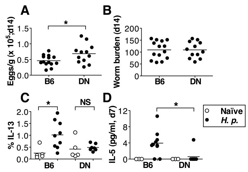
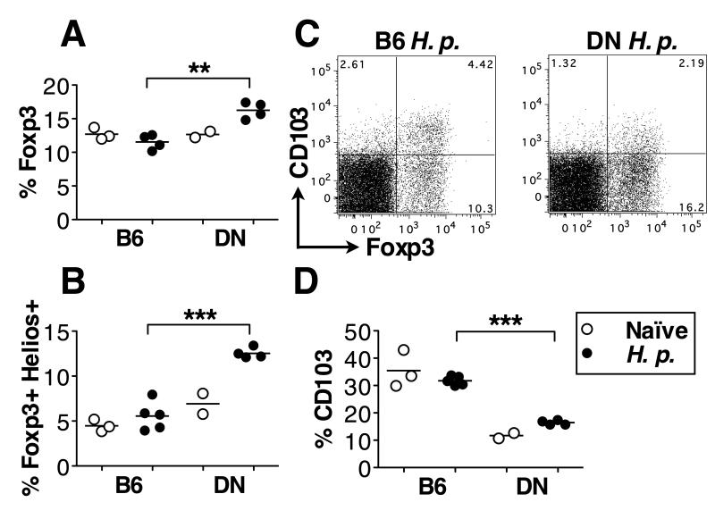
Multiple factors control susceptibility of C57BL/6 mice to infection with the helminth Heligmosomoides polygyrus, including TGF-β signaling, which inhibits immunity in vivo. However, mice expressing a T cell-specific dominant-negative TGF-β receptor II (TGF-βRII DN) show dampened Th2 immunity and diminished resistance to infection. Interestingly, H. polygyrus-infected TGF-βRII DN mice show greater frequencies of CD4(+)Foxp3(+)Helios(+) Tregs than infected wild-type mice, but levels of CD103 are greatly reduced on both these cells and on the CD4(+)Foxp3(+)Helios(-) population. Although Th9 and Th17 levels are comparable between infected TGF-βRII DN and wild-type mice, the former develop exaggerated CD4(+) and CD8(+) T cell IFN-γ responses. Increased susceptibility conferred by TGF-βRII DN expression was lost in IFN-γ-deficient mice, although they remained unable to completely clear infection. Hence, overexpression of IFN-γ negatively modulates immunity, and the presence of Helios(+) Tregs may maintain susceptibility on the C57BL/6 background.
Figures


Similar articles
-
Helminth secretions induce de novo T cell Foxp3 expression and regulatory function through the TGF-β pathway.J Exp Med. 2010 Oct 25;207(11):2331-41. doi: 10.1084/jem.20101074. Epub 2010 Sep 27. J Exp Med. 2010. PMID: 20876311 Free PMC article.
-
Role of T cell TGF-beta signaling in intestinal cytokine responses and helminthic immune modulation.Eur J Immunol. 2009 Jul;39(7):1870-8. doi: 10.1002/eji.200838956. Eur J Immunol. 2009. PMID: 19544487 Free PMC article.
-
Heligmosomoides polygyrus bakeri Infection Decreases Smad7 Expression in Intestinal CD4+ T Cells, Which Allows TGF-β to Induce IL-10-Producing Regulatory T Cells That Block Colitis.J Immunol. 2019 Apr 15;202(8):2473-2481. doi: 10.4049/jimmunol.1801392. Epub 2019 Mar 8. J Immunol. 2019. PMID: 30850474 Free PMC article.
-
TGFβ in T cell biology and tumor immunity: Angel or devil?Cytokine Growth Factor Rev. 2014 Aug;25(4):423-35. doi: 10.1016/j.cytogfr.2014.07.014. Epub 2014 Jul 29. Cytokine Growth Factor Rev. 2014. PMID: 25156420 Free PMC article. Review.
-
Review of the activation of TGF-beta in immunity.J Leukoc Biol. 2009 Jan;85(1):29-33. doi: 10.1189/jlb.0708415. Epub 2008 Sep 25. J Leukoc Biol. 2009. PMID: 18818372 Free PMC article. Review.
Cited by
-
Th9 cells: differentiation and disease.Immunol Rev. 2013 Mar;252(1):104-15. doi: 10.1111/imr.12028. Immunol Rev. 2013. PMID: 23405898 Free PMC article. Review.
-
Modification of hemodynamic and immune responses to exposure with a weak antigen by the expression of a hypomorphic BMPR2 gene.PLoS One. 2013;8(1):e55180. doi: 10.1371/journal.pone.0055180. Epub 2013 Jan 29. PLoS One. 2013. PMID: 23383100 Free PMC article.
-
IL-4 together with IL-1β induces antitumor Th9 cell differentiation in the absence of TGF-β signaling.Nat Commun. 2019 Mar 26;10(1):1376. doi: 10.1038/s41467-019-09401-9. Nat Commun. 2019. PMID: 30914642 Free PMC article.
-
Immunity to the model intestinal helminth parasite Heligmosomoides polygyrus.Semin Immunopathol. 2012 Nov;34(6):829-46. doi: 10.1007/s00281-012-0347-3. Epub 2012 Oct 11. Semin Immunopathol. 2012. PMID: 23053394 Free PMC article. Review.
-
Cultivation of Heligmosomoides polygyrus: an immunomodulatory nematode parasite and its secreted products.J Vis Exp. 2015 Apr 6;(98):e52412. doi: 10.3791/52412. J Vis Exp. 2015. PMID: 25867600 Free PMC article.
References
-
- Allen JE, Maizels RM. Diversity and dialogue in immunity to helminths. Nat Rev Immunol. 2011;11:375–388. - PubMed
-
- Grainger JR, Smith KA, Hewitson JP, McSorley HJ, Harcus Y, Filbey KJ, Finney CAM, Greenwood EJD, Knox DP, Wilson MS, Belkaid Y, Rudensky AY, Maizels RM. Helminth secretions induce de novo T cell Foxp3 expression and regulatory function through the TGF-β pathway. J Exp Med. 2010;207:2331–2341. - PMC - PubMed
Publication types
MeSH terms
Substances
Grants and funding
LinkOut - more resources
Full Text Sources
Molecular Biology Databases
Research Materials
