. 2012 Jan;45(1):45-52.
doi: 10.4103/0970-0358.96584.

Reverse peroneal artery flap for large defects of ankle and foot: A reliable reconstructive technique

Affiliations

Reverse peroneal artery flap for large defects of ankle and foot: A reliable reconstructive technique

Jose Tharayil et al. Indian J Plast Surg. 2012 Jan.

Abstract

Background: Large soft tissue defects around the lower third of the leg, ankle and foot always have been challenging to reconstruct. Reverse sural flaps have been used for this problem with variable success. Free tissue transfer has revolutionised management of these problem wounds in selected cases.

Materials and methods: Twenty-two patients with large defects around the lower third of the leg, ankle and foot underwent reconstruction with reverse peroneal artery flap (RPAF) over a period of 7 years. The mean age of these patients was 41.2 years.

Results: Of the 22 flaps, 21 showed complete survival without even marginal necrosis. One flap failed, where atherosclerotic occlusion of peroneal artery was evident on the table. Few patients had minor donor site problems that settled with conservative management.

Conclusions: RPAF is a very reliable flap for the coverage of large soft tissue defects of the heel, sole and dorsum of foot. This flap adds versatility in planning and execution of this extended reverse sural flap.

Keywords: Distally based peroneal flaps; extended reverse sural flaps; foot reconstruction; peroneal artery; reverse peroneal flaps.


Conflict of interest statement

Conflict of Interest: None declared.

Figures

Figure 1
Figure 1
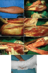
(a) Flap planning. Flap has been marked symmetrically around the posterior midline with pivot point around 5 cm above the lateral malleolus. Small skin paddle marked over the pedicle area helps in easy closure after transposition. (b) Flap dissection from the medial to the lateral side. Sural vessels are carefully included. (c) Pedicle area is also dissected just above the paratenon over tendoachillis and reflected laterally till good perforators are visible. (d) Perforators are tracked down to the peroneal artery through the posterior septum. (e) Dissection from the lateral side. Sub-periosteal dissection helps in easy retrieval of the peroneal system from behind the fibula. The peroneal vessels are clamped just above the perforator. (f) Dissection is continued along with the peroneal system as much as needed to reach the defect, without violating the lower 5 cm above the malleoli. (g) The post-operative position. The leg is well supported by padding above and below the pedicle area. A strong plaster is applied over this so that the pedicle area is completely off-loaded without disturbing the patient
Figure 2
Figure 2
(a–c) A 26-year-old male with traumatic forefoot amputation and degloving of the skin over the dorsum of the foot and anterolateral aspect of the lower leg and part of the sole distal to the heel. (d) Flap planned for complete cover of the defect. (e) Flap used to cover the defect. (f and g) Well-settled flap from the lateral and plantar aspects at the time of suture removal
Figure 3
Figure 3
(a and b) A 45-year-old male with avulsion of the heel and sole tissue. (c) One week following the injury, the non-viable tissue is well demarcated. (d) Following debridement, the raw area was extending from the posterior heel to the junction of the middle and distal thirds of the sole. (e) Large flap planned on the posterior calf. (f, g and h) Well-settled flap and the donor site few months following the surgery
Figure 4
Figure 4
(a) A 4-year-old boy with run-over injury just before debridement. (b) Following debridement, multiple bones and joints got exposed. (c) Reverse peroneal flap was used to cover this defect. (d) Two weeks post-operatively, the flap settled well except a small raw area over the pedicle. (e) Well-settled flap 1 month following the surgery

Similar articles

Cited by

References

    1. Benito-Ruiz J, Yoon T, Guisantes-Pintos E, Monner J, Serra-Renom JM. Reconstruction of soft tissue defects of the heel with local fasciocutaneous flaps. Ann Plast Surg. 2004;52:380–4. - PubMed
    1. Eren S, Ghofrani A, Reifenrath M. The distally pedicled peroneus brevis muscle flap: A new flap for the lower leg. Plast Reconstr Surg. 2001;107:1443–8. - PubMed
    1. Yang YL, Lin TM, Lee SS, Chang KP, Lai CS, Ruan HJ, Cai PH, et al. The extended peroneal artery perforator flap for lower extremity reconstruction. Ann Plast Surg. 2010;64:451–57. - PubMed
    1. The distally pedicled peroneus brevis muscle flap anatomic studies and clinical applications. J Foot Ankle Surg. 2005;44:259–64. - PubMed
    1. Donski PK, Fogdestam I. Distally based fasciocutaneous flap from the sural region: A preliminary report. Scand J Plast Reconstr Surg. 1983;17:191–6. - PubMed