. 2012;13(5):5628-5644.
doi: 10.3390/ijms13055628. Epub 2012 May 10.

Anti-TNF-α activity of Portulaca oleracea in vascular endothelial cells

Affiliations

Anti-TNF-α activity of Portulaca oleracea in vascular endothelial cells

An Sook Lee et al. Int J Mol Sci. 2012.

Abstract

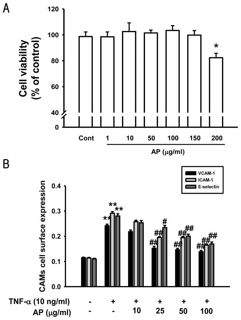
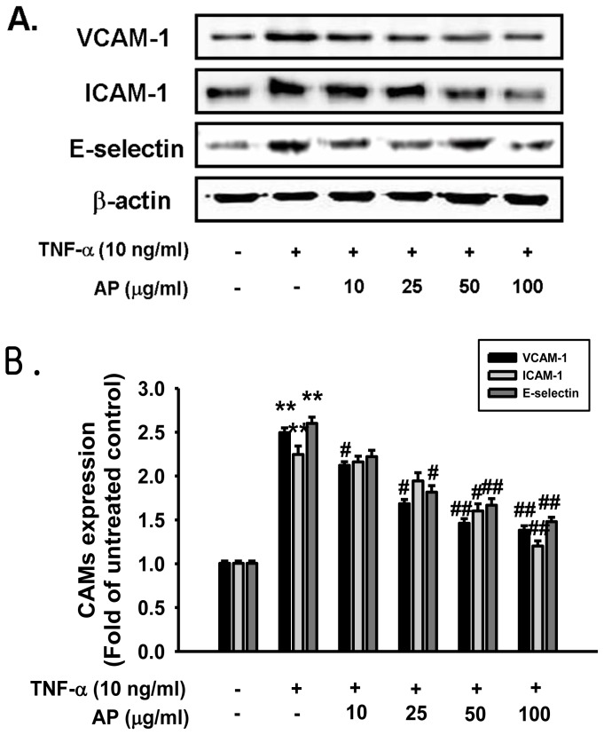
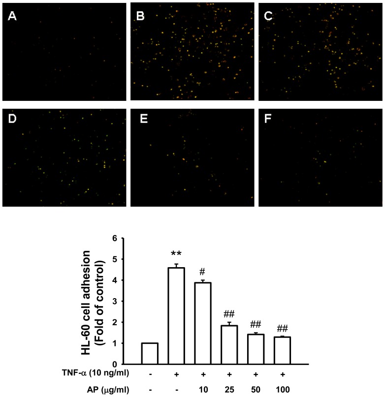
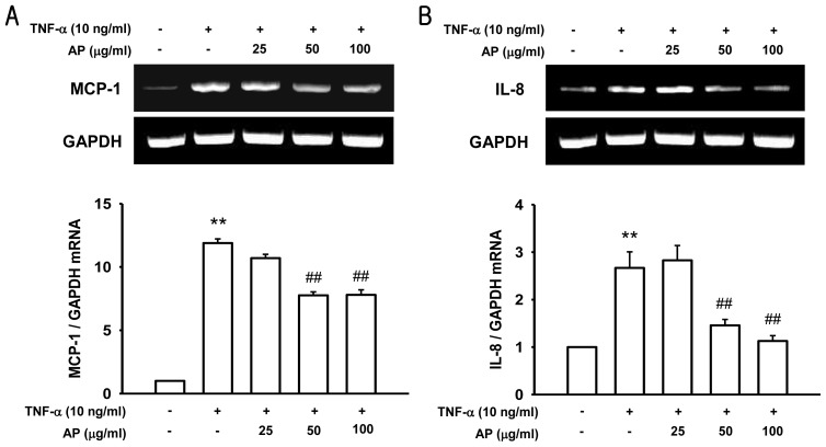
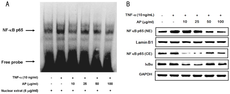
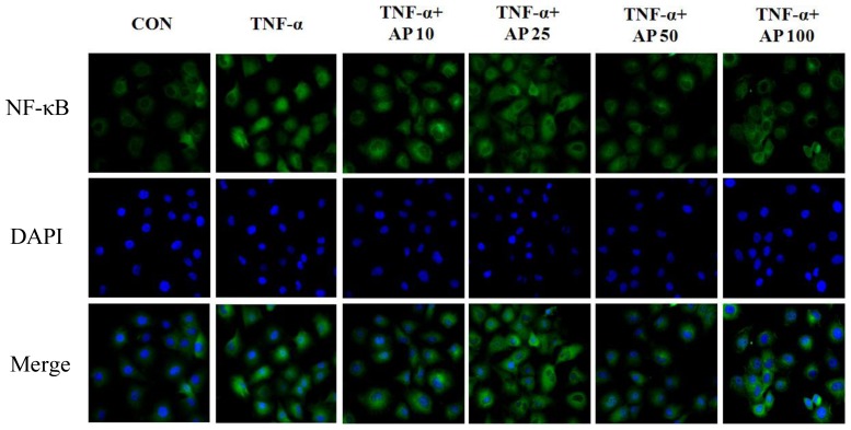
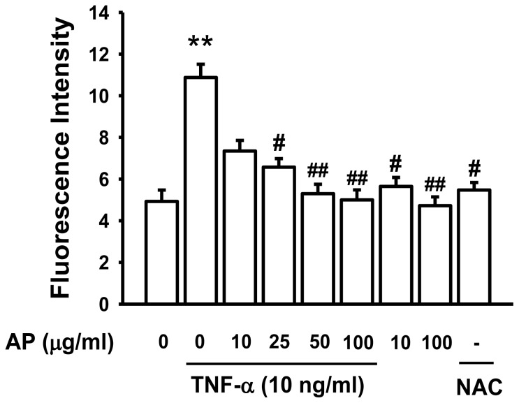
Vascular inflammation plays a key role in the pathogenesis and progression of atherosclerosis, a main complication of diabetes. The present study investigated whether an aqueous extract of Portulaca oleracea (AP) prevents the TNF-α-induced vascular inflammatory process in the human umbilical vein endothelial cell (HUVEC). The stimulation of TNF-α induced overexpression of adhesion molecules affects vascular cell adhesion molecule (VCAM)-1, intercellular adhesion molecule (ICAM)-1 and E-selectin for example. However, AP significantly suppressed TNF-α-induced over-expression of these adhesion molecules in a dose-dependent manner. In addition, pretreatment with AP dose-dependently reduced an increase of the adhesion of HL-60 cells to TNF-α-induced HUVEC. Furthermore, we observed that stimulation of TNF-α significantly increased intracellular reactive oxygen species (ROS) production. However, pretreatment with AP markedly blocked TNF-α-induced ROS production in a dose-dependent manner. The western blot and immunofluorescence analysis showed that AP inhibited the translocation of p65 NF-κB to the nucleus. In addition, AP suppressed the TNF-α-induced degradation of IκB-α and attenuated the TNF-α-induced NF-κB binding. AP also effectively reduced TNF-α-induced mRNA expressions of monocyte chemoattractant protein (MCP)-1 and interleukin (IL)-8 in a dose-dependent manner. Taken together, AP prevents the vascular inflammatory process through the inhibition of intracellular ROS production and NF-κB activation as well as the reduction of adhesion molecule expression in TNF-α-induced HUVEC. These results suggested that AP might have a potential therapeutic effect by inhibiting the vascular inflammation process in vascular diseases such as atherosclerosis.

Keywords: NF-κB; Portulaca oleracea; atherosclerosis; inflammation; reactive oxygen species (ROS).


Figures

Figure 1
Figure 1
Effects of an aqueous extract of Portulaca oleracea (AP) on cytotoxicity and endothelial cell surface expressions of vascular cell adhesion molecules (VCAM-1), intercellular adhesion molecules (ICAM-1), and endothelial cell selectin (E-selectin). (a) Confluent endothelial cell monolayers were incubated with various concentrations of AP for 24 h. Cell viability was determined by MTT assay; (b) The endothelial cells were pretreated with AP and then stimulated with pro-inflammatory cytokine, tumor necrosis factor (TNF)-α (10 ng/mL) for 6 h. Cell surface expressions of ICAM-1, VCAM-1, and E-selectin were analyzed by cell ELISA, as described in the Materials and Methods. Values are expressed as mean ± S.E. of three individual experiments. * p < 0.05, ** p < 0.01 vs. control, ## p < 0.01 vs. TNF-α alone.
Figure 2
Figure 2
Effect of AP on TNF-α-induced increases in protein expression of VCAM-1, ICAM-1, and E-selectin. (a) Primary cultured human umbilical vein endothelial cells (HUVECs) were pretreated with AP and then stimulated with TNF-α for 6 h. After treatment, protein was extracted from the cells. VCAM-1, ICAM-1 and E-selectin protein levels were determined by western blot analysis; (b) Quantitative data are expressed as VCAM-1 (black), ICAM-1 (gray), and E-selectin (dark gray) normalized to β-actin, and the results are expressed as the fold of the control. Each electrophoretogram is representative of the results from five independent experiments. * p < 0.05, ** p < 0.01 vs. control, # p < 0.05, ## p < 0.01 vs. TNF-α alone.
Figure 3
Figure 3
Effect of AP on TNF-α-induced adhesion of HL-60 cells to HUVEC. Adhesion of fluorescence-labeled HL-60 cells to HUVEC was determined as described in Materials and Methods. (a) Control; (b) TNF-α (10 ng/mL); (c) co-treated with TNF-α and AP (10 μg/mL); (d) co-treated with TNF-α and AP (25 μg/mL); (e) co-treated with TNF-α and AP (50 μg/mL); (f) co-treated with TNF-α and AP (100 μg/mL). The amounts of adherent HL-60 cells were monitored by fluorescence microscopy. Low panel represents ratio of fluorescence intensity. Values are expressed as mean ± S.E. of five independent experiments with triplicate dishes. ** p < 0.01 vs. control, # p < 0.05, ## p < 0.01 vs. TNF-α alone.
Figure 4
Figure 4
Effect of AP on TNF-α-induced increase of monocyte chemoattractant protein-1 (MCP-1) (a) and Interleukin-8 (IL-8) (b) mRNA expression. HUVEC were pretreated with AP and then stimulated with TNF-α. MCP-1 and IL-8 mRNA expression was determined by RT-PCR. Values are expressed as a fold of basal value and are the mean ± S.E. of five independent experiments. ** p < 0.01 vs. control, ## p < 0.01 vs. TNF-α alone.
Figure 5
Figure 5
Effect of AP on TNF-α-induced NF-κB activation in HUVECs. Cells were preincubated with AP and treated with 10 ng/mL of TNF-α. (a) Nuclear protein extracts were prepared, and electrophoretic mobility shift assay (EMSA) was performed using the biotin-labeled double-stranded oligonucleotide containing consensus NF-κB binding sequences, as described in Materials and Methods; (b) Effect of AP on TNF-α-induced NF-κB p65 translocation into nucleus and IκBα degradation in HUVEC. Each electrophoretogram is representative of the results from three independent experiments.
Figure 6
Figure 6
Effect of AP on TNF-α-induced NF-κB nuclear translocation. Subcellular localization of the p65 NF-κB subunit was assayed with the cell immunofluorescence technique. HUVEC were stained with an antibody versus NF-κB (FITC; green staining), and nuclei were counterstained with DAPI (blue staining). Each electrophoretogram is representative of the results from three individual experiments.
Figure 7
Figure 7
Effect of AP on TNF-α-induced ROS production. HUVECs were pretreated with AP (10–100 μg/mL) or NAC (50 μM) for 30 min and then stimulated with/without TNF-α (10 ng/mL) for 1 h. The fluorescence intensity of cells was measured using a fluorescence microplate. Values are expressed as a mean percentage of the fluorescence intensity ± S.E. of five individual experiments. ** p < 0.01 vs. control, # p < 0.05, ## p < 0.01 vs. TNF-α alone.

Similar articles

Cited by

References

    1. Libby P., Sukhova G., Lee R.T., Galis Z.S. Cytokines regulate vascular functions related to stability of the atherosclerotic plaque. J. Cardiovasc. Pharmacol. 1995;2:9–12. - PubMed
    1. Ross R. Atherosclerosis: An inflammatory disease. N. Engl. J. Med. 1999;340:115–126. - PubMed
    1. Quagliaro L., Piconi L., Assaloni R., da Ros R., Maier A., Zuodar G., Ceriello A. Intermittent HG enhances ICAM-1, VCAM-1 and E-selectin expression in human umbilical vein endothelial cells in culture: The distinct role of protein kinase C and mitochondrial superoxide production. Atherosclerosis. 2005;183:259–267. - PubMed
    1. Minami T., Aird W.C. Endothelial cell gene regulation. Trends Cardiovasc. Med. 2005;15:174–184. - PubMed
    1. Madge L.A., Pober J.S. TNF signaling in vascular endothelial cells. Exp. Mol. Pathol. 2001;70:317–325. - PubMed

Publication types

MeSH terms

LinkOut - more resources