. 2012;13(5):6220-6235.
doi: 10.3390/ijms13056220. Epub 2012 May 21.

Antioxidative characteristics of Anisomeles indica extract and inhibitory effect of ovatodiolide on melanogenesis

Affiliations

Antioxidative characteristics of Anisomeles indica extract and inhibitory effect of ovatodiolide on melanogenesis

Huey-Chun Huang et al. Int J Mol Sci. 2012.

Abstract

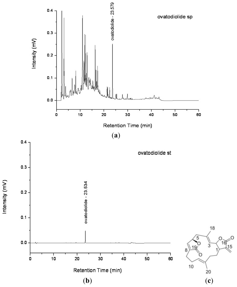
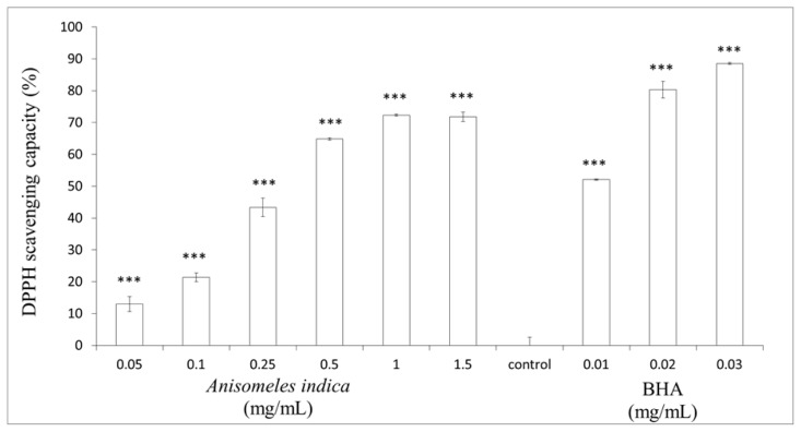
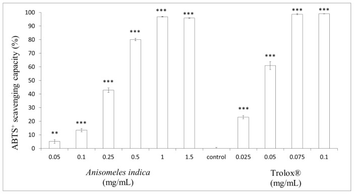
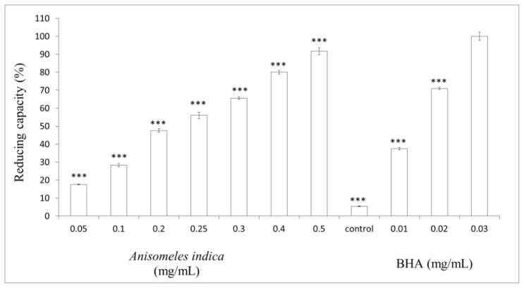
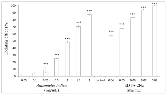
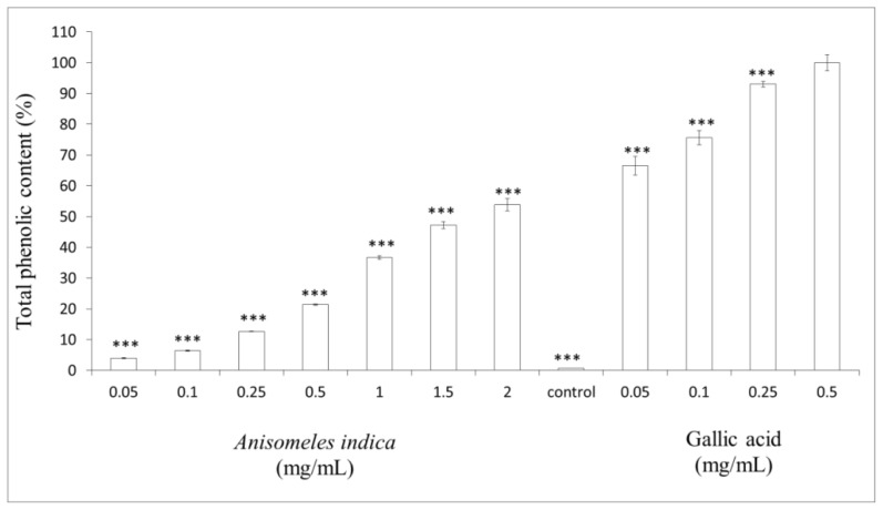
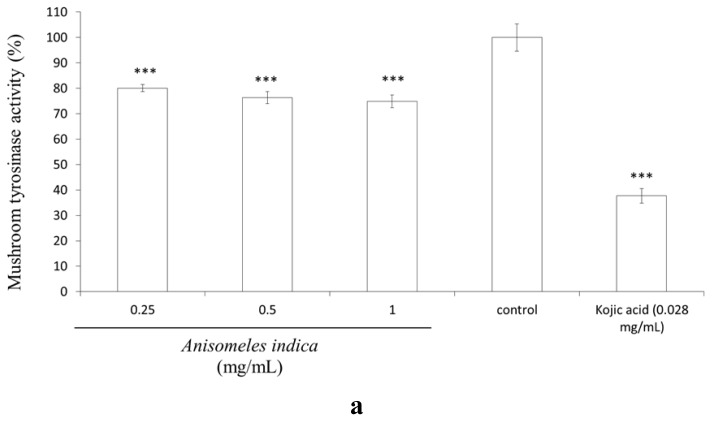
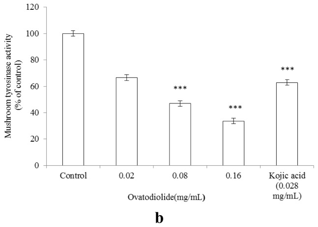
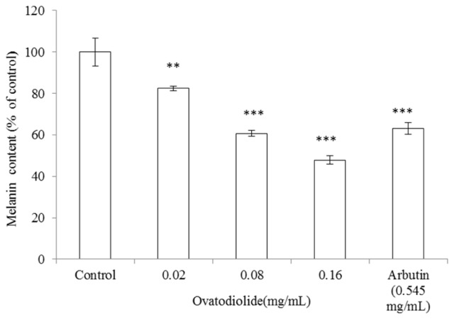
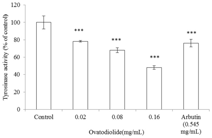
The purpose of the study was to investigate the antioxidant characteristics of Anisomeles indica methanol extract and the inhibitory effect of ovatodiolide on melanogenesis. In the study, the antioxidant capacities of A. indica methanol extract such as DPPH assay, ABTS radical scavenging assay, reducing capacity and metal ion chelating capacity as well as total phenolic content of the extract were investigated. In addition, the inhibitory effects of ovatodiolide on mushroom tyrosinase, B16F10 intracellular tyrosinase and melanin content were determined spectrophotometrically. Our results revealed that the antioxidant capacities of A. indica methanol extract increased in a dose-dependent pattern. The purified ovatodiolide inhibited mushroom tyrosinase activity (IC(50) = 0.253 mM), the compound also effectively suppressed intracellular tyrosinase activity (IC(50) = 0.469 mM) and decreased the amount of melanin (IC(50) = 0.435 mM) in a dose-dependent manner in B16F10 cells. Our results concluded that A. indica methanol extract displays antioxidant capacities and ovatodiolide purified from the extract inhibited melanogenesis in B16F10 cells. Hence, A. indica methanol extract and ovatodiolide could be applied as a type of dermatological whitening agent in skin care products.

Keywords: Anisomeles indica; antioxidant; melanin; melanogenesis; ovatodiolide; tyrosinase.


Figures

Figure 1
Figure 1
(a), (b) HPLC chromatogram and (c) chemical structure of ovatodiolide.
Figure 2
Figure 2
DPPH radical scavenging effects of Anisomeles indica methanol extract. Various concentrations of the extract (final concentration 0.05, 0.1, 0.25, 0.5, 1.0 and 1.5 mg/mL) or BHA (0.01, 0.02 and 0.03 mg/mL) were interacted with DPPH, respectively. Results are represented as percentages of control, and the data are mean ± SD for three separate experiments. Values are significantly different by comparison with control. *** p < 0.001.
Figure 3
Figure 3
ABTS+ radical scavenging effects of A. indica methanol extract. The extract (final concentration 0.05, 0.1, 0.25, 0.5, 1.0 and 1.5 mg/mL) or Trolox® (0.025, 0.05, 0.075 and 0.1 mg/mL) were interacted with ABTS. Results are represented as percentages of control, and the data are mean ± SD for three separate experiments. Values are significantly different by comparison with control. *** p < 0.001.
Figure 4
Figure 4
Reducing capacity of A. indica methanol extract. Different concentrations of the extract (0.05, 0.1, 0.2, 0.25, 0.3, 0.4 and 0.5 mg/mL) or BHA (0.01, 0.02, 0.03 mg/mL) were used in the assay. Results are represented as percentages of control, and the data are mean ± SD for three separate experiments. Values are significantly different by comparison with control. *** p < 0.001.
Figure 5
Figure 5
Metal-ion chelating activity of A. indica methanol extract. Different concentrations of the extract (0.05, 0.1, 0.25, 0.5, 1.0, 1.5 and 2.0 mg/mL) or EDTA (0.04–0.08 mg/mL) were used in the study. Results are represented as percentages of control, and the data are mean ± SD for three separate experiments. Values are significantly different by comparison with control. *** p < 0.001.
Figure 6
Figure 6
Determination of total phenolic content. Different concentrations of A. indica methanol extract (0.05, 0.1, 0.25, 0.5, 1.0, 1.5 and 2.0 mg/mL) and gallic acid (0.05, 0.1, 0.25, 0.5 mg/mL) were used in the assay. Results are represented as percentages of control, and the data are mean ± SD for three separate experiments. Values are significantly different by comparison with control. *** p < 0.001.
Figure 7
Figure 7
Inhibitory effect of A. indica methanol extract and ovatodiolide on mushroom tyrosinase activity. Different concentrations of (a) the methanol extract (0.25, 0.5, 1.0 mg/mL); (b) ovatodiolide (0.02, 0.08 and 0.16 mg/mL) or kojic acid (0.028 mg/mL) were incubated with the same units of mushroom tyrosinase. Results are represented as percentages of control, and data are presented as mean ± SD for three separate experiments. Values are significantly different by comparison with control. *** p < 0.001.
Figure 7
Figure 7
Inhibitory effect of A. indica methanol extract and ovatodiolide on mushroom tyrosinase activity. Different concentrations of (a) the methanol extract (0.25, 0.5, 1.0 mg/mL); (b) ovatodiolide (0.02, 0.08 and 0.16 mg/mL) or kojic acid (0.028 mg/mL) were incubated with the same units of mushroom tyrosinase. Results are represented as percentages of control, and data are presented as mean ± SD for three separate experiments. Values are significantly different by comparison with control. *** p < 0.001.
Figure 8
Figure 8
Effect of ovatodiolide on melanin synthesis in B16F10 cells. Melanin content assessment was performed as described in the “Experimental Section”. Briefly, cells were cultured with α-MSH (100 nM) for 24 h, and then the melanin content was measured after treatment with various concentrations of ovatodiolide (0.02, 0.08 and 0.16 mg/mL) or arbutin (0.545 mg/mL) for another 24 h. Results are represented as percentages of control, and data are presented as mean ± SD for three separate experiments. Values are significantly different by comparison with control. *** p < 0.001.
Figure 9
Figure 9
Effect of ovatodiolide on tyrosinase activity in B16F10 cells. Enzyme assay was performed as described in the “Experimental Section”. Briefly, B16F10 melanoma cells were stimulated with α-MSH (100 nM) for 24 h, and the cellular tyrosinase activity was assayed after treatment with ovatodiolide (0.02, 0.08 and 0.16 mg/mL) or arbutin (0.545 mg/mL) for another 24 h. Results are represented as percentages of control, and the data are mean ± SD for three separate experiments. Values are significantly different by comparison with control. *** p < 0.001.

Similar articles

Cited by

References

    1. Bergendi L., Benes L., Durackova Z., Ferencik M. Chemistry, physiology and pathology of free radicals. Life Sci. 1999;65:1865–1874. - PubMed
    1. Darr D., Fridovich I. Free radicals in cutaneous biology. J. Invest. Dermatol. 1994;102:671–675. - PubMed
    1. Young I.S., Woodside J.V. Antioxidants in health and disease. J. Clin. Pathol. 2001;54:176–186. - PMC - PubMed
    1. Sies H., Stahl W. Nutritional protection against skin damage from sunlight. Annu. Rev. Nutr. 2004;24:173–200. - PubMed
    1. Slominski A., Tobin D.J., Shibahara S., Wortsman J. Melanin pigmentation in mammalian skin and its hormonal regulation. Physiol. Rev. 2004;84:1155–1228. - PubMed

Publication types