. 2012 Jun 19:6:37.
doi: 10.3389/fnint.2012.00037. eCollection 2012.

Three-dimensional eye position signals shape both peripersonal space and arm movement activity in the medial posterior parietal cortex

Affiliations

Three-dimensional eye position signals shape both peripersonal space and arm movement activity in the medial posterior parietal cortex

K Hadjidimitrakis et al. Front Integr Neurosci. .

Abstract

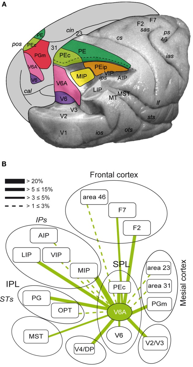
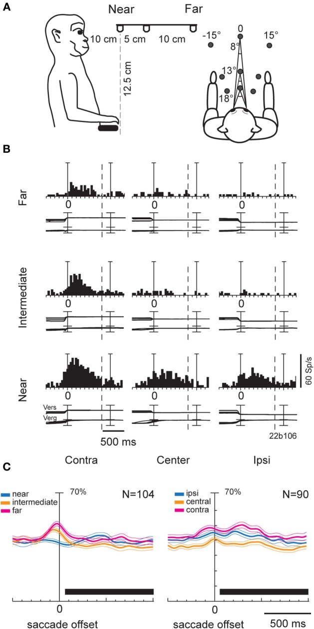
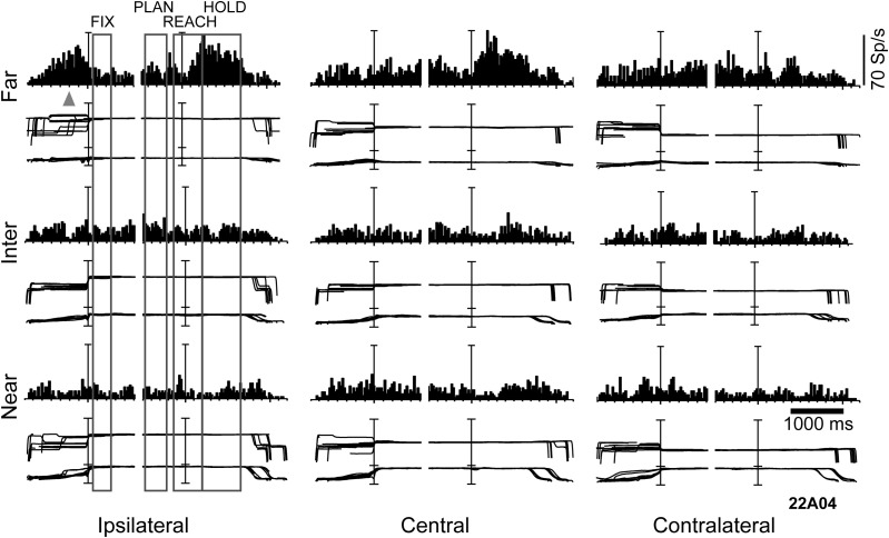
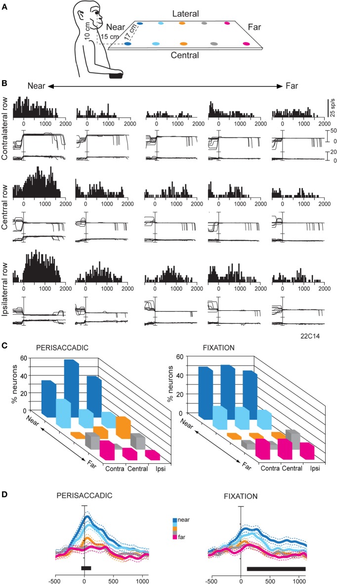
Research conducted over the last decades has established that the medial part of posterior parietal cortex (PPC) is crucial for controlling visually guided actions in human and non-human primates. Within this cortical sector there is area V6A, a crucial node of the parietofrontal network involved in arm movement control in both monkeys and humans. However, the encoding of action-in-depth by V6A cells had been not studied till recently. Recent neurophysiological studies show the existence in V6A neurons of signals related to the distance of targets from the eyes. These signals are integrated, often at the level of single cells, with information about the direction of gaze, thus encoding spatial location in 3D space. Moreover, 3D eye position signals seem to be further exploited at two additional levels of neural processing: (a) in determining whether targets are located in the peripersonal space or not, and (b) in shaping the spatial tuning of arm movement related activity toward reachable targets. These findings are in line with studies in putative homolog regions in humans and together point to a role of medial PPC in encoding both the vergence angle of the eyes and peripersonal space. Besides its role in spatial encoding also in depth, several findings demonstrate the involvement of this cortical sector in non-spatial processes.

Keywords: eye-hand coordination; fixation depth; gaze; reaching; sensorimotor transformation; vergence; version.


Figures

Figure 1
Figure 1
(A) Location and extent of the areas that form the superior parietal cortex of the macaque brain. Posterolateral view of a partially dissected macaque brain modified from Galletti et al. (1996). The occipital pole has been partially removed and inferior parietal lobule of the right hemisphere has been cut away at the level of the fundus of the intraparietal sulcus to show the cortex of the medial bank of this sulcus. The occipital lobe of the same hemisphere has been cut away at the level of the fundus of the parieto-occipital and lunate sulci to show the cortex of the anterior bank of the parieto-occipital sulcus. The medial surface of the left hemisphere is drawn to show the location on it of all areas that extend medially. pos, parieto-occipital sulcus; cal, calcarine sulcus; cin, cingulate sulcus; ips, intraparietal sulcus; ios, inferior-occipital sulcus; ots, occipitotemporal sulcus; sts, superior temporal sulcus; lf, lateral fissure; cs, central sulcus; sas, superior arcuate sulcus; ias, inferior arcuate sulcus; ps, principal sulcus; V6, area V6; V3, area V3; V2, area V2; V1, area V1; PEc, area caudal PE; PE, area PE; PEip, intraparietal area PE; MIP, medial intraparietal area, PGm, area medial PG; VIP, ventral intraparietal area; LIP, lateral intraparietal area; AIP, anterior intraparietal area; MT, middle temporal area; MST, medial superior temporal area; Brodmann's areas 23, 31, 46, and dorsal premotor areas F2 and F7 in are also shown. (B) Flow chart of the connections of V6A modified from Passarelli et al. (2011). Rostral/caudal brain areas are shown at the top/bottom part of the figure. The thickness of the lines is proportional to the strength of each connection. Areas in the ventral part of the parieto-occipital sulcus (V6, ventral V6A) are dominated by visual input, whereas as one proceeds toward the dorsal part of V6A sensory association and visuomotor/premotor connections prevail. Inferior parietal lobule areas Opt and PG and occipital areas V4/DP are also shown.
Figure 2
Figure 2
(A) Scheme of the experimental setup set up used for the fixation-in-depth and reaching-in-depth tasks. Exact distances and angles of the targets are indicated in the lateral view (left) and top view (right), respectively. (B) Example of a V6A neuron modulated by both version and vergence during fixation. The discharge to the nine LEDs arranged from near (bottom) to far (top) was aligned twice (at the start of the fixation and at the LED change; dashed line: point where trials were cut because of double alignment). From left to right, the behavioral events are: LED onset, saccade offset (first alignment marker), LED change (second alignment marker). The cell has a clear preference for the near-contralateral space. Bin size for spike histograms: 20 ms; scale for version and vergence traces is 100 and 20°, respectively. (C) Population activity of V6A neurons modulated during fixation. Population average spike density functions (SDF) were constructed by ranking the response for each tested row of fixation targets. The thick lines indicate average normalized SDF; the light lines indicate variability bands (SEM). The peak of the SDF of the row showing the maximum activity was set to 1 (100%) and was used to normalize the SDF curves of the other rows. Activity is aligned with the offset of the saccade. The black rectangles below each plot indicate the periods where the permutation test was run. In the left plot, no statistical difference was observed between the curves (permutation test, p > 0.05), whereas in the right, the central row is statistically different from the other two (permutation test, p < 0.05). Scale on abscissa: 100 ms/division; vertical scale: 70% of normalized activity (10% per division). (A–C) panels were modified from Breveglieri et al. (2012).
Figure 3
Figure 3
Example of a V6A neuron modulated by depth and direction signals during several epochs of a reaching-in-depth task. Target LEDs were arranged as in Figure 2B. Spike histograms and eye traces were aligned twice, at the fixation onset and at the start of movement. This cell prefers far space during movement planning, execution and holding of the target epochs. In the last two epochs it was also tuned for ipsilateral space. The gray triangle indicates the mean time of LED onset. Bin size for spike histograms; 20 ms; scale for version and vergence traces is 100 and 20°, respectively.
Figure 4
Figure 4
(A) Scheme of the set up used for the fixation-in-depth task below eye level. The monkey was fixating in darkness one of the LEDs embedded in each panel. The LEDs are depicted with different colors according to their distance from a frontal plane passing from monkey's eyes. (B) Example of a V6A neuron modulated by vergence during fixation below eye level. Top/Middle/Bottom: neural responses (peristimulus time histograms) and eye traces to the five LEDs of the contralateral/central/ipsilateral space arranged from near (left) to far (right), aligned at the end of the saccade. This cell prefers targets located at near and ipsilateral space. Other conventions as in Figure 3. (C) Frequency histogram of the positions that neurons preferred in perisaccadic (N = 91) and fixation (N = 167) epochs. Ipsi and Contra indicate fixation position with respect to the recording hemisphere. Center refers to the straight ahead of the monkey. In both epochs there is a clear preference for near, reachable targets across all space. (D) Population activity for each target position, illustrated with a different color, of V6A neurons with activity modulated during perisaccadic (left) and fixation (right) epochs. Activity is aligned on the saccade onset in both panels. Other conventions as in Figure 2C. (A–D): Adapted from Hadjidimitrakis et al. (2011b).
Figure 5
Figure 5
(A) Experimental set-up and time course of a frontal reaching task. Reaching movements were performed in darkness, from a home-button (black rectangle) toward one out of nine targets (open circles) located on a panel in front of the animal. (B) Incidence of V6A cells spatially modulated in the reaching task. Columns indicate the percentages of spatially modulated V6A cells during outward reaching movements (M1), and static position of the arm in the peripersonal space (HOLD). “Proper” activity = “raw” activity – FIX activity. (C) Example of spatially tuned modulation of reach-related activity. Neuron spatially tuned in M1, preferring rightward M1 movements. Neural activity and eye-traces have been aligned twice in each inset, with the onsets of outward (1st) and inward (2nd) reach movements. The mean duration of epochs FIX, M1, HOLD, M2 is indicated in the bottom left inset. Behavioral markers on rasters from left to right: LED color change, outward movement onset, outward movement end, LED offset, inward movement onset, inward movement end, end of trial. Bin size in peri-event time histograms: 15 ms; eye traces: scale bar, 60°. Other details as in Figure 2. Modified from Fattori et al. (2005).

Similar articles

Cited by

References

    1. Ahissar E., Vaadia E., Ahissar M., Bergman H., Arieli A., Abeles M. (1992). Dependence of cortical plasticity on correlated activity of single neurons and on behavioral context. Science 257, 1412–1415 10.1126/science.1529342 - DOI - PubMed
    1. Akao T., Kurkin S. A., Fukushima J., Fukushima K. (2005). Visual and vergence eye movement-related responses of pursuit neurons in the caudal frontal eye fields to motion-in-depth stimuli. Exp. Brain Res. 164, 92–108 10.1007/s00221-004-2213-6 - DOI - PubMed
    1. Andersen R. A., Bracewell R. M., Barash S., Gnadt J. W., Fogassi L. (1990). Eye position effects on visual, memory, and saccade-related activity in areas LIP and 7a of macaque. J. Neurosci. 10, 1176–1196 - PMC - PubMed
    1. Andersen R. A., Cui H. (2009). Intention, action planning, and decision making in parietal-frontal circuits. Neuron 63, 568–583 10.1016/j.neuron.2009.08.028 - DOI - PubMed
    1. Andersen R. A., Mountcastle V. B. (1983). The influence of the angle of gaze upon the excitability of the light-sensitive neurons of the posterior parietal cortex. J. Neurosci. 3, 532–548 - PMC - PubMed

LinkOut - more resources