

Neural activation differences in amputees during imitation of intact versus amputee movements
- PMID: 22754516
- PMCID: PMC3386563
- DOI: 10.3389/fnhum.2012.00182
Neural activation differences in amputees during imitation of intact versus amputee movements
Abstract
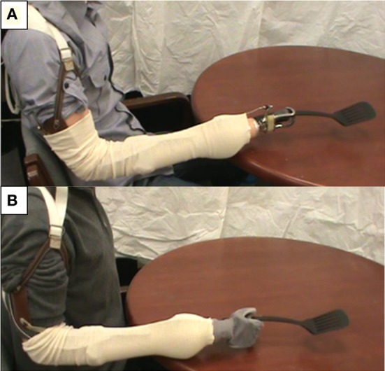
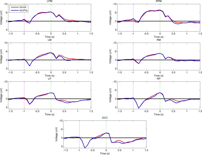
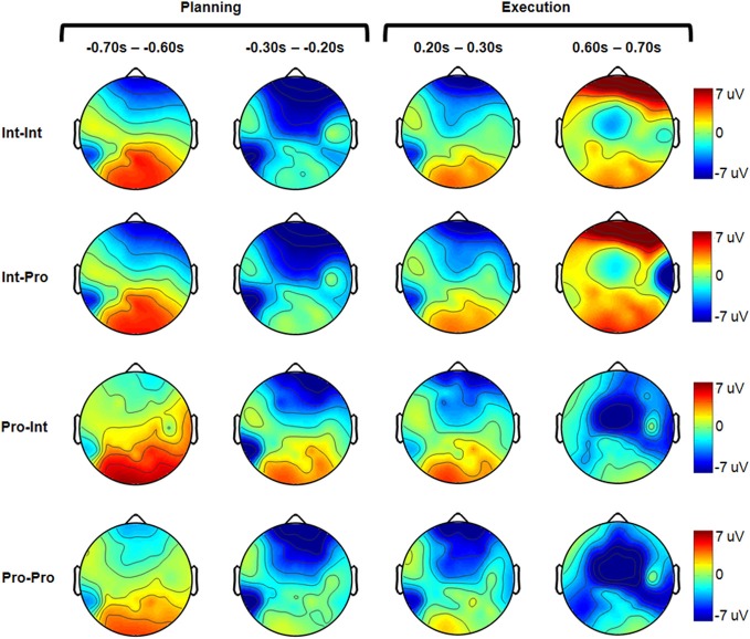
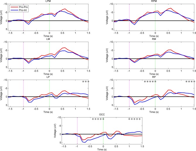
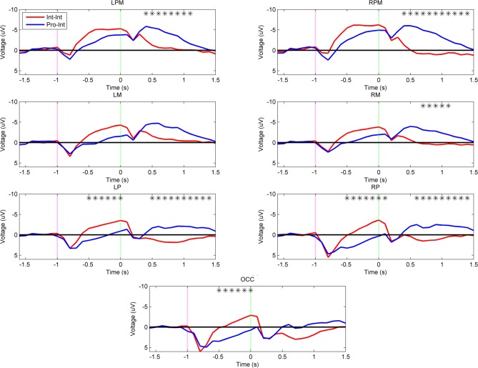
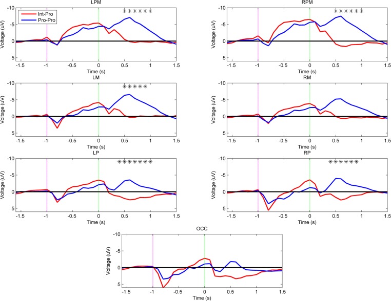
The mirror neuron system (MNS) has been attributed with increased activation in motor-related cortical areas upon viewing of another's actions. Recent work suggests that limb movements that are similar and dissimilar in appearance to that of the viewer equivalently activate the MNS. It is unclear if this result can be observed in the action encoding areas in amputees who use prosthetic devices. Intact subjects and upper extremity amputee prosthesis users were recruited to view video demonstrations of tools being used by an intact actor and a prosthetic device user. All subjects pantomimed the movements seen in the video while recording electroencephalography (EEG). Intact subjects showed equivalent left parietofrontal activity during imitation planning after watching the intact or prosthetic arm. Likewise, when prosthesis users imitated prosthesis demonstrations, typical left parietofrontal activation was observed. When prosthesis users imitated intact actors, an additional pattern was revealed which showed greater activity in right parietal and occipital regions that are associated with the mentalizing system. This change may be required for prosthesis users to plan imitation movements in which the limb states between the observed and the observer do not match. The finding that prosthesis users imitating other prosthesis users showed typical left parietofrontal activation suggests that these subjects engage normal planning related activity when they are able to imitate a limb matching their own. This result has significant implications on rehabilitation, as standard therapy involves training with an intact occupational therapist, which could necessitate atypical planning mechanisms in amputees when learning to use their prosthesis.
Keywords: EEG; amputee; mentalizing; mirror neuron; motor control; prosthesis; tool use; upper extremity.
Figures
Similar articles
-
Influence of Perspective of Action Observation Training on Residual Limb Control in Naïve Prosthesis Usage.J Mot Behav. 2016 Sep-Oct;48(5):446-54. doi: 10.1080/00222895.2015.1134432. Epub 2016 Jun 2. J Mot Behav. 2016. PMID: 27253208 Free PMC article.
-
Motor performance benefits of matched limb imitation in prosthesis users.Exp Brain Res. 2014 Jul;232(7):2143-54. doi: 10.1007/s00221-014-3904-2. Epub 2014 Mar 19. Exp Brain Res. 2014. PMID: 24643547
-
Enhanced Neurobehavioral Outcomes of Action Observation Prosthesis Training.Neurorehabil Neural Repair. 2016 Jul;30(6):573-82. doi: 10.1177/1545968315606992. Epub 2015 Oct 5. Neurorehabil Neural Repair. 2016. PMID: 26438442 Free PMC article.
-
Adaptations from the prosthetic and intact limb during standing on a sway-referenced support surface for transtibial prosthesis users.Disabil Rehabil Assist Technol. 2019 Oct;14(7):682-691. doi: 10.1080/17483107.2018.1498925. Epub 2018 Nov 8. Disabil Rehabil Assist Technol. 2019. PMID: 30409065
-
Outcome Measures Used to Assess Hand Activity in Amputee and Intact Populations: a Literature Review.Can Prosthet Orthot J. 2022 Dec 25;5(2):39023. doi: 10.33137/cpoj.v5i2.39023. eCollection 2022. Can Prosthet Orthot J. 2022. PMID: 37614636 Free PMC article. Review.
Cited by
-
A Structured Rehabilitation Protocol for Improved Multifunctional Prosthetic Control: A Case Study.J Vis Exp. 2015 Nov 6;(105):e52968. doi: 10.3791/52968. J Vis Exp. 2015. PMID: 26575620 Free PMC article.
-
Influence of Perspective of Action Observation Training on Residual Limb Control in Naïve Prosthesis Usage.J Mot Behav. 2016 Sep-Oct;48(5):446-54. doi: 10.1080/00222895.2015.1134432. Epub 2016 Jun 2. J Mot Behav. 2016. PMID: 27253208 Free PMC article.
-
Motor performance benefits of matched limb imitation in prosthesis users.Exp Brain Res. 2014 Jul;232(7):2143-54. doi: 10.1007/s00221-014-3904-2. Epub 2014 Mar 19. Exp Brain Res. 2014. PMID: 24643547
-
Virtual reality-based action observation facilitates the acquisition of body-powered prosthetic control skills.J Neuroeng Rehabil. 2020 Aug 20;17(1):113. doi: 10.1186/s12984-020-00743-w. J Neuroeng Rehabil. 2020. PMID: 32819412 Free PMC article.
-
Emergence of perceptuomotor relationships during paleolithic stone toolmaking learning: intersections of observation and practice.Commun Biol. 2021 Nov 11;4(1):1278. doi: 10.1038/s42003-021-02768-w. Commun Biol. 2021. PMID: 34764417 Free PMC article.
References
Grants and funding
LinkOut - more resources
Full Text Sources