

Plasmacytoid dendritic cells in atherosclerosis
- PMID: 22754539
- PMCID: PMC3385355
- DOI: 10.3389/fphys.2012.00230
Plasmacytoid dendritic cells in atherosclerosis
Abstract
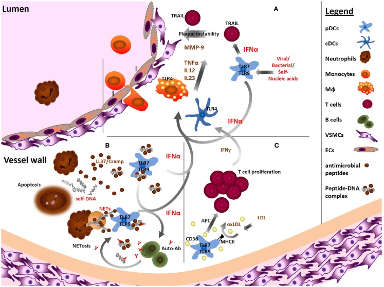
Atherosclerosis, a chronic inflammatory disease of the vessel wall and the underlying cause of cardiovascular disease, is initiated and maintained by innate and adaptive immunity. Accumulating evidence suggests an important contribution of autoimmune responses to this disease. Plasmacytoid dendritic cells (pDCs), a specialized cell type known to produce large amounts of type I interferons (IFNs) in response to bacterial and viral infections, have recently been revealed to play important roles in atherosclerosis. For example, the development of autoimmune complexes consisting of self-DNA and antimicrobial peptides, which trigger chronic type I IFN production by pDCs, promote early atherosclerotic lesion formation. pDCs and pDC-derived type I IFNs can also induce the maturation of conventional DCs and macrophages, and the development of autoreactive B cells and antibody production. These mechanisms, known to play a role in the pathogenesis of other autoimmune diseases such as systemic lupus erythematosus and psoriasis, may also affect the development and progression of atherosclerotic lesion formation. This review discusses emerging evidence showing a contribution of pDCs in the onset and progression of atherosclerosis.
Keywords: atherosclerosis; plasmacytoid dendritic cells; type I IFN.
Figures
Similar articles
-
Sequential activation of conventional and plasmacytoid dendritic cells in autoimmune pancreatitis and systemic lupus erythematosus: similarities and dissimilarities.Front Immunol. 2025 Feb 18;16:1554492. doi: 10.3389/fimmu.2025.1554492. eCollection 2025. Front Immunol. 2025. PMID: 40040712 Free PMC article. Review.
-
The role of plasmacytoid dendritic cells (pDCs) in immunity during viral infections and beyond.Cell Mol Immunol. 2024 Sep;21(9):1008-1035. doi: 10.1038/s41423-024-01167-5. Epub 2024 May 22. Cell Mol Immunol. 2024. PMID: 38777879 Free PMC article. Review.
-
Disease-Associated Plasmacytoid Dendritic Cells.Front Immunol. 2017 Oct 16;8:1268. doi: 10.3389/fimmu.2017.01268. eCollection 2017. Front Immunol. 2017. PMID: 29085361 Free PMC article. Review.
-
Auto-antigenic protein-DNA complexes stimulate plasmacytoid dendritic cells to promote atherosclerosis.Circulation. 2012 Apr 3;125(13):1673-83. doi: 10.1161/CIRCULATIONAHA.111.046755. Epub 2012 Mar 2. Circulation. 2012. PMID: 22388324
-
Update on the role of plasmacytoid dendritic cells in inflammatory/autoimmune skin diseases.Exp Dermatol. 2016 Jun;25(6):415-21. doi: 10.1111/exd.12957. Epub 2016 Apr 12. Exp Dermatol. 2016. PMID: 26837058 Review.
Cited by
-
Anti-atherogenic peptide Ep1.B derived from apolipoprotein E induces tolerogenic plasmacytoid dendritic cells.Clin Exp Immunol. 2014 Sep;177(3):732-42. doi: 10.1111/cei.12370. Clin Exp Immunol. 2014. PMID: 24784480 Free PMC article.
-
Neuroimmunology of the atherosclerotic plaque: a morphological approach.J Neuroimmune Pharmacol. 2013 Mar;8(1):15-27. doi: 10.1007/s11481-012-9421-9. Epub 2012 Nov 14. J Neuroimmune Pharmacol. 2013. PMID: 23150034 Review.
-
Neutrophil extracellular traps: a walk on the wild side of exercise immunology.Sports Med. 2015 May;45(5):625-40. doi: 10.1007/s40279-014-0296-1. Sports Med. 2015. PMID: 25504501 Review.
-
Dendritic cells immunotargeted therapy for atherosclerosis.Acta Pharm Sin B. 2025 Feb;15(2):792-808. doi: 10.1016/j.apsb.2024.12.029. Epub 2024 Dec 31. Acta Pharm Sin B. 2025. PMID: 40177571 Free PMC article. Review.
-
Evaluation of the BDCA2-DTR Transgenic Mouse Model in Chronic and Acute Inflammation.PLoS One. 2015 Aug 7;10(8):e0134176. doi: 10.1371/journal.pone.0134176. eCollection 2015. PLoS One. 2015. PMID: 26252890 Free PMC article.
References
-
- Ait-Oufella H., Salomon B. L., Potteaux S., Robertson A. K., Gourdy P., Zoll J., Merval R., Esposito B., Cohen J. L., Fisson S., Flavell R. A., Hansson G. K., Klatzmann D., Tedgui A., Mallat Z. (2006). Natural regulatory T cells control the development of atherosclerosis in mice. Nat. Med. 12, 178–18010.1038/nm1343 - DOI - PubMed
-
- Asselin-Paturel C., Brizard G., Pin J. J., Briere F., Trinchieri G. (2003). Mouse strain differences in plasmacytoid dendritic cell frequency and function revealed by a novel monoclonal antibody. J. Immunol. 171, 6466–6477 - PubMed
LinkOut - more resources
Full Text Sources