. 2012:2012:894815.
doi: 10.1155/2012/894815. Epub 2012 Jun 13.

In vivo assessment of osseous wound healing using a novel bone putty containing lidocaine in the surgical management of tooth extractions

Affiliations

In vivo assessment of osseous wound healing using a novel bone putty containing lidocaine in the surgical management of tooth extractions

Akshay Kumarswamy et al. Int J Dent. 2012.

Abstract

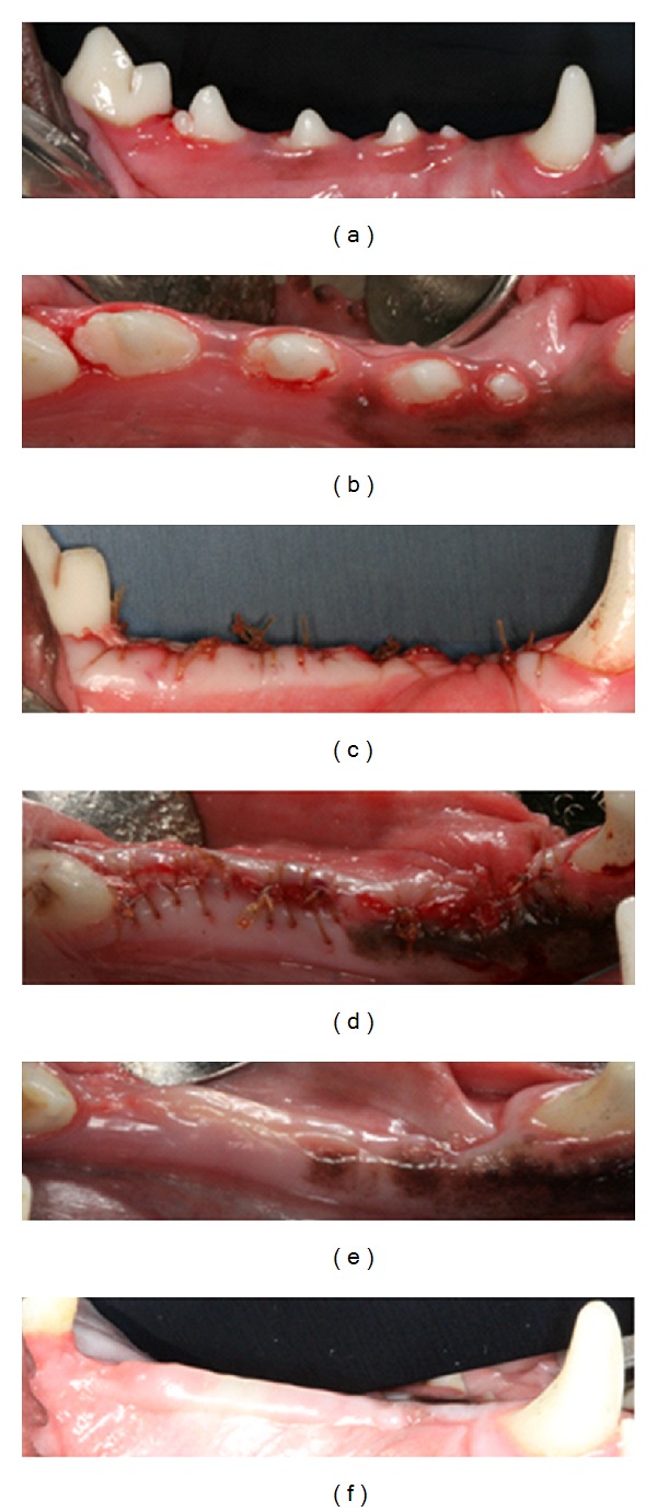
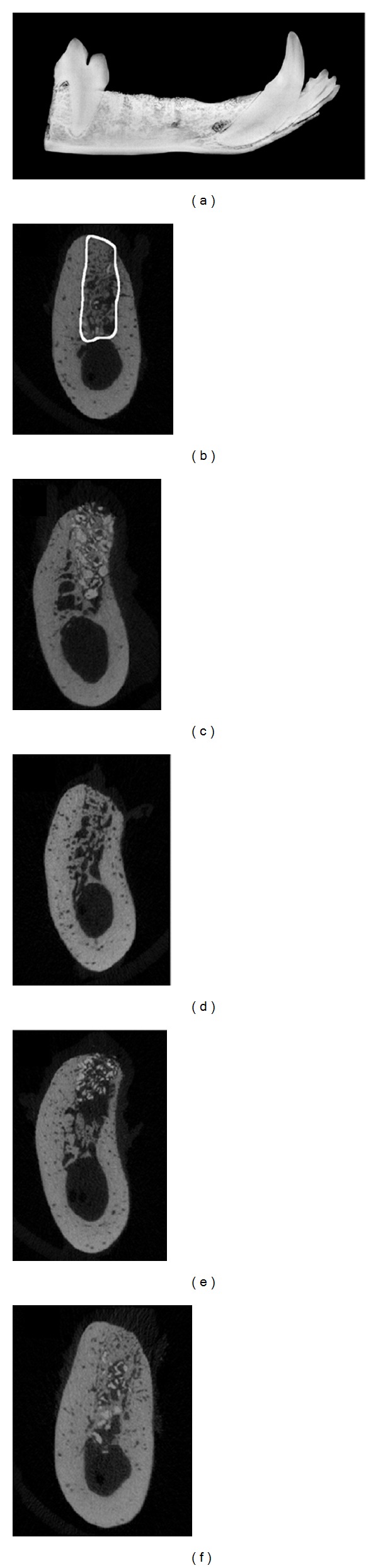
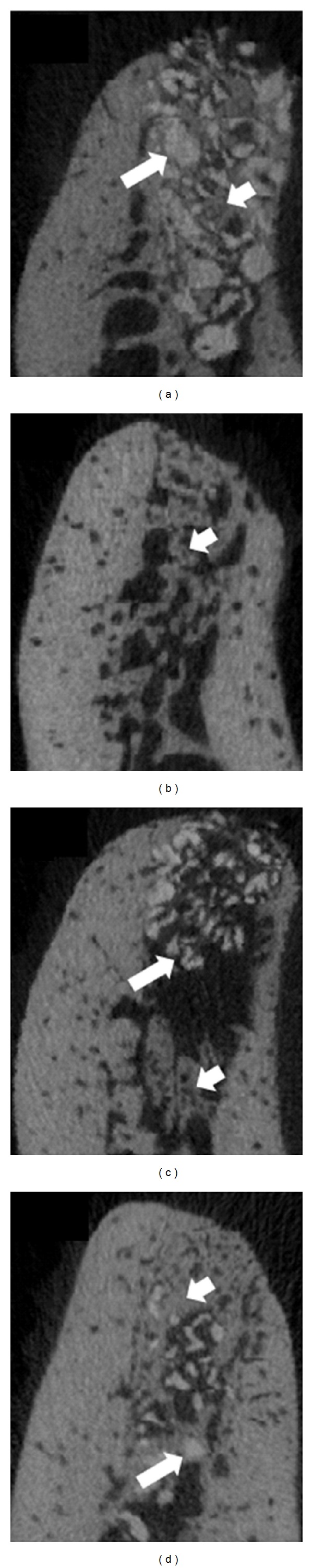
Objective. This preclinical pilot study evaluated the systemic, radiographic, and histological responses to bone putty containing lidocaine in a canine tooth extraction model. Methods. In five beagle dogs the right mandibular premolars were extracted and sockets grafted with (1) xenograft particulate bone and a collagen sponge plug (control), (2) bone putty alone, (3) bone putty mixed with xenograft (3 : 1), or (4) xenograft sandwiched between bone putty. At 6 weeks post-op, the systemic and local responses were evaluated using a blood chemistry panel, micro-CT, and histological analyses. Results. No significant differences in blood chemistries were noted at 6 weeks postgrafting compared to baseline. Sockets grafted with either bone putty formulation demonstrated comparable radiographic and histologic evidence of bone healing compared to control sockets. Conclusions. Our preclinical results indicate that this bone putty appears to be a safe biocompatible device that may be useful in the postoperative management of tooth extractions.


Figures

Figure 1
Figure 1
Representative images of preextraction (a) and (b), immediate postextraction (c) and (d), and six-week postextraction healing (e) and (f). Complete closure of extraction sockets and lack of clinical signs of inflammation are evident.
Figure 2
Figure 2
Representative image of a conventional mandibular radiograph (a) and μCT slices (b–f) within the middle of the distal root, 4th premolar extraction sockets after the six-week healing time. (b) μCT image of an extraction socket showing the region of interest analyzed for BV/TV and BMD. (c) Bovine xenograft particulate and collagen plug (X), (d) bone putty only (Bp), (e) xenograft particles sandwiched between layers of bone putty placed in the coronal and apical aspect, (Bp-X-Bp), and (f) a 3 : 1 mixture by weight of bone putty and xenograft particles, respectively (3Bp-X). Residual xenograft particles (appearing denser than the surround bone) can be seen in within the extraction sockets in panels (c), (e), and (f).
Figure 3
Figure 3
Enlarged representative images of μCT slices within the middle of the distal root, 4th premolar extraction sockets after the six-week healing time. (a) Bovine xenograft particulate and collagen plug (X), (b) bone putty only (Bp), (c) xenograft particles sandwiched between layers of bone putty placed in the coronal and apical aspect (Bp-X-Bp), and (d) a 3 : 1 mixture by weight of bone putty and xenograft particles, respectively (3Bp-X). Residual xenograft particles (appearing denser than the surround bone) can be seen in within the extraction sockets in panels (a), (c), and (d) (long arrows) interspersed between newly formed bone (short arrows, (a)–(d)).
Figure 4
Figure 4
Representative hematoxylin-eosin ((a)–(d)) and Gomori trichrome stain ((e)–(h)) histological images of extraction sockets from the 4th premolar after six weeks of healing time. (a) and (e), bovine xenograft particulate and collagen plug (X), (b) and (f), bone putty only (Bp), (c) and (g), xenograft particles sandwiched between layers of bone putty placed in the coronal and apical aspect, (Bp-X-Bp), and (d) and (h), a 3 : 1 mixture by weight of bone putty and xenograft particles, respectively (3Bp-X). Arrows highlight newly formed bone between xenograft particles or in Bp only grafted sockets (b) and (f). Scale bar = 400 microns, 20x magnification.
Figure 5
Figure 5
Representative hematoxylin-eosin (a)–(d) images of midsockets from the 4th premolar at six weeks following extractions and material placements. (a) bovine xenograft particulate and collagen plug (X), (b) bone putty only (Bp), (c) xenograft particles sandwiched between layers of bone putty placed in the coronal and apical aspect, (Bp-X-Bp), and (d) a 3 : 1 mixture by weight of bone putty and xenograft particles, respectively (3Bp-X). Arrows highlight newly formed bone between xenograft particles or in Bp only grafted sockets (b). Scale bar = 200 microns, 40x magnification.

Similar articles

Cited by

References

    1. Malamed SF. Local anesthetics: dentistry’s most important drugs, clinical update 2006. Journal of the California Dental Association. 2006;34(12):971–976. - PubMed
    1. Bouloux GF, Steed MB, Perciaccante VJ. Complications of third molar surgery. Oral and Maxillofacial Surgery Clinics of North America. 2007;19(1):117–128. - PubMed
    1. Scully C. Medical Problems in Dentistry. London, UK: Churchill Livingstone Elsevier; 2010. Haematology; pp. 177–233.
    1. Seymour AR, Meechan JG, Yates SM. Pharmacology and Dental Therapeutics. New York, NY, USA: Oxford University Press; 1999. Aspirin, other non-steroidal anti-inflammatory drugs, and paracetamol; pp. 89–94.
    1. Fischer LM, Schlienger RG, Matter CM, Jick H, Meier CR. Discontinuation of nonsteroidal anti-inflammatory drug therapy and risk of acute myocardial infarction. Archives of Internal Medicine. 2004;164(22):2472–2476. - PubMed

LinkOut - more resources