

Homeostasis of the apical plasma membrane during regulated exocytosis in the salivary glands of live rodents
- PMID: 22754613
- PMCID: PMC3384574
- DOI: 10.4161/bioa.18405
Homeostasis of the apical plasma membrane during regulated exocytosis in the salivary glands of live rodents
Abstract
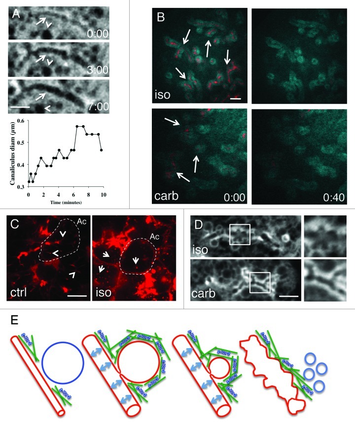
In exocrine organs such as the salivary glands, fluids and proteins are secreted into ductal structures by distinct mechanisms that are tightly coupled. In the acinar cells, the major secretory units of the salivary glands, fluids are secreted into the acinar canaliculi through paracellular and intracellular transport, whereas proteins are stored in large granules that undergo exocytosis and fuse with the apical plasma membranes releasing their content into the canaliculi. Both secretory processes elicit a remodeling of the apical plasma membrane that has not been fully addressed in in vitro or ex vivo models. Recently, we have studied regulated exocytosis in the salivary glands of live rodents, focusing on the role that actin and myosin plays in this process. We observed that during exocytosis both secretory granules and canaliculi are subjected to the hydrostatic pressure generated by fluid secretion. Furthermore, the absorption of the membranes of the secretory granules contributes to the expansion and deformation of the canaliculi. Here we suggest that the homeostasis of the apical plasma membranes during exocytosis is maintained by various strategies that include: (1) membrane retrieval via compensatory endocytosis, (2) increase of the surface area via membrane folds and (3) recruitment of a functional actomyosin complex. Our observations underscore the important relationship between tissue architecture and cellular response, and highlight the potential of investigating biological processes in vivo by using intravital microscopy.
Figures

Comment in
- Masedunskas A, Sramkova M, Parente L, Sales KU, Amornphimoltham P, Bugge TH, Weigert R. Role for the actomyosin complex in regulated exocytosis revealed by intravital microscopy. Proc Natl Acad Sci U S A. 2011;108:13552–7. doi: 10.1073/pnas.1016778108.
Similar articles
-
The Actomyosin Cytoskeleton Drives Micron-Scale Membrane Remodeling In Vivo Via the Generation of Mechanical Forces to Balance Membrane Tension Gradients.Bioessays. 2018 Sep;40(9):e1800032. doi: 10.1002/bies.201800032. Epub 2018 Aug 6. Bioessays. 2018. PMID: 30080263 Free PMC article. Review.
-
Linking differences in membrane tension with the requirement for a contractile actomyosin scaffold during exocytosis in salivary glands.Commun Integr Biol. 2012 Jan 1;5(1):84-7. doi: 10.4161/cib.18258. Commun Integr Biol. 2012. PMID: 22482019 Free PMC article.
-
Exocytosis by vesicle crumpling maintains apical membrane homeostasis during exocrine secretion.Dev Cell. 2021 Jun 7;56(11):1603-1616.e6. doi: 10.1016/j.devcel.2021.05.004. Dev Cell. 2021. PMID: 34102104 Free PMC article.
-
Role for the actomyosin complex in regulated exocytosis revealed by intravital microscopy.Proc Natl Acad Sci U S A. 2011 Aug 16;108(33):13552-7. doi: 10.1073/pnas.1016778108. Epub 2011 Aug 1. Proc Natl Acad Sci U S A. 2011. PMID: 21808006 Free PMC article.
-
Molecular Regulatory Mechanism of Exocytosis in the Salivary Glands.Int J Mol Sci. 2018 Oct 17;19(10):3208. doi: 10.3390/ijms19103208. Int J Mol Sci. 2018. PMID: 30336591 Free PMC article. Review.
Cited by
-
Multiple roles for the actin cytoskeleton during regulated exocytosis.Cell Mol Life Sci. 2013 Jun;70(12):2099-121. doi: 10.1007/s00018-012-1156-5. Epub 2012 Sep 18. Cell Mol Life Sci. 2013. PMID: 22986507 Free PMC article. Review.
-
Intravital microscopy: a practical guide on imaging intracellular structures in live animals.Bioarchitecture. 2012 Sep-Oct;2(5):143-57. doi: 10.4161/bioa.21758. Epub 2012 Sep 1. Bioarchitecture. 2012. PMID: 22992750 Free PMC article. Review.
-
The Actomyosin Cytoskeleton Drives Micron-Scale Membrane Remodeling In Vivo Via the Generation of Mechanical Forces to Balance Membrane Tension Gradients.Bioessays. 2018 Sep;40(9):e1800032. doi: 10.1002/bies.201800032. Epub 2018 Aug 6. Bioessays. 2018. PMID: 30080263 Free PMC article. Review.
-
Polyethylenimine-mediated expression of transgenes in the acinar cells of rats salivary glands in vivo.Front Cell Dev Biol. 2015 Jan 9;2:74. doi: 10.3389/fcell.2014.00074. eCollection 2014. Front Cell Dev Biol. 2015. PMID: 25621283 Free PMC article.
-
RNA-seq based transcriptomic map reveals new insights into mouse salivary gland development and maturation.BMC Genomics. 2016 Nov 16;17(1):923. doi: 10.1186/s12864-016-3228-7. BMC Genomics. 2016. PMID: 27852218 Free PMC article.
References
LinkOut - more resources
Full Text Sources