

Refilins: A link between perinuclear actin bundle dynamics and mechanosensing signaling
- PMID: 22754617
- PMCID: PMC3384578
- DOI: 10.4161/bioa.18246
Refilins: A link between perinuclear actin bundle dynamics and mechanosensing signaling
Abstract
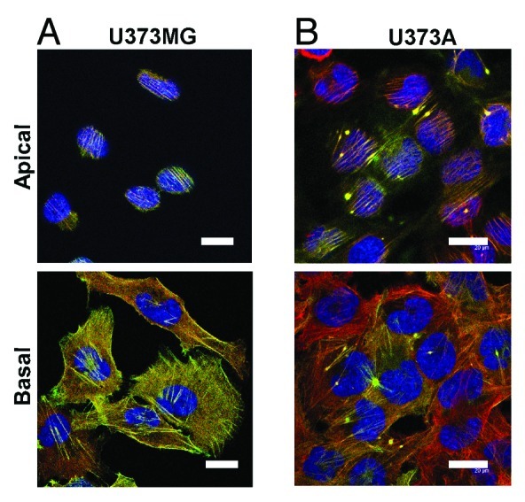
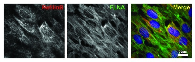
Actin cytoskeleton dynamics lie at the heart of cell mechanosensing signaling. In fibroblast cells, two perinuclear acto-myosin structures, the actin cap and the transmembrane actin-associated nuclear (TAN) line, are components of a physical pathway transducing extracellular physical signals to changes in nuclear shape and movements. We recently demonstrated the existence of a previously uncharacterized third apical perinuclear actin organization in epithelial cells that forms during epithelial-mesenchymal transition (EMT) mediated by TGFβ (TGFβ). A common regulatory mechanism for these different perinuclear actin architectures has emerged with the identification of a novel family of actin bundling proteins, the Refilins. Here we provide updates on some characteristics of Refilin proteins, and we discuss potential function of the Refilins in cell mechanosensing signaling.
Figures

Similar articles
-
Refilins are short-lived Actin-bundling proteins that regulate lamellipodium protrusion dynamics.Biol Open. 2016 Oct 15;5(10):1351-1361. doi: 10.1242/bio.019588. Biol Open. 2016. PMID: 27744291 Free PMC article.
-
Refilin holds the cap.Commun Integr Biol. 2011 Nov 1;4(6):791-5. doi: 10.4161/cib.17911. Commun Integr Biol. 2011. PMID: 22446558 Free PMC article.
-
RefilinB (FAM101B) targets filamin A to organize perinuclear actin networks and regulates nuclear shape.Proc Natl Acad Sci U S A. 2011 Jul 12;108(28):11464-9. doi: 10.1073/pnas.1104211108. Epub 2011 Jun 27. Proc Natl Acad Sci U S A. 2011. PMID: 21709252 Free PMC article.
-
The filamin-B-refilin axis - spatiotemporal regulators of the actin-cytoskeleton in development and disease.J Cell Sci. 2018 Apr 13;131(8):jcs213959. doi: 10.1242/jcs.213959. J Cell Sci. 2018. PMID: 29654161 Review.
-
The assembly and function of perinuclear actin cap in migrating cells.Protoplasma. 2017 May;254(3):1207-1218. doi: 10.1007/s00709-017-1077-0. Epub 2017 Jan 18. Protoplasma. 2017. PMID: 28101692 Review.
Cited by
-
Role of cellular cytoskeleton in epithelial-mesenchymal transition process during cancer progression.Biomed Rep. 2015 Sep;3(5):603-610. doi: 10.3892/br.2015.494. Epub 2015 Jul 27. Biomed Rep. 2015. PMID: 26405532 Free PMC article.
-
Refilins are short-lived Actin-bundling proteins that regulate lamellipodium protrusion dynamics.Biol Open. 2016 Oct 15;5(10):1351-1361. doi: 10.1242/bio.019588. Biol Open. 2016. PMID: 27744291 Free PMC article.
-
BioArchitecture: the organization and regulation of biological space.Bioarchitecture. 2012 Nov-Dec;2(6):200-3. doi: 10.4161/bioa.22726. Bioarchitecture. 2012. PMID: 23267413 Free PMC article. Review.
-
Risk assessment, disease prevention and personalised treatments in breast cancer: is clinically qualified integrative approach in the horizon?EPMA J. 2013 Feb 19;4(1):6. doi: 10.1186/1878-5085-4-6. EPMA J. 2013. PMID: 23418957 Free PMC article.
-
Biomechanical cell regulatory networks as complex adaptive systems in relation to cancer.Cancer Cell Int. 2017 Feb 1;17:16. doi: 10.1186/s12935-017-0385-y. eCollection 2017. Cancer Cell Int. 2017. PMID: 28167863 Free PMC article. Review.
References
LinkOut - more resources
Full Text Sources
Miscellaneous